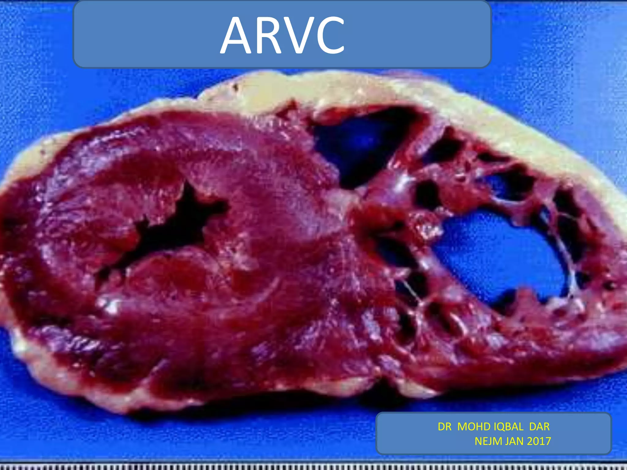
ARVC is a heritable heart muscle disorder that predominantly affects the right ventricle. It is caused by genetic defects in cardiac desmosomes, which are important for cell-to-cell adhesion. This leads to progressive loss of right ventricular myocardium and replacement by fibrofatty tissue. ARVC can cause dangerous ventricular arrhythmias and is a leading cause of sudden cardiac death in young people. Diagnosis involves imaging tests and electrocardiography to detect right ventricular structural abnormalities and arrhythmias.
Review of a rare but important cardiac dysplasia causing high mortality and morbidity in mostly young patients.
In this presentation, the epidemiology, pathophysiology, diagnosis and treatment of said disease is examines.
Review of a rare but important cardiac dysplasia causing high mortality and morbidity in mostly young patients.
In this presentation, the epidemiology, pathophysiology, diagnosis and treatment of said disease is examines.
Wolff–Parkinson–White syndrome (WPW) is one of several disorders of the conduction system of the heart that are commonly referred to as pre-excitation syndromes. WPW is caused by the presence of an abnormal accessory electrical conduction pathway between the atria and the ventricles. Electrical signals travelling down this abnormal pathway (known as the bundle of Kent) may stimulate the ventricles to contract prematurely, resulting in a unique type of supraventricular tachycardia referred to as an atrioventricular reciprocating tachycardia.The incidence of WPW is between 0.1% and 0.3% in the general population.Sudden cardiac death in people with WPW is rare (incidence of less than 0.6%), and is usually caused by the propagation of an atrial tachydysrhythmia (rapid and abnormal heart rate) to the ventricles by the abnormal accessory pathway.
Despite the advances in diagnostic methods and techniques for surgical treatment in the last two decades, aortic diseases remain a major cause of mortality and cardiovascular morbidity, challenging physicians and molecular biologists. It is believed that about 600 million years ago, during the Cambrian period, variant forms of life appeared, among them were the oxygen-producing cyano bacteria.
Lung Cancer: Artificial Intelligence, Synergetics, Complex System Analysis, S...Oleg Kshivets
RESULTS: Overall life span (LS) was 2252.1±1742.5 days and cumulative 5-year survival (5YS) reached 73.2%, 10 years – 64.8%, 20 years – 42.5%. 513 LCP lived more than 5 years (LS=3124.6±1525.6 days), 148 LCP – more than 10 years (LS=5054.4±1504.1 days).199 LCP died because of LC (LS=562.7±374.5 days). 5YS of LCP after bi/lobectomies was significantly superior in comparison with LCP after pneumonectomies (78.1% vs.63.7%, P=0.00001 by log-rank test). AT significantly improved 5YS (66.3% vs. 34.8%) (P=0.00000 by log-rank test) only for LCP with N1-2. Cox modeling displayed that 5YS of LCP significantly depended on: phase transition (PT) early-invasive LC in terms of synergetics, PT N0—N12, cell ratio factors (ratio between cancer cells- CC and blood cells subpopulations), G1-3, histology, glucose, AT, blood cell circuit, prothrombin index, heparin tolerance, recalcification time (P=0.000-0.038). Neural networks, genetic algorithm selection and bootstrap simulation revealed relationships between 5YS and PT early-invasive LC (rank=1), PT N0—N12 (rank=2), thrombocytes/CC (3), erythrocytes/CC (4), eosinophils/CC (5), healthy cells/CC (6), lymphocytes/CC (7), segmented neutrophils/CC (8), stick neutrophils/CC (9), monocytes/CC (10); leucocytes/CC (11). Correct prediction of 5YS was 100% by neural networks computing (area under ROC curve=1.0; error=0.0).
CONCLUSIONS: 5YS of LCP after radical procedures significantly depended on: 1) PT early-invasive cancer; 2) PT N0--N12; 3) cell ratio factors; 4) blood cell circuit; 5) biochemical factors; 6) hemostasis system; 7) AT; 8) LC characteristics; 9) LC cell dynamics; 10) surgery type: lobectomy/pneumonectomy; 11) anthropometric data. Optimal diagnosis and treatment strategies for LC are: 1) screening and early detection of LC; 2) availability of experienced thoracic surgeons because of complexity of radical procedures; 3) aggressive en block surgery and adequate lymph node dissection for completeness; 4) precise prediction; 5) adjuvant chemoimmunoradiotherapy for LCP with unfavorable prognosis.
Title: Sense of Smell
Presenter: Dr. Faiza, Assistant Professor of Physiology
Qualifications:
MBBS (Best Graduate, AIMC Lahore)
FCPS Physiology
ICMT, CHPE, DHPE (STMU)
MPH (GC University, Faisalabad)
MBA (Virtual University of Pakistan)
Learning Objectives:
Describe the primary categories of smells and the concept of odor blindness.
Explain the structure and location of the olfactory membrane and mucosa, including the types and roles of cells involved in olfaction.
Describe the pathway and mechanisms of olfactory signal transmission from the olfactory receptors to the brain.
Illustrate the biochemical cascade triggered by odorant binding to olfactory receptors, including the role of G-proteins and second messengers in generating an action potential.
Identify different types of olfactory disorders such as anosmia, hyposmia, hyperosmia, and dysosmia, including their potential causes.
Key Topics:
Olfactory Genes:
3% of the human genome accounts for olfactory genes.
400 genes for odorant receptors.
Olfactory Membrane:
Located in the superior part of the nasal cavity.
Medially: Folds downward along the superior septum.
Laterally: Folds over the superior turbinate and upper surface of the middle turbinate.
Total surface area: 5-10 square centimeters.
Olfactory Mucosa:
Olfactory Cells: Bipolar nerve cells derived from the CNS (100 million), with 4-25 olfactory cilia per cell.
Sustentacular Cells: Produce mucus and maintain ionic and molecular environment.
Basal Cells: Replace worn-out olfactory cells with an average lifespan of 1-2 months.
Bowman’s Gland: Secretes mucus.
Stimulation of Olfactory Cells:
Odorant dissolves in mucus and attaches to receptors on olfactory cilia.
Involves a cascade effect through G-proteins and second messengers, leading to depolarization and action potential generation in the olfactory nerve.
Quality of a Good Odorant:
Small (3-20 Carbon atoms), volatile, water-soluble, and lipid-soluble.
Facilitated by odorant-binding proteins in mucus.
Membrane Potential and Action Potential:
Resting membrane potential: -55mV.
Action potential frequency in the olfactory nerve increases with odorant strength.
Adaptation Towards the Sense of Smell:
Rapid adaptation within the first second, with further slow adaptation.
Psychological adaptation greater than receptor adaptation, involving feedback inhibition from the central nervous system.
Primary Sensations of Smell:
Camphoraceous, Musky, Floral, Pepperminty, Ethereal, Pungent, Putrid.
Odor Detection Threshold:
Examples: Hydrogen sulfide (0.0005 ppm), Methyl-mercaptan (0.002 ppm).
Some toxic substances are odorless at lethal concentrations.
Characteristics of Smell:
Odor blindness for single substances due to lack of appropriate receptor protein.
Behavioral and emotional influences of smell.
Transmission of Olfactory Signals:
From olfactory cells to glomeruli in the olfactory bulb, involving lateral inhibition.
Primitive, less old, and new olfactory systems with different path
These lecture slides, by Dr Sidra Arshad, offer a quick overview of physiological basis of a normal electrocardiogram.
Learning objectives:
1. Define an electrocardiogram (ECG) and electrocardiography
2. Describe how dipoles generated by the heart produce the waveforms of the ECG
3. Describe the components of a normal electrocardiogram of a typical bipolar leads (limb II)
4. Differentiate between intervals and segments
5. Enlist some common indications for obtaining an ECG
Study Resources:
1. Chapter 11, Guyton and Hall Textbook of Medical Physiology, 14th edition
2. Chapter 9, Human Physiology - From Cells to Systems, Lauralee Sherwood, 9th edition
3. Chapter 29, Ganong’s Review of Medical Physiology, 26th edition
4. Electrocardiogram, StatPearls - https://www.ncbi.nlm.nih.gov/books/NBK549803/
5. ECG in Medical Practice by ABM Abdullah, 4th edition
6. ECG Basics, http://www.nataliescasebook.com/tag/e-c-g-basics
Prix Galien International 2024 Forum ProgramLevi Shapiro
June 20, 2024, Prix Galien International and Jerusalem Ethics Forum in ROME. Detailed agenda including panels:
- ADVANCES IN CARDIOLOGY: A NEW PARADIGM IS COMING
- WOMEN’S HEALTH: FERTILITY PRESERVATION
- WHAT’S NEW IN THE TREATMENT OF INFECTIOUS,
ONCOLOGICAL AND INFLAMMATORY SKIN DISEASES?
- ARTIFICIAL INTELLIGENCE AND ETHICS
- GENE THERAPY
- BEYOND BORDERS: GLOBAL INITIATIVES FOR DEMOCRATIZING LIFE SCIENCE TECHNOLOGIES AND PROMOTING ACCESS TO HEALTHCARE
- ETHICAL CHALLENGES IN LIFE SCIENCES
- Prix Galien International Awards Ceremony
Knee anatomy and clinical tests 2024.pdfvimalpl1234
This includes all relevant anatomy and clinical tests compiled from standard textbooks, Campbell,netter etc..It is comprehensive and best suited for orthopaedicians and orthopaedic residents.
New Directions in Targeted Therapeutic Approaches for Older Adults With Mantl...i3 Health
i3 Health is pleased to make the speaker slides from this activity available for use as a non-accredited self-study or teaching resource.
This slide deck presented by Dr. Kami Maddocks, Professor-Clinical in the Division of Hematology and
Associate Division Director for Ambulatory Operations
The Ohio State University Comprehensive Cancer Center, will provide insight into new directions in targeted therapeutic approaches for older adults with mantle cell lymphoma.
STATEMENT OF NEED
Mantle cell lymphoma (MCL) is a rare, aggressive B-cell non-Hodgkin lymphoma (NHL) accounting for 5% to 7% of all lymphomas. Its prognosis ranges from indolent disease that does not require treatment for years to very aggressive disease, which is associated with poor survival (Silkenstedt et al, 2021). Typically, MCL is diagnosed at advanced stage and in older patients who cannot tolerate intensive therapy (NCCN, 2022). Although recent advances have slightly increased remission rates, recurrence and relapse remain very common, leading to a median overall survival between 3 and 6 years (LLS, 2021). Though there are several effective options, progress is still needed towards establishing an accepted frontline approach for MCL (Castellino et al, 2022). Treatment selection and management of MCL are complicated by the heterogeneity of prognosis, advanced age and comorbidities of patients, and lack of an established standard approach for treatment, making it vital that clinicians be familiar with the latest research and advances in this area. In this activity chaired by Michael Wang, MD, Professor in the Department of Lymphoma & Myeloma at MD Anderson Cancer Center, expert faculty will discuss prognostic factors informing treatment, the promising results of recent trials in new therapeutic approaches, and the implications of treatment resistance in therapeutic selection for MCL.
Target Audience
Hematology/oncology fellows, attending faculty, and other health care professionals involved in the treatment of patients with mantle cell lymphoma (MCL).
Learning Objectives
1.) Identify clinical and biological prognostic factors that can guide treatment decision making for older adults with MCL
2.) Evaluate emerging data on targeted therapeutic approaches for treatment-naive and relapsed/refractory MCL and their applicability to older adults
3.) Assess mechanisms of resistance to targeted therapies for MCL and their implications for treatment selection
Pulmonary Thromboembolism - etilogy, types, medical- Surgical and nursing man...VarunMahajani
Disruption of blood supply to lung alveoli due to blockage of one or more pulmonary blood vessels is called as Pulmonary thromboembolism. In this presentation we will discuss its causes, types and its management in depth.
Recomendações da OMS sobre cuidados maternos e neonatais para uma experiência pós-natal positiva.
Em consonância com os ODS – Objetivos do Desenvolvimento Sustentável e a Estratégia Global para a Saúde das Mulheres, Crianças e Adolescentes, e aplicando uma abordagem baseada nos direitos humanos, os esforços de cuidados pós-natais devem expandir-se para além da cobertura e da simples sobrevivência, de modo a incluir cuidados de qualidade.
Estas diretrizes visam melhorar a qualidade dos cuidados pós-natais essenciais e de rotina prestados às mulheres e aos recém-nascidos, com o objetivo final de melhorar a saúde e o bem-estar materno e neonatal.
Uma “experiência pós-natal positiva” é um resultado importante para todas as mulheres que dão à luz e para os seus recém-nascidos, estabelecendo as bases para a melhoria da saúde e do bem-estar a curto e longo prazo. Uma experiência pós-natal positiva é definida como aquela em que as mulheres, pessoas que gestam, os recém-nascidos, os casais, os pais, os cuidadores e as famílias recebem informação consistente, garantia e apoio de profissionais de saúde motivados; e onde um sistema de saúde flexível e com recursos reconheça as necessidades das mulheres e dos bebês e respeite o seu contexto cultural.
Estas diretrizes consolidadas apresentam algumas recomendações novas e já bem fundamentadas sobre cuidados pós-natais de rotina para mulheres e neonatos que recebem cuidados no pós-parto em unidades de saúde ou na comunidade, independentemente dos recursos disponíveis.
É fornecido um conjunto abrangente de recomendações para cuidados durante o período puerperal, com ênfase nos cuidados essenciais que todas as mulheres e recém-nascidos devem receber, e com a devida atenção à qualidade dos cuidados; isto é, a entrega e a experiência do cuidado recebido. Estas diretrizes atualizam e ampliam as recomendações da OMS de 2014 sobre cuidados pós-natais da mãe e do recém-nascido e complementam as atuais diretrizes da OMS sobre a gestão de complicações pós-natais.
O estabelecimento da amamentação e o manejo das principais intercorrências é contemplada.
Recomendamos muito.
Vamos discutir essas recomendações no nosso curso de pós-graduação em Aleitamento no Instituto Ciclos.
Esta publicação só está disponível em inglês até o momento.
Prof. Marcus Renato de Carvalho
www.agostodourado.com
Tom Selleck Health: A Comprehensive Look at the Iconic Actor’s Wellness Journeygreendigital
Tom Selleck, an enduring figure in Hollywood. has captivated audiences for decades with his rugged charm, iconic moustache. and memorable roles in television and film. From his breakout role as Thomas Magnum in Magnum P.I. to his current portrayal of Frank Reagan in Blue Bloods. Selleck's career has spanned over 50 years. But beyond his professional achievements. fans have often been curious about Tom Selleck Health. especially as he has aged in the public eye.
Follow us on: Pinterest
Introduction
Many have been interested in Tom Selleck health. not only because of his enduring presence on screen but also because of the challenges. and lifestyle choices he has faced and made over the years. This article delves into the various aspects of Tom Selleck health. exploring his fitness regimen, diet, mental health. and the challenges he has encountered as he ages. We'll look at how he maintains his well-being. the health issues he has faced, and his approach to ageing .
Early Life and Career
Childhood and Athletic Beginnings
Tom Selleck was born on January 29, 1945, in Detroit, Michigan, and grew up in Sherman Oaks, California. From an early age, he was involved in sports, particularly basketball. which played a significant role in his physical development. His athletic pursuits continued into college. where he attended the University of Southern California (USC) on a basketball scholarship. This early involvement in sports laid a strong foundation for his physical health and disciplined lifestyle.
Transition to Acting
Selleck's transition from an athlete to an actor came with its physical demands. His first significant role in "Magnum P.I." required him to perform various stunts and maintain a fit appearance. This role, which he played from 1980 to 1988. necessitated a rigorous fitness routine to meet the show's demands. setting the stage for his long-term commitment to health and wellness.
Fitness Regimen
Workout Routine
Tom Selleck health and fitness regimen has evolved. adapting to his changing roles and age. During his "Magnum, P.I." days. Selleck's workouts were intense and focused on building and maintaining muscle mass. His routine included weightlifting, cardiovascular exercises. and specific training for the stunts he performed on the show.
Selleck adjusted his fitness routine as he aged to suit his body's needs. Today, his workouts focus on maintaining flexibility, strength, and cardiovascular health. He incorporates low-impact exercises such as swimming, walking, and light weightlifting. This balanced approach helps him stay fit without putting undue strain on his joints and muscles.
Importance of Flexibility and Mobility
In recent years, Selleck has emphasized the importance of flexibility and mobility in his fitness regimen. Understanding the natural decline in muscle mass and joint flexibility with age. he includes stretching and yoga in his routine. These practices help prevent injuries, improve posture, and maintain mobilit
Report Back from SGO 2024: What’s the Latest in Cervical Cancer?bkling
Are you curious about what’s new in cervical cancer research or unsure what the findings mean? Join Dr. Emily Ko, a gynecologic oncologist at Penn Medicine, to learn about the latest updates from the Society of Gynecologic Oncology (SGO) 2024 Annual Meeting on Women’s Cancer. Dr. Ko will discuss what the research presented at the conference means for you and answer your questions about the new developments.
2. Arrhythmogenic right ventricular cardiomyopathy
(ARVC), also known as arrhythmogenic right
ventricular dysplasia, is a heritable heart muscle
disorder that predominantly affects the right ventricle.
Progressive loss of right ventricular myocardium and
its replacement by fibrofatty tissue is the pathological
hallmark of the disease.
3. ARVC is one of the leading causes of arrhythmic cardiac arrest
in young people and athletes.
Since the original report by Marcus and colleagues was
published in 1982, describing 24 affected patients, there have
been substantial advances in our understanding of the
pathogenesis, clinical manifestations, and long-term outcome
of the disorder.
The disease was initially designated as a dysplasia because it
was thought to be a congenital defect in the development of
the right ventricular myocardium.
4. The subsequent discovery that the disease is caused
by a genetic defect in the cardiac desmosomes has
led to its recognition as a cardiomyopathy and its
inclusion in the classification of cardiomyopathies by
the American Heart Association
5. The distinctive histopathological feature of ARVC is the loss of
right ventricular myocardium, with the substitution of fibrous
and fatty tissue.
Patchy inflammatory infiltrates (mainly T lymphocytes) are often
observed in association with dying myocytes, suggesting that the
pathologic process may be immunologically mediated.
Pathological Features
6. The fibrofatty scar tissue progresses from the epicardium
toward the endocardium and predominantly involves the right
ventricular free wall, resulting in wall thinning and aneurysmal
dilatation, which are typically localized in the inflow tract
(subtricuspid region), outflow tract (infundibular region), and
apex (“triangle of dysplasia”).In the typical form of ARVC, the
left ventricle is affected to a lesser extent than the right
ventricle; however, there are disease variants characterized by
equivalent or even predominant involvement of the left
ventricle.
7.
8. Mutations in the genes encoding desmosomal proteins, which
are important in cell-to-cell adhesion, play a key role in the
pathogenesis of ARVC.
The characterization of ARVC as a cell-adhesion disorder was
first suggested by a molecular genetic study involving patients
with Naxos disease.
This disease is an autosomal recessive cardiocutaneous
syndrome characterized by cosegregation of abnormalities
of the heart (ARVC), skin (palmoplantar keratosis), and hair
(woolly hair).
Molecular Genetic Features
10. The concurrence of epidermal and myocardial abnormalities
in Naxos disease is explained by the fact that these two types of
tissue have similar junction structures.
Mutations in the gene encoding plakoglobin (JUP) were the
first disease-causing variants to be identified in patients with
Naxos disease.
Plakoglobin is a major constituent of desmosomal complexes.
Mutations in genes encoding other desmosomal proteins were
subsequently shown to cause the more common
(nonsyndromic) autosomal dominant form of ARVC
11. These proteins include
Desmoplakin (DSP),
Plakophilin 2 (PKP2),
Desmoglein 2 (DSG2), and
Desmocollin 2 (DSC2).
A recessive mutation in the DSP gene was shown to cause
another cardiocutaneous syndrome, the Carvajal
syndrome(Keratoderma ,woolly hair and left sided
cardiomyopathy)
Autosomal dominant ARVC has also been linked to rare
pathogenic mutations in genes unrelated to cell-to-cell
junctional apparatus.
12.
13. Ultrastructural studies of myocardial-biopsy samples obtained from
patients with ARVC have shown intercalated disk remodeling with
abnormalities and loss of desmosomes. These findings support the
theory that genetically abnormal desmosomes lead to disruption of
intercellular junctions, with myocyte detachment and cell
death .
Mechanical uncoupling of myocytes may be aggravated by physical
exercise, which increases pressure, afterload, and wall stress to a
disproportionately greater extent in the right ventricle than in the left
Pathophysiological Features
14. In addition to being specialized structures that provide
mechanical cell attachment, desmosomes are important
mediators of intracellular and intercellular signal transduction.
Elegant immunohistochemical studies have shown that
the mutant form of the plakoglobin protein fails to integrate
into desmosomes and shifts from intercalated disks to cytosol
and nuclear pools, where it causes changes in nuclear signaling
and transcriptional activity, in particular through pathways
regulated by the protein β-catenin. Studies in DSP-deficient
mice indicate that the inhibition of the canonical Wnt–β-
catenin signaling pathway induced by nuclear translocation of
plakoglobin may increase the expression of adipogenic and
fibrogenic genes and contribute to the development of
fibrofatty myocardial scarring
15. The fibrofatty tissue that replaces myocardium in ARVC is thought to
contribute to the development of ventricular arrhythmias by slowing
intraventricular conduction and acting as a substrate for arrhythmias
through a scar-related macroreentry mechanism, similar to that
observed after myocardial infarction.
Life-threatening ventricular arrhythmias in ARVC may also be the
result of mechanisms operating at the molecular and cellular levels.
Desmosomes, sodium channels, and gap-junction proteins interact
synergistically to regulate adhesion, excitability, and coupling of
myocytes; this coordinated network of proteins located at the
intercalated disks has been termed the “connexome.”
Loss of expression of desmosomal proteins may cause (or contribute
to) potentially fatal arrhythmias by inducing gap-junction remodeling,
with reduction of total content and substantial redistribution of the
gap-junction protein connexin , and decreasing the amplitude and
kinetics of the sodium current.
16. Sports activity increases the risk of sudden cardiac death
among adolescents and young adults with ARVC. Physical
exercise may aggravate mechanical uncoupling of myocytes; it
may also trigger malignant ventricular arrhythmias and is a
critical environmental factor in the promotion of the
development and progression of the disease. The key role of
exercise as a disease modifier was suggested by both
experimental studies of transgenic plakoglobin-deficient mice
and clinical studies of affected patients with and those without
desmosomal gene mutations.
17. The prevalence of ARVC is estimated to range from 1 case in 5000 persons in
the general population to 1 in 2000 in some European countries
such as Italy and Germany.
Approximately 50% of affected patients have a positive family history, but both
incomplete penetrance and limited phenotypic expression are common and
probably account for underestimation of the prevalence of familial disease.
The disease is typically transmitted with an autosomal dominant pattern of
inheritance, although rare auto-somal recessive forms have been described.
The disease is more malignant in men than in women, a finding that can be
explained either by a direct influence of sex hormones on the mechanisms
involved in the phenotypic expression of the disease or by sex-based
differences in the amount or intensity of exercise.
Epidemiology
18. Clinical Presentation and Natural History
ARVC typically becomes clinically apparent between the second and fourth
decades of life. Clinically overt disease is preceded by a preclinical
phase, which is characterized by minimal or no structural abnormalities
(“concealed disease”).
Sudden cardiac death may be the first clinical manifestation of the disease. In
a study in the Veneto region of Italy, 20% of deaths in young people and
athletes were caused by previously undiagnosed ARVC.
19. The most common clinical presentation is palpitations or effort-induced
syncope in an adolescent or young adult, with T-wave inversion in
the right precordial leads (V1 through V4) on the electrocardiogram,
ventricular arrhythmias with a left bundle-branch block pattern, and right
ventricular abnormalities on imaging tests.
Electrocardiographic depolarization abnormalities, which reflect defective
conduction through the diseased right ventricular myocardium, may also be
present .
Ventricular arrhythmias range from frequent premature ventricular beats to
ventricular tachycardia, which may degenerate into ventricular fibrillation;
the arrhythmias are characteristically triggered or worsened by adrenergic
stimulation.
20. The 12-lead standard electrocardiogram in Panel A shows a repolarization abnormality that is characteristic of ARVC, with negative T waves
in leads V1 through V4 and depolarization changes, including low QRS voltages (<0.5 mV) in the limb leads and prolongation of the right
precordial QRS complex, with a delayed S-wave upstroke. The terminal activation duration (TAD), which is the interval between the nadir of
the S wave and the end of all depolarization deflections, is prolonged, at 80 msec, in lead V1 (inset); the normal value is less than 55 msec.
Panel B shows an example of “epsilon waves” (i.e., small-amplitude distinct potentials between the end of the QRS complex and the beginning
of the T wave) in leads V1 and V2. This is a highly specific electrocardiographic abnormality that is seen in a minority of patients
with advanced disease. The 12-lead standard electrocardiogram in Panel C shows ventricular tachycardia (160 beats per minute) with a
left bundle-branch block pattern.
21. Diagnostic alterations of the right ventricle on imaging studies consist
of global dilatation and dysfunction and regional wall-motion abnormalities
such as systolic akinesia or dyskinesia or diastolic bulging
The left ventricle and the septum are usually involved to a lesser extent, if at
all.
Cardiac magnetic resonance imaging (MRI) has become the preferred
imaging technique because it combines the evaluation of structural and
functional ventricular abnormalities with noninvasive tissue
characterization with the use of late gadolinium enhancement, which
provides information about the presence and amount of fibrofatty
myocardial scarring.
22. The two-dimensional echocardiogram, parasternal long-axis view (PLAX), in Panel D shows dilatation
of the right ventricular outflow tract (RVOT), at 38 mm (normal value, <32 mm).
The cardiac MRI scan (systolic frame of right ventricular two-chamber long-axis view on cine sequences) in
Panel E shows an aneurysm of the RVOT (solid arrows) and multiple sacculations of the inferior and apical
regions (open arrows). RA denotes right atrium, and RV right ventricle.
23. End-stage right ventricular or biventricular pump failure may develop in
patients with longstanding disease. Genotype–phenotype correlation
studies and the increasing use of cardiac MRI have identified clinical variants
characterized by early left ventricular involvement, especially among
patients with DSP gene mutations, which may either parallel or exceed the
severity of right ventricular disease.
Clinical features of left-sided variants include inverted T waves in the
inferolateral leads, ventricular arrhythmias with a right bundle-branch block,
left ventricular dilatation and dysfunction, and late gadolinium
enhancement of the left ventricular wall with a subepicardial or
midmyocardial distribution.
These findings support the concept that ARVC can be a biventricular muscle
disease (i.e., a disease involving the myocardium of both ventricles) and
have led some to use the broader term “arrhythmogenic cardiomyopathy.”
24. Clinical Diagnosis
To standardize the clinical diagnosis of ARVC, in 1994 an international task force
proposed guidelines in the form of a qualitative scoring system with major and
minor criteria.
In 2010, the task force revised the guidelines to improve diagnostic sensitivity,
mostly for the clinical screening of family members, by providing quantitative
criteria for diagnosing right ventricular abnormalities and adding molecular
genetic criteria (Table 1).
However, the diagnosis remains problematic because of the low specificity of
electrocardiographic abnormalities, multiple causes of right ventricular
arrhythmias, difficulties in the use of imaging to assess right ventricular structure
and function, and the sometimes puzzling
results of genetic testing.
25.
26. The diagnosis is particularly challenging in children, because clinical
manifestations of early ARVC are subtle. Cardiac MRI has proved to be
more sensitive than echocardiography for detecting early ventricular
dilatation and dysfunction in children. Conditions that may be difficult to
differentiate from ARVC include
• idiopathic right ventricular outflow-tract tachycardia,
• cardiac sarcoidosis, and
• congenital heart disease leading to right ventricular volume overload.
Biventricular variants of the disease with severe left ventricular involvement
may be indistinguishable from dilated cardiomyopathy.
The difficult differential diagnosis, together with referral bias, may account
for the discrepancies in the reported incidence of heart failure in patients
with ARVC.
27. On the basis of pooled data from major studies of molecular genetic
screening for desmosomal gene mutations, the estimated overall rate of
successful genotyping among patients meeting the diagnostic criteria for
ARVC established by the international task force is approximately
50%.32,33,53,54 The most commonly affected gene is PKP2 (in 10 to 45% of
patients), followed by DSP (10 to 15%), DSG2 (7 to 10%), and DSC2 (2%).
Screening of nondesmosomal genes only marginally affects the detection
rate for mutations.
Preclinical Diagnosis by Genotyping
28. Clinically, genotyping is most often used to identify a mutation that is
considered likely to be causal in a pro-band who fulfills phenotypic
diagnostic criteria, and thus to identify gene carriers among family members
by means of mutation specific genetic testing. Genotyping to confirm the
diagnosis in an isolated patient with a borderline or questionable phenotype
is not indicated on a routine basis.
The true prevalence of disease causing mutations has yet to be determined.
Therefore, a negative genetic test does not exclude the possibility that the
phenotype is due to a mutation of an unknown disease-causing gene.
29. On the other hand, the interpretation of an apparently positive genetic test
for ARVC is made more challenging by the difficulty in differentiating
causative mutations, especially missense mutations, from nonpathogenetic
variants and polymorphisms, mainly because of the lack of
functional or biologic studies that confirm the pathogenetic effects of gene
variants.
It has been reported that 16% of healthy persons have missense
mutations in one of the major ARVC susceptibility genes.
The prognostic value of genotyping also remains to be elucidated. Data
indicate that patients with multiple desmosomal gene mutations are likely to
have a more severe phenotype and may have an increased lifetime risk of
malignant arrhythmias and sudden cardiac death.
30. The results of commercially available genetic tests for ARVC should be evaluated
cautiously, and genetic counseling is recommended for assistance in test
interpretation.Otherwise, critical information regarding the complexity of
genetic data and their limitations for clinical diagnosis and prognosis may not be
made clear when the results of such tests are reported.
31. Risk Stratification
The clinical course of ARVC is characterized by the occurrence of arrhythmic
events, which can cause
sudden death, and
the impairment of biventricular systolic function, which can lead to death from
heart failure.
The estimated overall mortality varies among studies, ranging from 0.08 to 3.6%
per year.
The mortality was initially overestimated because it was based on studies at
tertiary referral centers, which predominantly included high-risk patients.
Recent studies of community-based patient cohorts have shown that the long-
term outcome for treated index patients and family members is favorable (annual
mortality, <1%).
Prognosis and Treatment
32. The prognosis for patients with ARVC depends largely on the severity of
arrhythmias and ventricular dysfunction .
Prior cardiac arrest due to ventricular fibrillation and sustained ventricular
tachycardia are the most important predictors of life-threatening arrhythmic
events during follow-up.
Major risk factors include unexplained syncope, nonsustained ventricular
tachycardia on ambulatory monitoring or exercise testing, and severe systolic
dysfunction of the right ventricle, left ventricle, or both.
Several minor risk factors have been identified, but their association with an
unfavorable out-come is based on either limited scientific evidence or conflicting
data.
Although intra-cardiac electrophysiological testing has traditionally been used to
assess the risk of ventricular arrhythmias, the prognostic value of ventricular
tachycardia or ventricular fibrillation induced by programmed ventricular
stimulation in patients with asymptomatic ARVC remains unclear.
33.
34. The aims of clinical management of ARVC are to reduce the risk of sudden cardiac death and improve
the quality of life by alleviating arrhythmic and heart-failure symptoms.
Restriction from intense sports activity is regarded as an important preventive tool for both healthy gene
carriers and clinically affected persons in order to protect them from the risk of exercise-related malignant
arrhythmic events and disease development or progression.
The available evidence indicates that family members with a negative phenotype (either healthy gene
carriers or those with an unknown genotype) do not need any specific treatment other than sports
restriction; however, lifelong clinical assessment with the use of noninvasive tests at least every 2 years is
warranted because of the age-related penetrance and progressive nature of ARVC.
Therapy
35. Despite limited supportive data, beta-blockers are currently recommended for all
clinically affected persons, for both prevention of arrhythmias and reduction of
right ventricular wall stress.
In patients with ventricular arrhythmias, antiarrhythmic drug therapy offers the
potential to ameliorate symptoms, although there is no proof that it confers
protection against sudden cardiac death.
Amiodarone, alone or in association with beta-blockers, and sotalol are the most
effective drugs, combining the synergistic effects of class III antiarrhythmic
properties and betaadrenergic blockade.
The potential for serious cumulative toxic effects precludes long-term therapy
with amiodarone, especially in younger patients.
36. Catheter ablation is a therapeutic option for patients who have episodes of
sustained, monomorphic ventricular tachycardia (Fig. 4). However,
it should be regarded as a palliative rather than curative therapeutic approach
because of the high frequency of subsequent recurrences of ventricular
tachycardia and the unproven efficacy of ablation as a means of preventing
sudden cardiac death.
37. The poor long-term outcome has been attributed to the progressive nature of
ARVC, which leads to the development of multiple arrhythmogenic
foci over time. The epicardial location of some ventricular tachycardia reentry
circuits, which reflects the propensity of ARVC lesions to originate and progress
from the epicardium, may also explain the failure of conventional
endocardial mapping and catheter ablation.Several studies have shown the
feasibility and efficacy of epicardial catheter ablation for patients in whom one or
more endocardial procedures have been unsuccessful.
38.
39. Although randomized trials of defibrillator therapy have not been performed,
data from observational studies have consistently shown that it is effective and
safe. Patients who benefit most from defibrillators are those who have had an
episode of ventricular fibrillation or sustained ventricular tachycardia. It remains
uncertain whether defibrillator therapy is appropriate for primary prevention of
sudden cardiac death among patients with one or more risk factors and no prior
major arrhythmic events.
40. In asymptomatic patients with no risk factors and in healthy gene carriers, there
is generally no indication for prophylactic defibrillator implantation
because of the low risk of arrhythmias and the significant risk of device- and
electrode-related complications during long-term follow-up (estimated rate, 3.7%
per year).
It has become apparent that defibrillators may be inappropriately implanted in
patients with a false diagnosis of ARVC based on misinterpretation of cardiac MRI
studies.
41. Patients in whom right or left heart failure develops are treated with standard
pharmacologic therapy, including angiotensin-converting– enzyme inhibitors,
angiotensin II receptor blockers, beta-blockers, and diuretics.Therapy
with oral anticoagulants is reserved for patients with atrial fibrillation or
thromboembolic complications. Cardiac transplantation is the ultimate
therapy for patients with untreatable arrhythmias(e.g., incessant storms of
ventricular tachycardia or fibrillation) or congestive heart failure that is refractory
to pharmacologic and nonpharmacologic therapies.
42. Current therapeutic approaches to ARVC are palliative and partially alleviate
symptoms and the risk of sudden cardiac death but do not prevent
the development or progression of the disease process. A definitive curative
treatment will require a deeper knowledge of the biologic mechanisms and
environmental factors involved in the pathogenesis of ARVC.
A recent observation concerns a small molecule designated SB216763, which is
an activator of the Wnt signaling pathway. This molecule has been shown to
prevent or reverse phenotypic manifestations of ARVC induced by overexpression
of defective plakoglobin in a zebrafish model, as well as in rat cardiac myocytes.
Although this drug is of interest as a potential mechanism-based therapy of ARVC,
it has not yet been studied in humans.