This document summarizes an artificial neural network (ANN) based system called ANN-C3 for cancer cell classification using medical images. The system performs image pre-processing, segmentation using Harris corner detection and region growing, feature extraction of Tamura texture features, and classification using a neural network ensemble. Segmentation detects threshold points using Harris corner detection and performs region growing from these seed points. Feature extraction converts the image data into numerical form using Tamura texture features that capture variations in illumination and surfaces that human vision and surgeons use to differentiate cancerous and non-cancerous cells. The neural network is trained on a large set of labeled data to accurately classify cells.
Lung Cancer Detection on CT Images by using Image Processingijtsrd
This project is mainly based on image processing technique. In this work MATLAB have been used through every procedure made. Image processing techniques are widely use in bio-medical sector. The objective of our work is noise removal operation, thresholding, gray scale imaging, histogram equalization, texture segmentation, and morphological operation. Detection of lung cancer from computed tomography (CT) images is done by using MATLAB software. By using these methods the work has been done on CT images and the final tumor area has been shown with pixel values. Bindiya Patel | Dr. Pankaj Kumar Mishra | Prof. Amit Kolhe"Lung Cancer Detection on CT Images by using Image Processing" Published in International Journal of Trend in Scientific Research and Development (ijtsrd), ISSN: 2456-6470, Volume-2 | Issue-3 , April 2018, URL: http://www.ijtsrd.com/papers/ijtsrd11674.pdf http://www.ijtsrd.com/engineering/electronics-and-communication-engineering/11674/lung-cancer-detection-on-ct-images-by-using-image-processing/bindiya-patel
Lung Cancer Detection using Machine Learningijtsrd
Modern three dimensional 3 D medical imaging offers the potential and promise for major advances in science and medicine as higher fidelity images are produced. Due to advances in computer aided diagnosis and continuous progress in the field of computerized medical image visualization, there is need to develop one of the most important fields within scientific imaging. From the early basis report on cancer patients it has been seen that a greater number of people die of lung cancer than from other cancers such as colon, breast and prostate cancers combined. Lung cancer are related to smoking or secondhand smoke , or less often to exposure to radon or other environmental factors that’s why this can be prevented. But still it is not yet clear if these cancers can be prevented or not. In this research work, approach of segmentation, feature extraction and Convolution Neural Network CNN will be applied for locating, characterizing cancer portion. Harpreet Singh | Er. Ravneet Kaur | "Lung Cancer Detection using Machine Learning" Published in International Journal of Trend in Scientific Research and Development (ijtsrd), ISSN: 2456-6470, Volume-4 | Issue-6 , October 2020, URL: https://www.ijtsrd.com/papers/ijtsrd33659.pdf Paper Url: https://www.ijtsrd.com/computer-science/computer-architecture/33659/lung-cancer-detection-using-machine-learning/harpreet-singh
This document discusses brain tumor segmentation from MRI images using fuzzy c-means clustering. It begins with an introduction to brain tumors and MRI imaging. Next, it reviews existing methods for brain tumor segmentation such as thresholding, region growing, and clustering. It then discusses preprocessing MRI images, including converting images to grayscale and filtering. Finally, it describes fuzzy c-means clustering, which is an unsupervised learning technique used to segment and classify pixels in MRI images to detect tumor regions. The goal is to develop an accurate and automated method for brain tumor segmentation to assist medical experts.
Lung Cancer Detection using Image Processing TechniquesIRJET Journal
This document presents a technique for detecting lung cancer in x-ray images using image processing. It involves enhancing images using Gabor filtering, segmenting images using marker-controlled watershed segmentation, and extracting features using binarization and masking. The key steps are collecting lung x-ray images, enhancing quality using Gabor filtering, segmenting regions of interest using watershed segmentation, extracting pixel counts and mask features, and classifying images as normal or abnormal based on these features. The goal is to enable early detection of lung cancer through automated analysis of medical images.
IRJET- Lung Diseases using Deep Learning: A Review PaperIRJET Journal
This document reviews research on using deep learning techniques to detect lung diseases from medical images. It first provides background on lung anatomy and common lung diseases like pneumonia, tuberculosis, and lung cancer. Feature extraction and classification algorithms are important for automated detection. Several studies are summarized that used techniques like convolutional neural networks (CNNs) and support vector machines (SVMs) with shape, texture, and focal features to classify chest x-rays and CT scans as normal or abnormal. Deep learning approaches achieved higher accuracy than traditional "bag of features" methods. Overall, CNNs showed potential for developing high-performance automated lung disease detection systems.
IJRET : International Journal of Research in Engineering and Technology is an international peer reviewed, online journal published by eSAT Publishing House for the enhancement of research in various disciplines of Engineering and Technology. The aim and scope of the journal is to provide an academic medium and an important reference for the advancement and dissemination of research results that support high-level learning, teaching and research in the fields of Engineering and Technology. We bring together Scientists, Academician, Field Engineers, Scholars and Students of related fields of Engineering and Technology.
IJRET : International Journal of Research in Engineering and Technology is an international peer reviewed, online journal published by eSAT Publishing House for the enhancement of research in various disciplines of Engineering and Technology. The aim and scope of the journal is to provide an academic medium and an important reference for the advancement and dissemination of research results that support high-level learning, teaching and research in the fields of Engineering and Technology. We bring together Scientists, Academician, Field Engineers, Scholars and Students of related fields of Engineering and Technology
Brain tumor detection and localization in magnetic resonance imagingijitcs
A tumor also known as neoplasm is a growth in the abnormal tissue which can be differentiated from the
surrounding tissue by its structure. A tumor may lead to cancer, which is a major leading cause of death and
responsible for around 13% of all deaths world-wide. Cancer incidence rate is growing at an alarming rate
in the world. Great knowledge and experience on radiology are required for accurate tumor detection in
medical imaging. Automation of tumor detection is required because there might be a shortage of skilled
radiologists at a time of great need. We propose an automatic brain tumor detectionand localization
framework that can detect and localize brain tumor in magnetic resonance imaging. The proposed brain
tumor detection and localization framework comprises five steps: image acquisition, pre-processing, edge
detection, modified histogram clustering and morphological operations. After morphological operations,
tumors appear as pure white color on pure black backgrounds. We used 50 neuroimages to optimize our
system and 100 out-of-sample neuroimages to test our system. The proposed tumor detection and localization
system was found to be able to accurately detect and localize brain tumor in magnetic resonance imaging.
The preliminary results demonstrate how a simple machine learning classifier with a set of simple
image-based features can result in high classification accuracy. The preliminary results also demonstrate the
efficacy and efficiency of our five-step brain tumor detection and localization approach and motivate us to
extend this framework to detect and localize a variety of other types of tumors in other types of medical
imagery.
Application of-image-segmentation-in-brain-tumor-detectionMyat Myint Zu Thin
This document discusses applications of image segmentation in brain tumor detection. It begins by defining brain tumors and different types. It then discusses various image segmentation methods that can be used for brain tumor segmentation, including k-means clustering, region-based watershed algorithm, region growing, and active contour methods. It demonstrates how these methods can be implemented in Python for segmenting tumors from MRI images. The document also discusses computer-aided diagnosis systems and the roles of artificial intelligence and machine learning in medical image analysis and cancer diagnosis using image processing.
CANCER CELL DETECTION USING DIGITAL IMAGE PROCESSINGkajikho9
The document presents a lung cancer detection system using digital image processing techniques. It discusses lung anatomy and types of lung cancer. The system involves image capture, pre-processing using enhancement filters like Gabor and FFT, segmentation using thresholding and watershed approaches. Feature extraction is done using binarization and masking to detect cancer presence. The system helps in early detection of lung cancer to reduce mortality.
Image segmentation is still an active reason of research, a relevant research area
in computer vision and hundreds of image segmentation techniques have been proposed by
the researchers. All proposed techniques have their own usability and accuracy. In this paper
we are going present a review of some best lung nodule existing detection and segmentation
techniques. Finally, we conclude by focusing one of the best methods that may have high
level accuracy and can be used in detection of lung very small nodules accurately.
Image Segmentation and Identification of Brain Tumor using FFT Techniques of ...IDES Editor
The image processing tools are extensively used on
the development of new algorithms and mathematical tools
for the advanced processing of medical and biological images.
Given an MRI scan, first segment the tumor region in the
MRI brain image and study the pixel intensity values. A
detailed procedure using Matlab script is written to extract
tumor region in CT scan Brain Image and MRI Scan Brain
Image. MRI Scan has higher resolution and easier
identification compare to CT scan Brain image. Fast Fourier
Transform is used here to study the tumor region of MRI
Brain Image in terms of its pixel intensity. Types of FFT like
Zero padded FFT, Windowed FFT are used to study the signal
converted from the MRI Brain Image. It is found that lesser
spectral leakage for Zero Padded Windowed FFT than other
Types of FFT and hence the tumor cell identification is easier
than other methods. Finally higher pixel intensity values of
the cells gives identification of presence and activeness of
tumor cells.
DETECTING BRAIN TUMOUR FROM MRI IMAGE USING MATLAB GUI PROGRAMMEIJCSES Journal
Engineers have been actively developing tools to detect tumors and to process medical images. Medical image segmentation is a powerful tool that is often used to detect tumors. Many scientists and researchers are working to develop and add more features to this tool. This project is about detecting Brain tumors from MRI images using an interface of GUI in Matlab. Using the GUI, this program can use various combinations of segmentation, filters, and other image processing algorithms to achieve the best results.
We start with filtering the image using Prewitt horizontal edge-emphasizing filter. The next step for detecting tumor is "watershed pixels." The most important part of this project is that all the Matlab programs work with GUI “Matlab guide”. This allows us to use various combinations of filters, and other
image processing techniques to arrive at the best result that can help us detect brain tumors in their early stages.
Performance analysis of automated brain tumor detection from MR imaging and CT scan using basic image processing techniques based on various hard and soft computing has been performed in our work. Moreover, we applied six traditional classifiers to detect brain tumor in the images. Then we applied CNN for brain tumor detection to include deep learning method in our work. We compared the result of the traditional one having the best accuracy (SVM) with the result of CNN. Furthermore, our work presents a generic method of tumor detection and extraction of its various features.
An overview of automatic brain tumor detection frommagnetic resonance imagesMangesh Lingampalle
The document discusses several techniques for automatically detecting brain tumors from magnetic resonance (MR) images. It begins with an overview of MR imaging and challenges of manual tumor detection. Several existing techniques are then summarized, including thresholding-based methods, fuzzy classification with deformable models, using wavelets and statistics to segment tissues, feature extraction with Adaboost classification, and color-converted k-means clustering. The document proposes a technique using undecimated wavelet transform (UDWT) and Gabor filters for preprocessing, followed by morphological operations and parameter analysis to detect tumors. Automatic detection techniques could help address limitations of manual detection and improve diagnosis of brain tumors.
Brain tumor classification using artificial neural network on mri imageseSAT Journals
Abstract
In this paper, an attempt has been made to summarize the multi-resolution transformation and the different classifiers useful to
analyze the brain tumor using MRI. X-ray, MRI, Ultrasound etc. are different techniques used to scan brain tumor images.
Radiologist prefers MRI to get detail information about tumor to help him diagnoses. In this paper we have used MRI of brain
tumor for analysis. We have used Digital image processing tool for detection of the tumor. The identification, detection and
classification of brain tumor have been done by extracting features from MRI with the help of wavelet transformation. The MRI of
brain tumor is classified into two categories normal and abnormal brain. In this work Digital image processing has been used as
a tool for getting clear and exact details about tumor in earlier stages. This helps the physicians and practitioners for diagnoses.
Key word – Brain tumor, Wavelet transform, segmentation.
IRJET - Classification of Cancer Images using Deep LearningIRJET Journal
This document presents a methodology for classifying breast cancer histopathology images using deep learning. Specifically, it aims to classify images as either invasive ductal carcinoma (IDC) or non-IDC using a convolutional neural network (CNN) model. The proposed methodology involves preprocessing the images, building a CNN with convolutional, pooling and fully connected layers, training the model on labeled image data, and using the trained model to classify new images as IDC or non-IDC. The goal is to develop an automated system for early and accurate detection of breast cancer subtypes to improve diagnosis and patient outcomes.
IRJET- Image Processing based Lung Tumor Detection System for CT ImagesIRJET Journal
This document presents a method for detecting lung tumors in CT scan images using image processing techniques. The proposed method involves preprocessing images using median filtering for noise removal and contrast adjustment for enhancement. The lungs are then segmented from the images using mathematical morphology. Geometric and textural features are extracted from the segmented region of interest and used as input for an SVM classifier to detect lung cancer. The methodology was tested on a dataset from The Cancer Imaging Archive and was able to successfully detect lung tumors in CT images.
The document describes a study that used convolutional neural networks (CNNs) to detect brain tumors in MRI images. Three CNN models were developed and their performance was evaluated using metrics like accuracy, precision, recall, F1-score, and confusion matrices. Model 3 achieved the highest test accuracy of 94% for tumor detection. In total, over 2000 MRI images were used in the study after data augmentation. The CNN models incorporated convolution, pooling, and fully connected layers to analyze image features and classify tumors. This research demonstrates that CNNs can accurately detect brain tumors in medical images.
A Survey on Segmentation Techniques Used For Brain Tumor DetectionEditor IJMTER
In recent years Brain tumor is one of the most commonly found causes for death among
children and adults. Early detection of tumor is a must in order to reduce the death rate. For tumor
detection various image techniques can be used. In this paper we mainly concentrate on the images
obtained from MRI scans. In MRI images, the tumor may appear clearly, but for further treatment
the physician need to be a qualified and well experienced person. In order to help the radiologist in
detection computer-aided diagnosis was developed. The generation of a CAD system consists of
several processes and among them segmentation is considered to the most important process. Image
Segmentation is a process of partitioning an image into multiple segments. The main objective of
segmentation is to represent the image into a simplified form so as to increase the efficiency and
accuracy of the system. Therefore the segmentation of brain tumor can be considered as an important
role in the medical image process. Hence in this paper we concentrate on the recently used
segmentation techniques for the detection of tumor using MRI images.
IRJET - Lung Cancer Detection using GLCM and Convolutional Neural NetworkIRJET Journal
This document summarizes a research paper that proposes a method for detecting lung cancer using CT scan images with convolutional neural networks. The method involves preprocessing images using median filtering to remove noise, segmenting images using k-means clustering, extracting features using gray-level co-occurrence matrix, and classifying images using convolutional neural networks. The researchers achieved 96% accuracy in classifying tumors as malignant or benign, which is more accurate than traditional neural network methods.
Brain Tumor Segmentation and Extraction of MR Images Based on Improved Waters...IOSR Journals
This document summarizes a research paper about segmenting and extracting brain tumors from MR images using an improved watershed transform technique. It first preprocesses the MR images using techniques like edge enhancement to improve image quality. It then applies a marker-controlled watershed segmentation using foreground and background markers to avoid oversegmentation. The watershed transform is further improved by removing noise, adjusting pixel values, and introducing neighborhood relations between boundaries. Finally, mathematical morphology operations like erosion, dilation, opening and closing are used to get clear edges of the extracted brain tumor in the MR image.
Neural Network Based Brain Tumor Detection using MR ImagesAisha Kalsoom
This document outlines various techniques for detecting brain tumors using neural networks and magnetic resonance imaging (MRI). It discusses how Hopfield neural networks, multiparameter feature blocks, Markov random field segmentation, and adaptive spatial fuzzy clustering algorithms can be used for tumor detection and segmentation. The proposed research work involves preprocessing MRI images using adaptive filters, analyzing the images through segmentation, feature extraction and enhancement, and then using an artificial neural network for tumor detection.
Tumor Detection from Brain MRI Image using Neural Network Approach: A ReviewIRJET Journal
This document reviews using neural networks to detect tumors in brain MRIs. It discusses how MRI is commonly used to diagnose soft tissue issues and analyze conditions like trauma and strokes. The paper proposes a methodology for brain tumor detection that includes image acquisition, pre-processing, enhancement, thresholding, and morphological operations using MATLAB. A neural network approach is also presented. The conclusions state that neural networks can help detect, classify, segment, and visualize brain tumors in MRI images with ease and accuracy.
IRJET - Detection of Brain Tumor from MRI Images using MATLABIRJET Journal
This document presents a method for detecting brain tumors in MRI images using MATLAB. It involves pre-processing the MRI images to reduce noise and enhance contrast. Thresholding and watershed segmentation are then used to segment the images and isolate the tumor region. Morphological operations like erosion and dilation are applied post-segmentation to extract the tumor boundaries. The algorithm is tested on sample MRI images and is able to accurately detect tumors in all cases. The automated method provides faster and more consistent tumor detection compared to manual segmentation and reduces processing time.
The document describes a study that aims to detect brain tumors and edema in MRI images using MATLAB. It discusses how MRI is commonly used to identify brain anomalies. The proposed methodology uses basic image processing techniques in MATLAB, including preprocessing, enhancement, segmentation, and morphological operations to detect and segment tumors and edema. The final output highlights the boundaries between tumors and edema superimposed on the original MRI image to aid physicians in diagnosis and surgical planning.
IRJET- A New Strategy to Detect Lung Cancer on CT ImagesIRJET Journal
This document presents a new strategy for detecting lung cancer on CT images using image processing techniques. It involves acquiring CT scan images, preprocessing the images through techniques like grayscale conversion and Gabor filtering, segmenting the images using adaptive thresholding, extracting regions of interest through feature extraction methods like GLCM, and classifying images as cancerous or normal using support vector machines (SVM) and backpropagation neural networks (BPNN). The methodology achieves 96.32% accuracy for SVM and 83.07% accuracy for BPNN in detecting lung cancer from CT images.
Yes the robots in Ex Machina are a decade or two away (we believe). But artificial intelligence is all around us. Our world is architected by algorithms we don't understand, and as marketers, few of us use. This presentation looks at the modern approach to AI, with examples from everyday life. It digs into the algorithms that run our lives, and that shape our perceptions and priorities. It looks at the five tribes of machine learning to help you gain a better understanding of what Artificial Intelligence is, going beyond the wiki definition.
This document discusses using an artificial neural network to forecast power loads by taking the University of Lagos as a sample space. It involves gathering and arranging historical load data, determining an appropriate network type and topology, training the network using an algorithm, and analyzing the results to test the network's accuracy in predicting loads. The methodology includes randomizing and tagging the training data, experimenting to determine the network topology, training with cross-validation, and performing sensitivity and mean squared error analysis on the network.
This document discusses artificial intelligence and robotics. It covers:
- AI is entering the third and final stage of technological evolution involving automation and replicating human senses.
- Video games are a major area of experimentation for AI and what happens in this industry should be closely watched.
- Those controlling large data sets, like Google and Facebook, stand to be the likely winners in AI unless new business models are invented, as data is critical for training AI systems.
Artificial Neural Network Based Closed Loop Control of Multilevel InverterIJMTST Journal
Multi level inverters are gaining attraction because of the inherent advantages like low switching losses and less voltage stress which results in low filter cost. The common techniques that are available for switching the multi level inverters are based on sinusoidal pulse width modulation and using conventional PI based controllers, hysteresis based controllers. These controllers suffer with slow response time this makes usage of multi level inverters in custom power devices difficult. Because custom power devices require fast acting controller action which can be achieved by intelligent controllers. In this project artificial neural network based modulation scheme is designed and implemented for a cascaded H bridge inverter. The response time of controller for different operating power factors of the load are compared with conventional PI controllers and are presented. The developed control technique is developed by using Sim Power Systems Block set of MATLAB/SIMULINK Release R2015a.
Artificial inteligence and neural networksMireya Mendez
El documento describe la inteligencia artificial y las redes neuronales artificiales. Define la inteligencia artificial como una rama de la computación que desarrolla programas basados en la eficiencia humana para comprender mejor el conocimiento humano. Explica que la inteligencia artificial diseña procesos que producen resultados óptimos cuando se ejecutan en hardware. También describe las redes neuronales artificiales como simulaciones abstractas de sistemas nerviosos biológicos compuestos de neuronas conectadas, y explica que pueden aprender, auto-organizarse y ser tolerantes
Artificial intelligence is the study and design of intelligent agents, where an intelligent agent is a system that perceives its environment and takes actions that maximize its chances of success. Some key subfields of AI include knowledge representation, machine learning, program synthesis, automated reasoning, automated planning, and combinatorial search. Cognitive science is a related field that studies intelligence and cognitive functions in humans and other animals.
A short presentation that I made for a philosophy of mind course taken through the Continuing Education Department at Oxford University. This presentation explores the concept of Extended Mind in Artificial Intelligence through an examination of machine learning and neural networks.
Artificial Intelligence and Stock Marketingijsrd.com
Business sagacity is turning into a significant pattern in money related world. One such range is securities exchange knowledge that makes utilization of information mining strategies, for example, affiliation, grouping, fake neural systems, choice tree, hereditary calculation, master frameworks and fuzzy rationale. These strategies could be utilized to anticipate stock value or exchanging indicator naturally with adequate exactness. In spite of the fact that there has been a loads of exploration done here , still there are numerous issues that have not been investigated yet furthermore it is not clear to new analysts where and how to begin . Information mining could be connected on over a significant time span monetary information to create examples and choice making framework. This paper gives concise review of a few endeavors made via scientists for stock expectation by concentrating on securities exchange dissection furthermore characterizes another exploration space to comprehend the sagacity of stock exchange. This alludes as stock exchange brainpower, which is to create information mining strategies to help all parts of algorithmic exchanging furthermore recommend various exploration issues in stock knowledge identified with guaging& its exactness.
Artificial Intelligence (AI) is essential to provide value added Internet of Things (IoT) services by finding the patterns, correlations and anomalies in user behaviors for autonomous context-aware actions of the IoT system surrounding the user. Patents can provide insights regarding the state of the art and technical details of the AI innovation for the IoT applications.
ARTIFICIAL NEURAL NETWORK FOR DIAGNOSIS OF PANCREATIC CANCERIJCI JOURNAL
Cancer is malignant growth or tumour which forms due to an uncontrolled division of cells in a part of
body which may even lead to death. These are of different types depending upon the part of body affected.
If it is Pancreas then the disease is termed as Pancreatic Cancer. This paper presents an Artificial Neural
Network model to diagnose pancreatic cancer based on a set of symptoms. An ANN model is created after
analysing the actual procedure of disease diagnosis by the doctor. An approach to detect various stages of
cancer affected in pancreas is presented in the paper. Results of the study suggest the advantage of using
ANN model instead of manual disease diagnosis.
Artificial Neural Networks Jntu Model Paper{Www.Studentyogi.Com}guest3f9c6b
This document contains 4 sets of question papers for a JNTU-IV B.Tech Artificial Neural Networks exam from February 2008 and February 2007. Each set contains 8 multiple choice or short answer questions testing students' knowledge of topics like perceptrons, learning algorithms, radial basis networks, associative memory, applications of neural networks, etc. The questions are similar across the 4 sets, with some variations, and include diagrams or calculations in some cases.
ARTIFICIAL NEURAL NETWORK FOR DIAGNOSIS OF PANCREATIC CANCERIJCI JOURNAL
Cancer is malignant growth or tumour which forms due to an uncontrolled division of cells in a part of
body which may even lead to death. These are of different types depending upon the part of body affected.
If it is Pancreas then the disease is termed as Pancreatic Cancer. This paper presents an Artificial Neural
Network model to diagnose pancreatic cancer based on a set of symptoms. An ANN model is created after
analysing the actual procedure of disease diagnosis by the doctor. An approach to detect various stages of
cancer affected in pancreas is presented in the paper. Results of the study suggest the advantage of using
ANN model instead of manual disease diagnosis.
Neural networks are modeled after the human brain and consist of interconnected nodes that process information using activation functions. They can be trained to recognize patterns in data and make predictions. The network is initialized with random weights and biases then trained via backpropagation to minimize an error function by adjusting the weights. Issues that can arise include overfitting, choosing the number of hidden layers and units, and multiple local minima. Bayesian neural networks place prior distributions over the weights to better model uncertainty. Ensemble methods like bagging and boosting can improve performance.
The document discusses several types of artificial neural network architectures:
- The Perceptron network classifies inputs into categories by adjusting weights between input and output units.
- The Adaline network receives multiple inputs and one bias input, with weights that are positive or negative. It compares actual and predicted outputs.
- The Madaline network contains input, Adaline, and output layers. It is used in communication systems for equalization and noise cancellation.
- The Backpropagation network is a multilayer feedforward network that calculates outputs from inputs and uses backward signals in learning.
- The Autoassociative memory network trains inputs and outputs to be the same, connecting input and output layers with weights.
- Maxnet and
Neural networks in accounting and auditing slidecastm13chan
This document provides an overview of neural networks and their potential applications in accounting and auditing. It discusses how neural networks work, their history of use since the 1990s, and current applications in areas like continuous auditing, fraud detection, and improving auditor decisions. While neural networks have seen limited adoption in accounting and auditing so far, the document argues they could benefit the field by identifying patterns in large datasets that humans may miss. It recommends auditing professionals implement neural network models with a full-time commitment to help direct their work.
Artificial intelligence approaches to thermal imaging have several advantages and disadvantages. AI can help thermal imaging detect infrared radiation autonomously without human interference, similarly to how AI uses reasoning to solve problems. This allows thermal imaging to be used for surveillance, search and rescue, and military applications. However, AI replacing human jobs in thermal imaging and other fields could lead to issues like unemployment and poverty. While thermal imaging aided by AI is useful, it also has limitations like not detecting all types of moisture or damage.
IRJET - Detection of Heamorrhage in Brain using Deep LearningIRJET Journal
This document presents a method for detecting hemorrhage in brain CT scans using deep learning. It begins with an introduction to brain hemorrhage and the need for automated detection. Previous related work using various segmentation and classification methods is summarized. Deep learning is identified as a promising technique due to its ability to extract complex features from images. The proposed method uses a convolutional neural network model with several convolutional, max pooling, dropout and dense layers to classify brain CT scans as either normal or hemorrhagic. The model is trained on 180 images and tested on 20 images, achieving an accuracy of 94.4% at predicting hemorrhage. The method provides a fast and automated way to detect hemorrhage in brain CT scans to help
PADDY CROP DISEASE DETECTION USING SVM AND CNN ALGORITHMIRJET Journal
- The document discusses a study on detecting diseases in paddy/rice crops using deep learning algorithms like convolutional neural networks (CNN) and support vector machines (SVM).
- A dataset of rice leaf images was created and a CNN model using transfer learning with MobileNet was developed and trained on the dataset to classify rice diseases.
- The proposed method aims to automatically classify rice disease images to help farmers more accurately identify diseases, as manual identification can be difficult and inaccurate. This could help improve treatment and support farmers.
DIRECTIONAL CLASSIFICATION OF BRAIN TUMOR IMAGES FROM MRI USING CNN-BASED DEE...IRJET Journal
This document presents research on using a convolutional neural network (CNN) model for the detection and classification of brain tumors from MRI images. The CNN model improves the accuracy of tumor detection and can serve as a useful tool for physicians. The researchers trained and tested several CNN architectures, including CNN, ResNet50, MobileNetV2, and VGG19 on an MRI brain image database. Their proposed model uses a modified Residual U-Net architecture with residual blocks and attention gates to better segment tumors and extract local features from MRI images. Evaluation results found their model achieved better accuracy than existing methods like U-Net and CNN for brain tumor segmentation tasks.
International Journal of Engineering Research and Applications (IJERA) is an open access online peer reviewed international journal that publishes research and review articles in the fields of Computer Science, Neural Networks, Electrical Engineering, Software Engineering, Information Technology, Mechanical Engineering, Chemical Engineering, Plastic Engineering, Food Technology, Textile Engineering, Nano Technology & science, Power Electronics, Electronics & Communication Engineering, Computational mathematics, Image processing, Civil Engineering, Structural Engineering, Environmental Engineering, VLSI Testing & Low Power VLSI Design etc.
Overview of convolutional neural networks architectures for brain tumor segm...IJECEIAES
Due to the paramount importance of the medical field in the lives of people, researchers and experts exploited advancements in computer techniques to solve many diagnostic and analytical medical problems. Brain tumor diagnosis is one of the most important computational problems that has been studied and focused on. The brain tumor is determined by segmentation of brain images using many techniques based on magnetic resonance imaging (MRI). Brain tumor segmentation methods have been developed since a long time and are still evolving, but the current trend is to use deep convolutional neural networks (CNNs) due to its many breakthroughs and unprecedented results that have been achieved in various applications and their capacity to learn a hierarchy of progressively complicated characteristics from input without requiring manual feature extraction. Considering these unprecedented results, we present this paper as a brief review for main CNNs architecture types used in brain tumor segmentation. Specifically, we focus on researcher works that used the well-known brain tumor segmentation (BraTS) dataset.
Automatic Diagnosis of Abnormal Tumor Region from Brain Computed Tomography I...ijcseit
The research work presented in this paper is to achieve the tissue classification and automatically
diagnosis the abnormal tumor region present in Computed Tomography (CT) images using the wavelet
based statistical texture analysis method. Comparative studies of texture analysis method are performed
for the proposed wavelet based texture analysis method and Spatial Gray Level Dependence Method
(SGLDM). Our proposed system consists of four phases i) Discrete Wavelet Decomposition (ii)
Feature extraction (iii) Feature selection (iv) Analysis of extracted texture features by classifier. A
wavelet based statistical texture feature set is derived from normal and tumor regions. Genetic Algorithm
(GA) is used to select the optimal texture features from the set of extracted texture features. We construct
the Support Vector Machine (SVM) based classifier and evaluate the performance of classifier by
comparing the classification results of the SVM based classifier with the Back Propagation Neural network
classifier(BPN). The results of Support Vector Machine (SVM), BPN classifiers for the texture analysis
methods are evaluated using Receiver Operating Characteristic (ROC) analysis. Experimental results
show that the classification accuracy of SVM is 96% for 10 fold cross validation method. The system
has been tested with a number of real Computed Tomography brain images and has achieved satisfactory
results.
Comparative performance analysis of segmentation techniquesIAEME Publication
This document compares the performance of several image segmentation techniques: global thresholding, adaptive thresholding, region growing, and level set segmentation. It applies these techniques to medical and synthetic images corrupted with noise and evaluates the segmentation results using binary classification metrics like sensitivity, specificity, accuracy, and precision. The results show that level set segmentation best preserves object boundaries, adaptive thresholding captures most image details, and global thresholding has the highest success rate at extracting regions of interest. Overall, the study aims to determine the optimal segmentation method for medical images from CT scans.
Evaluation of deep neural network architectures in the identification of bone...TELKOMNIKA JOURNAL
This document evaluates the performance of three deep neural network architectures - ResNet, DenseNet, and NASNet - in identifying bone fissures in radiological images. The networks were trained on a dataset of 1000 labeled images of fissured and seamless bones. NASNet achieved the best performance with 75% accuracy, outperforming ResNet and DenseNet. While all networks reduced classification errors, NASNet did so with the fewest parameters. The document concludes NASNet is the best solution for this bone fissure identification task.
Performance Analysis of SVM Classifier for Classification of MRI ImageIRJET Journal
This document discusses using support vector machines (SVM) to classify MRI brain images as normal, benign tumor, or malignant tumor. Key steps include preprocessing images using median and Gaussian filters, extracting features using gray level co-occurrence matrix (GLCM) analysis, and training and testing an SVM classifier on the extracted features to classify new MRI images. The methodology first segments regions of interest in the images using k-means clustering, then extracts GLCM texture features from those regions to train and test the SVM for tumor classification.
Comparitive study of brain tumor detection using morphological operatorseSAT Journals
Abstract
Segmentation divides an image into foreground object and the background object. In our case foreground object is brain tumor and background is CSF, white matter, and grey matter. Aim of our study is to detect the tumor and remove the background completely and compare the morphological operations that can be used for this purpose. Segmentation remains a challenging area for researchers since many segmentation methods results in over segmentation or under segmentation and hence, leads to the false interpretation of the results. The proposed work is the comparative study of the morphological segmentation methods for segmenting brain tumor from MRI images. Before segmentation, filtration process is carried out using two method, Non Local mean filter and median filter and their results are compared using MSE and PSNR. NL mean filter preserves sharp edges and fine details in an image hence, preferred over median filter. Also tumor location is identified, to get an approximate idea about the position of the tumor in the brain i.e. in which part the brain tumor is located. The tumor is identified by using different algorithms which are based on morphology such as watershed segmentation, morphological erosion, and hole filling algorithm and comparison between them is carried out based on parameters like accuracy, sensitivity and elapsed time. Each of the segmentation results are compared with the tumor obtained using interactive tool present in MATLAB R2013b.
Keywords: Brain tumor, MRI images, Image segmentation, Morphology, Erosion, Thresholding, Hole filling, Watershed segmentation
This document describes a project report submitted by three students for their Bachelor of Engineering degree. The project involves developing a system for classifying brain images using machine learning techniques. It discusses challenges in detecting brain tumors and the need for automated classification methods. It also provides an overview of techniques for image segmentation, clustering, and feature extraction that will be used in the project.
Segmentation of unhealthy region of plant leaf using image processing techniq...eSAT Publishing House
1. This document discusses various image segmentation techniques that can be used to segment diseased regions of plant leaves for disease identification.
2. Common segmentation techniques discussed include K-means clustering, Fuzzy C-means clustering, Penalized Fuzzy C-means, and unsupervised segmentation.
3. After segmentation, texture and color features are extracted from the diseased regions to identify the plant disease using classification methods. The choice of segmentation technique depends on factors like noise levels and boundary definitions in the image.
IRJET- An Efficient Brain Tumor Detection System using Automatic Segmenta...IRJET Journal
This document presents a proposed method for an efficient brain tumor detection system using automatic segmentation with convolutional neural networks. The proposed method uses median filtering for noise removal, Otsu's thresholding for segmentation, and morphological operations for filtering. A convolutional neural network is then used for tumor classification. The methodology is tested on a brain MRI dataset, with evaluations of performance metrics like accuracy, precision, recall, and processing time. The goal is to develop an automated system for early detection of brain tumors using deep learning techniques for analysis of medical images.
Survey of various methods used for integrating machine learning into brain tu...Drjabez
This document surveys various machine learning methods used for integrating machine learning into brain tumor detection and classification from MRI images. It discusses preprocessing techniques like median filtering, Gaussian high pass filtering, and morphology dilation to enhance images. Segmentation techniques covered include thresholding, edge detection, region-based, watershed, Berkeley wavelet transform, K-means clustering, and neural networks. Feature extraction calculates correlation, skewness. Classification algorithms discussed are multi-layer perceptron, naive Bayes, and support vector machines. The document provides an overview of key steps and methods for machine learning-based brain tumor detection and segmentation from MRI images.
Development of Computational Tool for Lung Cancer Prediction Using Data MiningEditor IJCATR
The requirement for computerization of detection of lung cancer disease arises ever since recent-techniques which involve
manual-examination of the blood smear as the first step toward diagnosis. This is quite time-consuming, and their accurateness depends
upon the ability of operator's. So, prevention of lung cancer is very essential. This paper has surveyed various techniques used by previous
authors like ANN (Artificial Neural Network), image processing, LDA (Linear Dependent Analysis), SOM (Self Organizing Map) etc.
Performance Evaluation of Basic Segmented Algorithms for Brain Tumor DetectionIOSR Journals
This document evaluates and compares the performance of various segmentation algorithms for detecting brain tumors in MRI images, including hierarchical self-organizing mapping (HSOM), region growing, Otsu, K-means, and fuzzy C-means. It finds that HSOM performs best according to evaluation metrics like segmentation accuracy, Rand index, global consistency error, and variation of information. HSOM is able to segment brain tumor images with higher accuracy and consistency compared to other algorithms like region growing, Otsu, K-means and fuzzy C-means.
Performance Evaluation of Basic Segmented Algorithms for Brain Tumor DetectionIOSR Journals
In the field of computers segmentation of image plays a very important role. By this method the required
portion of object is traced from the image. In medical image segmentation, clustering is very famous
method . By clustering, an image is divided into a number of various groups or can also be called as clusters.
There are various methods of clustering and thresholding which have been proposed in this paper such as otsu
, region growing , K Means , fuzzy c means and Hierarchical self organizing mapping algorithm. Fuzzy c-means
(FCM) is a method of clustering which allows one piece of data to belong to two or more clusters. This method
(developed by Dunn in 1973 and improved by Bezdek in 1981) is frequently used in pattern recognition. As
process of fuzzy c mean is too slow, this drawback is then removed. In this paper by experimental analysis and
performance parameters the segmentation of hierarchical self organizing mapping method is done in a better
way as compared to other algorithms. The various parameters used for the evaluation of the performance are as follows: segmentation accuracy (Sa) , area (A), rand index (Ri),and global consistency error (Gce)
Segmentation of unhealthy region of plant leaf using image processing techniqueseSAT Journals
Abstract A segmentation technique is used to segment the diseased portion of a leaf. Based on the segmented area texture and color feature, disease can be identified by classification technique. There are many segmentation techniques such as Edge detection, Thresholding, K-Means clustering, Fuzzy C-Means clustering, Penalized Fuzzy C-Means, Unsupervised segmentation. Segmentation of diseased area of a plant leaf is the first step in disease detection and identification which plays crucial role in agriculture research. This paper provides different segmentation techniques that are used to segment diseased leaf of a plant. Keywords: Fuzzy C-Means, K-Means, Penalized FCM, Unsupervised Fuzzy Clustering
A Review On Gender Recognition Using Human Brain ImagesIRJET Journal
This document reviews various methods for identifying gender from human brain MRI images, including WHGO descriptor with SVM, multi-layer 3D CNN with ELM, 3D CNN, HSRC, 3D CNN multitask learning, and CNN+LSTM network. The multi-layer 3D CNN with ELM approach achieved the highest accuracy at 98%, outperforming other methods like 3D CNN and HSRC that obtained 97% and 96.77% accuracy respectively. In general, machine learning and deep learning techniques can reliably extract gender-related features from brain images and identify a person's gender.
Similar to Artificial neural network based cancer cell classification (20)
Abnormalities of hormones and inflammatory cytokines in women affected with p...Alexander Decker
Women with polycystic ovary syndrome (PCOS) have elevated levels of hormones like luteinizing hormone and testosterone, as well as higher levels of insulin and insulin resistance compared to healthy women. They also have increased levels of inflammatory markers like C-reactive protein, interleukin-6, and leptin. This study found these abnormalities in the hormones and inflammatory cytokines of women with PCOS ages 23-40, indicating that hormone imbalances associated with insulin resistance and elevated inflammatory markers may worsen infertility in women with PCOS.
A usability evaluation framework for b2 c e commerce websitesAlexander Decker
This document presents a framework for evaluating the usability of B2C e-commerce websites. It involves user testing methods like usability testing and interviews to identify usability problems in areas like navigation, design, purchasing processes, and customer service. The framework specifies goals for the evaluation, determines which website aspects to evaluate, and identifies target users. It then describes collecting data through user testing and analyzing the results to identify usability problems and suggest improvements.
A universal model for managing the marketing executives in nigerian banksAlexander Decker
This document discusses a study that aimed to synthesize motivation theories into a universal model for managing marketing executives in Nigerian banks. The study was guided by Maslow and McGregor's theories. A sample of 303 marketing executives was used. The results showed that managers will be most effective at motivating marketing executives if they consider individual needs and create challenging but attainable goals. The emerged model suggests managers should provide job satisfaction by tailoring assignments to abilities and monitoring performance with feedback. This addresses confusion faced by Nigerian bank managers in determining effective motivation strategies.
A unique common fixed point theorems in generalized dAlexander Decker
This document presents definitions and properties related to generalized D*-metric spaces and establishes some common fixed point theorems for contractive type mappings in these spaces. It begins by introducing D*-metric spaces and generalized D*-metric spaces, defines concepts like convergence and Cauchy sequences. It presents lemmas showing the uniqueness of limits in these spaces and the equivalence of different definitions of convergence. The goal of the paper is then stated as obtaining a unique common fixed point theorem for generalized D*-metric spaces.
A trends of salmonella and antibiotic resistanceAlexander Decker
This document provides a review of trends in Salmonella and antibiotic resistance. It begins with an introduction to Salmonella as a facultative anaerobe that causes nontyphoidal salmonellosis. The emergence of antimicrobial-resistant Salmonella is then discussed. The document proceeds to cover the historical perspective and classification of Salmonella, definitions of antimicrobials and antibiotic resistance, and mechanisms of antibiotic resistance in Salmonella including modification or destruction of antimicrobial agents, efflux pumps, modification of antibiotic targets, and decreased membrane permeability. Specific resistance mechanisms are discussed for several classes of antimicrobials.
A transformational generative approach towards understanding al-istifhamAlexander Decker
This document discusses a transformational-generative approach to understanding Al-Istifham, which refers to interrogative sentences in Arabic. It begins with an introduction to the origin and development of Arabic grammar. The paper then explains the theoretical framework of transformational-generative grammar that is used. Basic linguistic concepts and terms related to Arabic grammar are defined. The document analyzes how interrogative sentences in Arabic can be derived and transformed via tools from transformational-generative grammar, categorizing Al-Istifham into linguistic and literary questions.
A time series analysis of the determinants of savings in namibiaAlexander Decker
This document summarizes a study on the determinants of savings in Namibia from 1991 to 2012. It reviews previous literature on savings determinants in developing countries. The study uses time series analysis including unit root tests, cointegration, and error correction models to analyze the relationship between savings and variables like income, inflation, population growth, deposit rates, and financial deepening in Namibia. The results found inflation and income have a positive impact on savings, while population growth negatively impacts savings. Deposit rates and financial deepening were found to have no significant impact. The study reinforces previous work and emphasizes the importance of improving income levels to achieve higher savings rates in Namibia.
A therapy for physical and mental fitness of school childrenAlexander Decker
This document summarizes a study on the importance of exercise in maintaining physical and mental fitness for school children. It discusses how physical and mental fitness are developed through participation in regular physical exercises and cannot be achieved solely through classroom learning. The document outlines different types and components of fitness and argues that developing fitness should be a key objective of education systems. It recommends that schools ensure pupils engage in graded physical activities and exercises to support their overall development.
A theory of efficiency for managing the marketing executives in nigerian banksAlexander Decker
This document summarizes a study examining efficiency in managing marketing executives in Nigerian banks. The study was examined through the lenses of Kaizen theory (continuous improvement) and efficiency theory. A survey of 303 marketing executives from Nigerian banks found that management plays a key role in identifying and implementing efficiency improvements. The document recommends adopting a "3H grand strategy" to improve the heads, hearts, and hands of management and marketing executives by enhancing their knowledge, attitudes, and tools.
This document discusses evaluating the link budget for effective 900MHz GSM communication. It describes the basic parameters needed for a high-level link budget calculation, including transmitter power, antenna gains, path loss, and propagation models. Common propagation models for 900MHz that are described include Okumura model for urban areas and Hata model for urban, suburban, and open areas. Rain attenuation is also incorporated using the updated ITU model to improve communication during rainfall.
A synthetic review of contraceptive supplies in punjabAlexander Decker
This document discusses contraceptive use in Punjab, Pakistan. It begins by providing background on the benefits of family planning and contraceptive use for maternal and child health. It then analyzes contraceptive commodity data from Punjab, finding that use is still low despite efforts to improve access. The document concludes by emphasizing the need for strategies to bridge gaps and meet the unmet need for effective and affordable contraceptive methods and supplies in Punjab in order to improve health outcomes.
A synthesis of taylor’s and fayol’s management approaches for managing market...Alexander Decker
1) The document discusses synthesizing Taylor's scientific management approach and Fayol's process management approach to identify an effective way to manage marketing executives in Nigerian banks.
2) It reviews Taylor's emphasis on efficiency and breaking tasks into small parts, and Fayol's focus on developing general management principles.
3) The study administered a survey to 303 marketing executives in Nigerian banks to test if combining elements of Taylor and Fayol's approaches would help manage their performance through clear roles, accountability, and motivation. Statistical analysis supported combining the two approaches.
A survey paper on sequence pattern mining with incrementalAlexander Decker
This document summarizes four algorithms for sequential pattern mining: GSP, ISM, FreeSpan, and PrefixSpan. GSP is an Apriori-based algorithm that incorporates time constraints. ISM extends SPADE to incrementally update patterns after database changes. FreeSpan uses frequent items to recursively project databases and grow subsequences. PrefixSpan also uses projection but claims to not require candidate generation. It recursively projects databases based on short prefix patterns. The document concludes by stating the goal was to find an efficient scheme for extracting sequential patterns from transactional datasets.
A survey on live virtual machine migrations and its techniquesAlexander Decker
This document summarizes several techniques for live virtual machine migration in cloud computing. It discusses works that have proposed affinity-aware migration models to improve resource utilization, energy efficient migration approaches using storage migration and live VM migration, and a dynamic consolidation technique using migration control to avoid unnecessary migrations. The document also summarizes works that have designed methods to minimize migration downtime and network traffic, proposed a resource reservation framework for efficient migration of multiple VMs, and addressed real-time issues in live migration. Finally, it provides a table summarizing the techniques, tools used, and potential future work or gaps identified for each discussed work.
A survey on data mining and analysis in hadoop and mongo dbAlexander Decker
This document discusses data mining of big data using Hadoop and MongoDB. It provides an overview of Hadoop and MongoDB and their uses in big data analysis. Specifically, it proposes using Hadoop for distributed processing and MongoDB for data storage and input. The document reviews several related works that discuss big data analysis using these tools, as well as their capabilities for scalable data storage and mining. It aims to improve computational time and fault tolerance for big data analysis by mining data stored in Hadoop using MongoDB and MapReduce.
1. The document discusses several challenges for integrating media with cloud computing including media content convergence, scalability and expandability, finding appropriate applications, and reliability.
2. Media content convergence challenges include dealing with the heterogeneity of media types, services, networks, devices, and quality of service requirements as well as integrating technologies used by media providers and consumers.
3. Scalability and expandability challenges involve adapting to the increasing volume of media content and being able to support new media formats and outlets over time.
This document surveys trust architectures that leverage provenance in wireless sensor networks. It begins with background on provenance, which refers to the documented history or derivation of data. Provenance can be used to assess trust by providing metadata about how data was processed. The document then discusses challenges for using provenance to establish trust in wireless sensor networks, which have constraints on energy and computation. Finally, it provides background on trust, which is the subjective probability that a node will behave dependably. Trust architectures need to be lightweight to account for the constraints of wireless sensor networks.
This document discusses private equity investments in Kenya. It provides background on private equity and discusses trends in various regions. The objectives of the study discussed are to establish the extent of private equity adoption in Kenya, identify common forms of private equity utilized, and determine typical exit strategies. Private equity can involve venture capital, leveraged buyouts, or mezzanine financing. Exits allow recycling of capital into new opportunities. The document provides context on private equity globally and in developing markets like Africa to frame the goals of the study.
This document discusses a study that analyzes the financial health of the Indian logistics industry from 2005-2012 using Altman's Z-score model. The study finds that the average Z-score for selected logistics firms was in the healthy to very healthy range during the study period. The average Z-score increased from 2006 to 2010 when the Indian economy was hit by the global recession, indicating the overall performance of the Indian logistics industry was good. The document reviews previous literature on measuring financial performance and distress using ratios and Z-scores, and outlines the objectives and methodology used in the current study.
Observability Concepts EVERY Developer Should Know -- DeveloperWeek Europe.pdfPaige Cruz
Monitoring and observability aren’t traditionally found in software curriculums and many of us cobble this knowledge together from whatever vendor or ecosystem we were first introduced to and whatever is a part of your current company’s observability stack.
While the dev and ops silo continues to crumble….many organizations still relegate monitoring & observability as the purview of ops, infra and SRE teams. This is a mistake - achieving a highly observable system requires collaboration up and down the stack.
I, a former op, would like to extend an invitation to all application developers to join the observability party will share these foundational concepts to build on:
Full-RAG: A modern architecture for hyper-personalizationZilliz
Mike Del Balso, CEO & Co-Founder at Tecton, presents "Full RAG," a novel approach to AI recommendation systems, aiming to push beyond the limitations of traditional models through a deep integration of contextual insights and real-time data, leveraging the Retrieval-Augmented Generation architecture. This talk will outline Full RAG's potential to significantly enhance personalization, address engineering challenges such as data management and model training, and introduce data enrichment with reranking as a key solution. Attendees will gain crucial insights into the importance of hyperpersonalization in AI, the capabilities of Full RAG for advanced personalization, and strategies for managing complex data integrations for deploying cutting-edge AI solutions.
AI 101: An Introduction to the Basics and Impact of Artificial IntelligenceIndexBug
Imagine a world where machines not only perform tasks but also learn, adapt, and make decisions. This is the promise of Artificial Intelligence (AI), a technology that's not just enhancing our lives but revolutionizing entire industries.
UiPath Test Automation using UiPath Test Suite series, part 6DianaGray10
Welcome to UiPath Test Automation using UiPath Test Suite series part 6. In this session, we will cover Test Automation with generative AI and Open AI.
UiPath Test Automation with generative AI and Open AI webinar offers an in-depth exploration of leveraging cutting-edge technologies for test automation within the UiPath platform. Attendees will delve into the integration of generative AI, a test automation solution, with Open AI advanced natural language processing capabilities.
Throughout the session, participants will discover how this synergy empowers testers to automate repetitive tasks, enhance testing accuracy, and expedite the software testing life cycle. Topics covered include the seamless integration process, practical use cases, and the benefits of harnessing AI-driven automation for UiPath testing initiatives. By attending this webinar, testers, and automation professionals can gain valuable insights into harnessing the power of AI to optimize their test automation workflows within the UiPath ecosystem, ultimately driving efficiency and quality in software development processes.
What will you get from this session?
1. Insights into integrating generative AI.
2. Understanding how this integration enhances test automation within the UiPath platform
3. Practical demonstrations
4. Exploration of real-world use cases illustrating the benefits of AI-driven test automation for UiPath
Topics covered:
What is generative AI
Test Automation with generative AI and Open AI.
UiPath integration with generative AI
Speaker:
Deepak Rai, Automation Practice Lead, Boundaryless Group and UiPath MVP
In the rapidly evolving landscape of technologies, XML continues to play a vital role in structuring, storing, and transporting data across diverse systems. The recent advancements in artificial intelligence (AI) present new methodologies for enhancing XML development workflows, introducing efficiency, automation, and intelligent capabilities. This presentation will outline the scope and perspective of utilizing AI in XML development. The potential benefits and the possible pitfalls will be highlighted, providing a balanced view of the subject.
We will explore the capabilities of AI in understanding XML markup languages and autonomously creating structured XML content. Additionally, we will examine the capacity of AI to enrich plain text with appropriate XML markup. Practical examples and methodological guidelines will be provided to elucidate how AI can be effectively prompted to interpret and generate accurate XML markup.
Further emphasis will be placed on the role of AI in developing XSLT, or schemas such as XSD and Schematron. We will address the techniques and strategies adopted to create prompts for generating code, explaining code, or refactoring the code, and the results achieved.
The discussion will extend to how AI can be used to transform XML content. In particular, the focus will be on the use of AI XPath extension functions in XSLT, Schematron, Schematron Quick Fixes, or for XML content refactoring.
The presentation aims to deliver a comprehensive overview of AI usage in XML development, providing attendees with the necessary knowledge to make informed decisions. Whether you’re at the early stages of adopting AI or considering integrating it in advanced XML development, this presentation will cover all levels of expertise.
By highlighting the potential advantages and challenges of integrating AI with XML development tools and languages, the presentation seeks to inspire thoughtful conversation around the future of XML development. We’ll not only delve into the technical aspects of AI-powered XML development but also discuss practical implications and possible future directions.
Removing Uninteresting Bytes in Software FuzzingAftab Hussain
Imagine a world where software fuzzing, the process of mutating bytes in test seeds to uncover hidden and erroneous program behaviors, becomes faster and more effective. A lot depends on the initial seeds, which can significantly dictate the trajectory of a fuzzing campaign, particularly in terms of how long it takes to uncover interesting behaviour in your code. We introduce DIAR, a technique designed to speedup fuzzing campaigns by pinpointing and eliminating those uninteresting bytes in the seeds. Picture this: instead of wasting valuable resources on meaningless mutations in large, bloated seeds, DIAR removes the unnecessary bytes, streamlining the entire process.
In this work, we equipped AFL, a popular fuzzer, with DIAR and examined two critical Linux libraries -- Libxml's xmllint, a tool for parsing xml documents, and Binutil's readelf, an essential debugging and security analysis command-line tool used to display detailed information about ELF (Executable and Linkable Format). Our preliminary results show that AFL+DIAR does not only discover new paths more quickly but also achieves higher coverage overall. This work thus showcases how starting with lean and optimized seeds can lead to faster, more comprehensive fuzzing campaigns -- and DIAR helps you find such seeds.
- These are slides of the talk given at IEEE International Conference on Software Testing Verification and Validation Workshop, ICSTW 2022.
UiPath Test Automation using UiPath Test Suite series, part 5DianaGray10
Welcome to UiPath Test Automation using UiPath Test Suite series part 5. In this session, we will cover CI/CD with devops.
Topics covered:
CI/CD with in UiPath
End-to-end overview of CI/CD pipeline with Azure devops
Speaker:
Lyndsey Byblow, Test Suite Sales Engineer @ UiPath, Inc.
Maruthi Prithivirajan, Head of ASEAN & IN Solution Architecture, Neo4j
Get an inside look at the latest Neo4j innovations that enable relationship-driven intelligence at scale. Learn more about the newest cloud integrations and product enhancements that make Neo4j an essential choice for developers building apps with interconnected data and generative AI.
Driving Business Innovation: Latest Generative AI Advancements & Success StorySafe Software
Are you ready to revolutionize how you handle data? Join us for a webinar where we’ll bring you up to speed with the latest advancements in Generative AI technology and discover how leveraging FME with tools from giants like Google Gemini, Amazon, and Microsoft OpenAI can supercharge your workflow efficiency.
During the hour, we’ll take you through:
Guest Speaker Segment with Hannah Barrington: Dive into the world of dynamic real estate marketing with Hannah, the Marketing Manager at Workspace Group. Hear firsthand how their team generates engaging descriptions for thousands of office units by integrating diverse data sources—from PDF floorplans to web pages—using FME transformers, like OpenAIVisionConnector and AnthropicVisionConnector. This use case will show you how GenAI can streamline content creation for marketing across the board.
Ollama Use Case: Learn how Scenario Specialist Dmitri Bagh has utilized Ollama within FME to input data, create custom models, and enhance security protocols. This segment will include demos to illustrate the full capabilities of FME in AI-driven processes.
Custom AI Models: Discover how to leverage FME to build personalized AI models using your data. Whether it’s populating a model with local data for added security or integrating public AI tools, find out how FME facilitates a versatile and secure approach to AI.
We’ll wrap up with a live Q&A session where you can engage with our experts on your specific use cases, and learn more about optimizing your data workflows with AI.
This webinar is ideal for professionals seeking to harness the power of AI within their data management systems while ensuring high levels of customization and security. Whether you're a novice or an expert, gain actionable insights and strategies to elevate your data processes. Join us to see how FME and AI can revolutionize how you work with data!
GraphRAG for Life Science to increase LLM accuracyTomaz Bratanic
GraphRAG for life science domain, where you retriever information from biomedical knowledge graphs using LLMs to increase the accuracy and performance of generated answers
Dr. Sean Tan, Head of Data Science, Changi Airport Group
Discover how Changi Airport Group (CAG) leverages graph technologies and generative AI to revolutionize their search capabilities. This session delves into the unique search needs of CAG’s diverse passengers and customers, showcasing how graph data structures enhance the accuracy and relevance of AI-generated search results, mitigating the risk of “hallucinations” and improving the overall customer journey.
Threats to mobile devices are more prevalent and increasing in scope and complexity. Users of mobile devices desire to take full advantage of the features
available on those devices, but many of the features provide convenience and capability but sacrifice security. This best practices guide outlines steps the users can take to better protect personal devices and information.
Essentials of Automations: The Art of Triggers and Actions in FMESafe Software
In this second installment of our Essentials of Automations webinar series, we’ll explore the landscape of triggers and actions, guiding you through the nuances of authoring and adapting workspaces for seamless automations. Gain an understanding of the full spectrum of triggers and actions available in FME, empowering you to enhance your workspaces for efficient automation.
We’ll kick things off by showcasing the most commonly used event-based triggers, introducing you to various automation workflows like manual triggers, schedules, directory watchers, and more. Plus, see how these elements play out in real scenarios.
Whether you’re tweaking your current setup or building from the ground up, this session will arm you with the tools and insights needed to transform your FME usage into a powerhouse of productivity. Join us to discover effective strategies that simplify complex processes, enhancing your productivity and transforming your data management practices with FME. Let’s turn complexity into clarity and make your workspaces work wonders!
Why You Should Replace Windows 11 with Nitrux Linux 3.5.0 for enhanced perfor...SOFTTECHHUB
The choice of an operating system plays a pivotal role in shaping our computing experience. For decades, Microsoft's Windows has dominated the market, offering a familiar and widely adopted platform for personal and professional use. However, as technological advancements continue to push the boundaries of innovation, alternative operating systems have emerged, challenging the status quo and offering users a fresh perspective on computing.
One such alternative that has garnered significant attention and acclaim is Nitrux Linux 3.5.0, a sleek, powerful, and user-friendly Linux distribution that promises to redefine the way we interact with our devices. With its focus on performance, security, and customization, Nitrux Linux presents a compelling case for those seeking to break free from the constraints of proprietary software and embrace the freedom and flexibility of open-source computing.
GraphSummit Singapore | The Art of the Possible with Graph - Q2 2024Neo4j
Neha Bajwa, Vice President of Product Marketing, Neo4j
Join us as we explore breakthrough innovations enabled by interconnected data and AI. Discover firsthand how organizations use relationships in data to uncover contextual insights and solve our most pressing challenges – from optimizing supply chains, detecting fraud, and improving customer experiences to accelerating drug discoveries.
GraphSummit Singapore | The Art of the Possible with Graph - Q2 2024
Artificial neural network based cancer cell classification
1. Computer Engineering and Intelligent Systems www.iiste.org
ISSN 2222-1719 (Paper) ISSN 2222-2863 (Online)
Vol 3, No.2, 2012
Artificial Neural Network based Cancer Cell Classification
(ANN – C3)
Guruprasad Bhat1* Vidyadevi G Biradar2 H Sarojadevi2 Nalini N2
1. Cisco Systems, SEZ Unit, Cessna business park, Marathahalli-Sarjapur outer ring road, Bangalore,
Karnataka, India – 560 103
2. Nitte Meenakshi Insitute of Technology, Bangalore , Karnataka, India – 560 064
*guruprasadbharatibhat@gmail.com,
vgb2011@gmail.com,hsarojadevi@gmail.com,nalinaniranjan@hotmail.com
Abstract
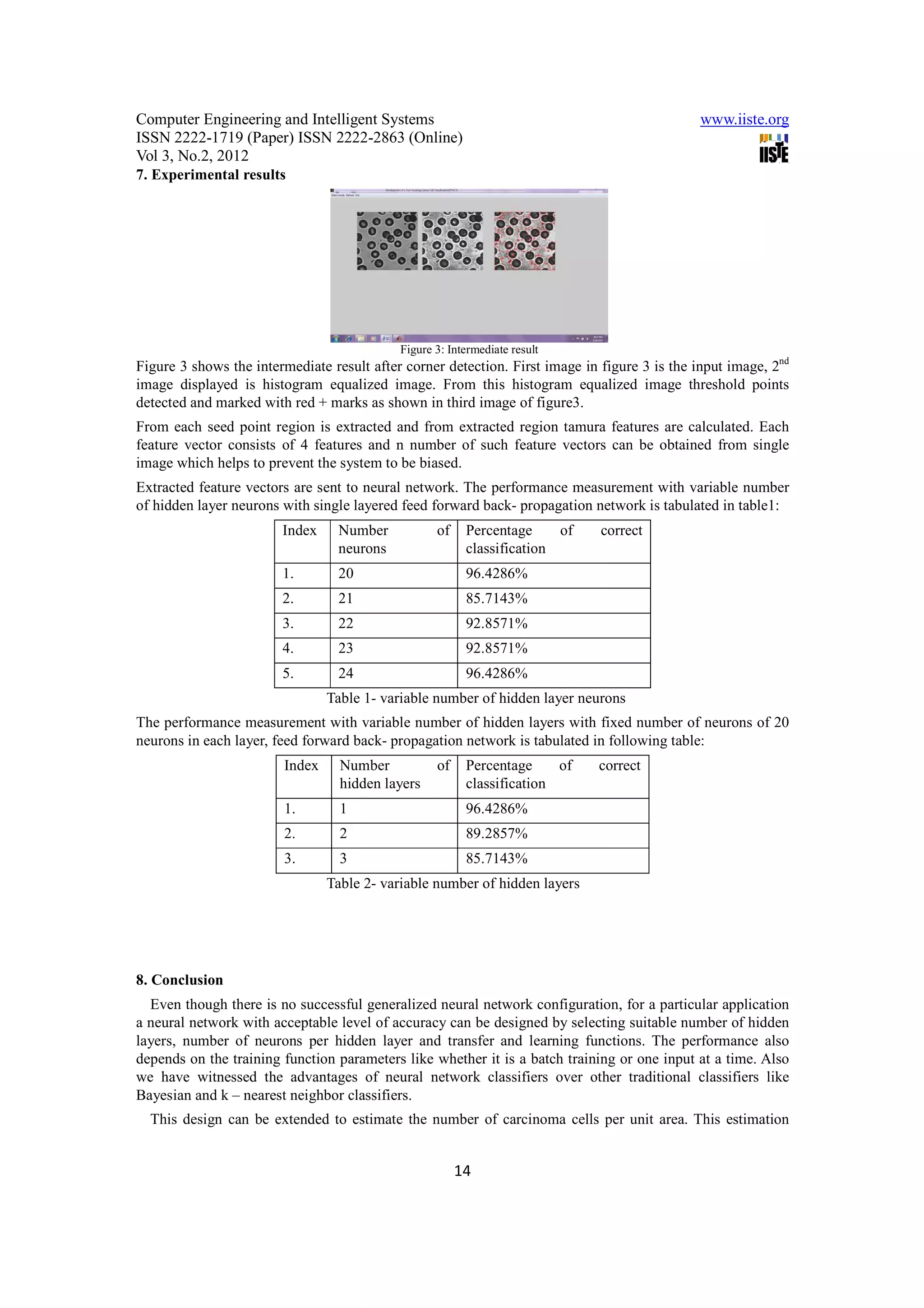
This paper addresses the system which achieves auto-segmentation and cell characterization for prediction
of percentage of carcinoma (cancerous) cells in the given image with high accuracy. The system has been
designed and developed for analysis of medical pathological images based on hybridization of syntactic
and statistical approaches, using Artificial Neural Network as a classifier tool (ANN) [2]. This system
performs segmentation and classification as is done in human vision system [1] [9] [10] [12], which
recognize objects; perceives depth; identifies different textures, curved surfaces, or a surface inclination by
texture information and brightness.
In this paper, an attempt has been made to present an approach for soft tissue characterization utilizing
texture-primitive features and segmentation with Artificial Neural Network (ANN) classifier tool. The
present approach directly combines second, third, and fourth steps into one algorithm. This is a semi-
supervised approach in which supervision is involved only at the level of defining structure of Artificial
Neural Network; afterwards, algorithm itself scans the whole image and performs the segmentation and
classification in unsupervised mode. Finally, algorithm was applied to selected pathological images for
segmentation and classification. Results were in agreement with those with manual segmentation and were
clinically correlated [18] [21].
Keywords: Grey scale images, Histogram equalization, Gausian filtering, Haris corner detector, Threshold,
Seed point, Region growing segmentation, Tamura texture feature extraction, Artificial Neural
Network(ANN), Artificial Neuron, Synapses, Weights, Activation function, Learning function,
Classification matrix.
1. Introduction
In the modern age of computerized fully automated trend of living, the field of automated diagnostic
systems plays an important and vital role. Automated diagnostic system designs in Medical Image
processing are one such field where numerous systems are proposed and still many more under conceptual
design due explosive growth of the technology today. From the past decades, we have witnessed an
explosive growth of Digital image processing for analysis of the data that can be captured by digital images
and artificial neural networks are used to aggregate the analyzed data from these images to produce a
diagnosis prediction with high accuracy instantaneously where digital images serve as tool for input data
[20] [21]. Hence in the process of surgery these automated systems help the surgeon to identify the infected
parts or tumors in case of cancerous growth of cells to be removed with high accuracy hence by increasing
the probability of survival of a patient. In this proposal one of such an automated system for cancer cell
classification which helps as a tool assisting surgeon to differentiate cancerous cells from those normal
cells i.e. percentage of carcinoma cells, instantaneously during the surgery. Here the pathological images
serve as input data. The analysis of these pathological images is directly based on four steps: 1) image
filtering or enhancement, 2) segmentation, 3) feature extraction, and 4) analysis of extracted features by
pattern recognition system or classifier [21]. Since neural network ensembles are used as decision makers
7
2. Computer Engineering and Intelligent Systems www.iiste.org
ISSN 2222-1719 (Paper) ISSN 2222-2863 (Online)
Vol 3, No.2, 2012
even though network takes more time to adapt behavior, once it is trained it classifies almost
instantaneously due to electrical signal communication of nodes in the network.
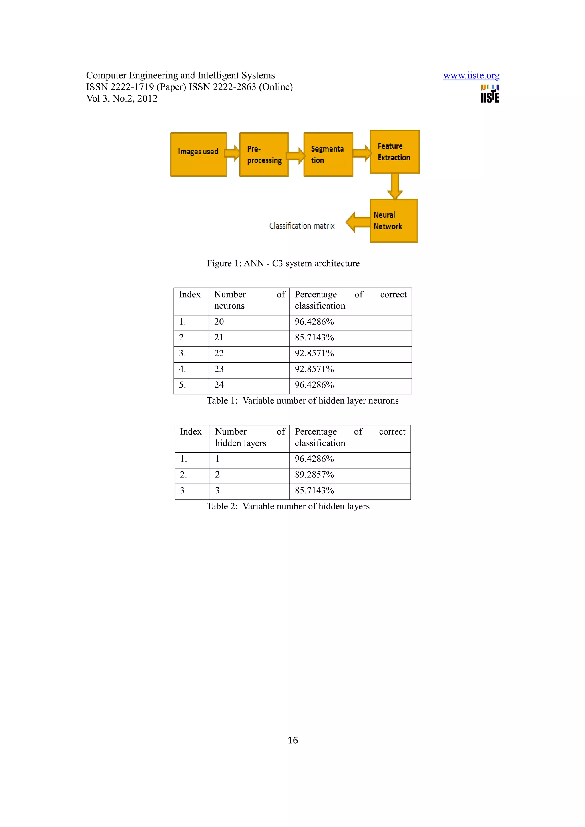
2. System architecture
The ANN – C3 architecture is shown in figure 1. It comprises of five distinct components, as show below.
Each component is described briefly in subsequent sections.
Figure 1: ANN - C3 system architecture
2.1 Images used
This system is designed and verified to take grey scale pathological images as input. Grey scale
pathological images help to identify affected cells makes these images for analysis of cancerous growth of
cells.
2.2 Pre-processing
Grey scale pathological imaging process may be dirtied by various noises. Perform an image pre processing
task to remove noise in a pathological image first. To remove the noise the Histogram equalization or
Gaussian filter based median filtering is done [5] [6] [8] [19].
2.3 Segmentation
Segmentation includes two phases. First phase deals with threshold detection and the later one with similar
region identification. For threshold detection various methods like GUI selection, graphical method or
corner detectors can be used. GUI selection reduces automation and graphical method fails when multiple
objects are present in an input data. Since this design mainly deals with multiple objects (cells) in an input
image, Haris corner detectors are used to find threshold. In second phase, threshold points detected by
corners serve as seed point for segmentation. Four neighborhood based region growing segmentation is
used increase the speed compare to eight neighborhood and increase the accuracy compared to region split
and merge i.e. trade off between accuracy and speed. A brief discussion of Haris corner detector and 4-
neighborhood region growing Segmentation is done in section III [11] [13].
2.4 Feature Extraction
Neural network classifiers are those differ from traditional classifiers like Bayesian and k – nearest
neighborhood classifiers in various aspects from type of input data to output representation. Since the
neural networks are used as classifiers in this design which takes only numerical data as input rather than
any kind of data as input by Bayesian and k – nearest neighbor classifiers, the input image data has to be
converted to numerical form. This conversion is done by extracting tamura texture features. A brief
discussion of tamura texture feature is done in section IV.
Tamura texture features:
The human vision system (HVS) permits scene interpretation ‘at a glance’ i.e. the human eye ‘sees’ not
scenes but sets of objects in various relations to each other, in spite of the fact that the ambient illumination
8
3. Computer Engineering and Intelligent Systems www.iiste.org
ISSN 2222-1719 (Paper) ISSN 2222-2863 (Online)
Vol 3, No.2, 2012
is likely to vary from one object to another—and over the various surfaces of each object—and in spite of
the fact that there will be secondary illumination from one object to another. These variations in the
captured images are referred as tamura texture features, even the same texture features are observed by
surgeon to differentiate carcinoma cells and non-carcinoma cells.
2.5 Neural Network
Supervised feed-forward back-propagation neural network ensemble used as a classifier tool. As discussed
previously, neural network differs in various ways from traditional classifiers like Bayesian and k – nearest
neighbor classifiers. One of the main differences is linearity of data. Traditional classifiers like Bayesian
and k – nearest neighbor requires linear data to work correctly. But neural network works as well for non-
linear data because it is simulated on the observation of biological neurons and network of neurons. Wide
range of input data for training makes neural network to work with higher accuracy, in other words a small
set of data or large set of similar data makes system to be biased [22]. Thus neural network classifier
requires a large set of data for training and also long time to train to reach the stable state. But once the
network is trained it works as fast as biological neural networks by propagating signals as fast as electrical
signals.
3. Haris corner detector and 4-neighborhood region growing segmentation
3.1 Haris corner detector
A corner can be defined as the intersection of two edges. A corner can also be defined as points for which
there are two dominant and different edge directions in a local neighborhood of the point. An interest point
is a point in an image which has a well-defined position and can be robustly detected. This means that an
interest point can be a corner but it can also be, for example, an isolated point of local intensity maximum
or minimum, line endings, or a point on a curve where the curvature is locally maximal.
In practice, most so-called corner detection methods detect interest points in general, rather than corners in
particular. As a consequence, if only corners are to be detected it is necessary to do a local analysis of
detected interest points to determine which of these real corners are.
A simple approach to corner detection in images is using correlation, but this gets very computationally
expensive and suboptimal. Haris corner detector is one such corner detector, which uses differential of the
corner score with respect to direction directly, instead of using shifted patches. This corner score is often
referred to as autocorrelation.
The algorithm of haris corner detector as follows:
Without loss of generality, we will assume a grayscale 2-dimensional image is used. Let this image be
given by I. Consider taking an image patch over the area (u,v) and shifting it by (x,y). The weighted sum of
squared differences (SSD) between these two patches, denoted S, is given by:
(1)
I(u + x,v + y) can be approximated by a Taylor expansion . Let Ix and Iy be the partial derivatives of I, such
that
(2)
This produces the approximation
(3)
This can be written in matrix form:
(4)
Where A is the structure tensor,
9
4. Computer Engineering and Intelligent Systems www.iiste.org
ISSN 2222-1719 (Paper) ISSN 2222-2863 (Online)
Vol 3, No.2, 2012
(5)
This matrix (5) is a Harris matrix, and angle brackets denote averaging (i.e. summation over (u,v)). If a
circular window is used, then the response will be isotropic [16].
A corner (or in general an interest point) is characterized by a large variation of S in all directions of the
vector (x,y). By analyzing the eigenvalues of A, this characterization can be expressed in the following
way: A should have two "large" eigenvalues for an interest point. Based on the magnitudes of the
eigenvalues, the following inferences can be made based on this argument:
If λ1≈0 and λ2≈0 then this pixel (x , y) has no features of interest.
If λ1≈0 and λ2 has some large positive value, then an edge is found.
If λ1 and λ2 have large positive values, then a corner is found.
Haris and Stephens noted that exact computation of the eigenvalues is computationally expensive, since it
requires the computation of a Square root, and instead suggest the following function Mc, where κ is a
tunable sensitivity parameter:
Mc= λ1λ2 – k(λ1+λ2)2=det(A) – k trace2(A) (6)
Therefore, the algorithm does not have to actually compute the Eigen value decomposition of the matrix A
and instead it is sufficient to evaluate the determinant and trace of A to find corners, or rather interest points
in general.
The value of κ has to be determined empirically, and in the literature values in the range 0.04 - 0.15 have
been reported as feasible.
The covariance matrix for the corner position is A − 1, i.e.
(7)
Compute x and y derivatives of image
(8)
(9)
Compute product of derivatives of each image
(10)
(11)
3. Compute the sums of products of derivatives at each pixel
(12)
(13)
(14)
Define at each pixel (x, y) the matrix
(15)
Compute the response of the detector at each pixel
R=Det(H) – k(Trace(H))2 (16)
Threshold on value R. Compute nonmax suppression.
3.2 4-neigbourhood region growing Segmentation
Segmentation is the process of identifying the region of interest from the input image. Considering an input
image I being read and converted to the greyscale image .let’s assume the seed point to be (x , y). If the
10
5. Computer Engineering and Intelligent Systems www.iiste.org
ISSN 2222-1719 (Paper) ISSN 2222-2863 (Online)
Vol 3, No.2, 2012
seed point is provided by the GUI then a function getpts() will make sure the x and y axes values have been
fetched. To create a mask we’ll convert all the pixels in the image I’ to 0 and call the image J.
In order to discover the neighbors we will use four pixel connectivity [14]. Starting with the seed point the
algorithm looks for the 4 pixels surrounding the pixel in consideration. Every time a surrounding pixel is
considered, the region mean is calculated and checked with that of the pixel in consideration and added to
the region. Similarly as the pixel is added to the region corresponding pixel in the image J is highlighted to
1 which would result in the highest intensity hence illuminating the pixel. As the segmentation continues
the region into consideration is intensified in the image J resulting in the segmentation of the affected area,
which later can be combined with the original image and displayed to the user.
5. Tamura Textutre feature extraction
Tamura texture feature concepts proposed by Tamura et al in 1978. These tamura texture features
corresponding to human perception and these features examined by 6 different constituent features. Six
features are: [15]
Coarseness – Coarseness is the numerical value describing whether texture is coarse or fine.
Contrast – Contrast defines whether texture contrast is high or low.
Directionality – Directionality defines whether texture pallets are oriented in single direction or not i.e.
directional or non-directional.
Line-likeness – Line-likeness correspond to pattern elements i.e. whether texture formed by lines i.e. line-
like or blob-like.
Regularity – Regularity defines the interval in which patterns repeated. If patterns are repeated in regular
interval then the texture is regular else it is said to be Irregular.
Roughness – Roughness defines the whether the surface is rough or smooth.
In these six features, Coarseness, Contrast and Directionality correspond to strong human perception and
these features are calculated pixel-wise by creating 3-D histogram of these three features. Estimation of
these three features are described in subsequent sections.
Coarseness relates to distances of notable spatial variations of grey levels, that is, implicitly, to the size of
the primitive elements (texels) forming the texture. The proposed computational procedure accounts for
differences between the average signals for the non-overlapping windows of different size:
At each pixel (x,y), compute six averages for the windows of size 2k × 2k, k=0,1,...,5, around the pixel.
At each pixel, compute absolute differencesEk(x,y) between the pairs of nonoverlapping averages in the
horizontal and vertical directions.
At each pixel, find the value of k that maximises the difference Ek(x,y) in either direction and set the best
size Sbest(x,y)=2k.
Compute the coarseness feature Fcrs by averaging Sbest(x,y) over the entire image. Instead of the average of
Sbest(x,y, an improved coarseness feature to deal with textures having multiple coarseness properties is a
histogram characterising the whole distribution of the best sizes over the image.
Contrast measures how grey levels q; q = 0, 1, ..., qmax, vary in the image g and to what extent their
distribution is biased to black or white. The second-order and normalised fourth-order central moments of
the grey level histogram (empirical probability distribution), that is, the variance, σ2, and kurtosis, α4, are
used to define the contrast:
(17)
Where,
(18)
11
6. Computer Engineering and Intelligent Systems www.iiste.org
ISSN 2222-1719 (Paper) ISSN 2222-2863 (Online)
Vol 3, No.2, 2012
(19)
(20)
and m is the mean grey level, i.e. the first order moment of the grey level probability distribution. The value
n=0.25 is recommended as the best for discriminating the textures.
Degree of directionality is measured using the frequency distribution of oriented local edges against their
directional angles. The edge strength e(x,y) and the directional angle a(x,y) are computed using the Sobel
edge detector approximating the pixel-wise x- and y-derivatives of the image:
e(x,y)=0.5(|∆x(x,y)|+ |∆y(x,y)|) (21)
-1
a(x,y)=tan (∆x(x,y)/ ∆y(x,y)) (22)
where ∆x(x,y) and ∆y(x,y) are the horizontal and vertical grey level differences between the neighbouring
pixels, respectively. The differences are measured using the following 3 × 3 moving window operators:
−1 0 1 1 1 1
−1 0 1 0 0 0
−1 0 1 −1 −1 −1
A histogram Hdir(a) of quantised direction values a is constructed by counting numbers of the edge pixels
with the corresponding directional angles and the edge strength greater than a predefined threshold. The
histogram is relatively uniform for images without strong orientation and exhibits peaks for highly
directional images. The degree of directionality relates to the sharpness of the peaks:
(23)
where np is the number of peaks, ap is the position of the pth peak, wp is the range of the angles attributed to
the pth peak (that is, the range between valleys around the peak), r denotes a normalising factor related to
quantising levels of the angles a, and a is the quantised directional angle (cyclically in modulo 180o). Three
other features are highly correlated with the above three features and do not add much to the effectiveness
of the texture description.
The linelikeness feature Flin is defined as an average coincidence of the edge directions (more precisely,
coded directional angles) that co-occurred in the pairs of pixels separated by a distance d along the edge
direction in every pixel. The edge strength is expected to be greater than a given threshold eliminating
trivial "weak" edges. The coincidence is measured by the cosine of difference between the angles, so that
the co-occurrences in the same direction are measured by +1 and those in the perpendicular directions by -
1. The regularity feature is defined as Freg=1-r(scrs+scon+sdir + slin) where r is a normalising factor and each
s... means the standard deviation of the corresponding feature F... in each subimage the texture is partitioned
into. The roughness feature is given by simply summing the coarseness and contrast measures:
Frgh=Fcrs+Fcon . These features capture the high-level perceptual attributes of a texture well and are useful
for image browsing. However, they are not very effective for finer texture discrimination.
6. Artificial Neural Network
A neural network is a massively parallel distributed processor that has a natural propensity for storing
experiential knowledge and making it available for use. It resembles the brain in two respects [3] [4] [7]:
1. Knowledge is acquired by the network through a learning process.
2. Interneuron connection strengths known as synaptic weights are used to store the knowledge.
Benefits of neural network
Nonlinearity.
Input-output mapping.
12
7. Computer Engineering and Intelligent Systems www.iiste.org
ISSN 2222-1719 (Paper) ISSN 2222-2863 (Online)
Vol 3, No.2, 2012
Adaptivity.
Contextual information.
Fault tolerance.
VLSI implementability.
Uniformity of analysis and design.
Neurobiological analogy.
Model of a neuron
A neuron is an information-processing unit that is fundamental to the operation of a neural network. We
may identify three basic elements of the neuron model: [17] [18]
Figure 2:Non-linear model of a neuron.
A set of synapses, each of which is characterized by a weight or strength of its own. Specifically, a signal xj
at the input of synapse j connected to neuron k is multiplied by the synaptic weight wkj. It is important to
make a note of the manner in which the subscripts of the synaptic weight wkj are written. The first subscript
refers to the neuron in question and the second subscript refers to the input end of the synapse to which the
weight refers. The weight wkj is positive if the associated synapse is excitatory; it is negative if the synapse
is inhibitory.
An adder for summing the input signals, weighted by the respective synapses of the neuron.
An activation function for limiting the amplitude of the output of a neuron. The activation function is also
referred to in the literature as a squashing function in that it squashes (limits) the permissible amplitude
range of the output signal to some finite value.
Typically, the normalized amplitude range of the output of a neuron is written as the closed unit interval [0,
1] or alternatively [-1, 1].
The model of a neuron also includes an externally applied bias (threshold) wk0 = bk that has the effect of
lowering or increasing the net input of the activation function.
Since after feature tamura feature extraction data is in the form of numerical values, Artificial Neural
Network classifier suits well for classification. Also non – linearity of the data makes other traditional
classifiers like Bayesian and kth – nearest neighbor classifier inefficient compared to ANN classifier. Thus
in this system ANN classifier is used as classification tool.
13
8. Computer Engineering and Intelligent Systems www.iiste.org
ISSN 2222-1719 (Paper) ISSN 2222-2863 (Online)
Vol 3, No.2, 2012
7. Experimental results
Figure 3: Intermediate result
Figure 3 shows the intermediate result after corner detection. First image in figure 3 is the input image, 2nd
image displayed is histogram equalized image. From this histogram equalized image threshold points
detected and marked with red + marks as shown in third image of figure3.
From each seed point region is extracted and from extracted region tamura features are calculated. Each
feature vector consists of 4 features and n number of such feature vectors can be obtained from single
image which helps to prevent the system to be biased.
Extracted feature vectors are sent to neural network. The performance measurement with variable number
of hidden layer neurons with single layered feed forward back- propagation network is tabulated in table1:
Index Number of Percentage of correct
neurons classification
1. 20 96.4286%
2. 21 85.7143%
3. 22 92.8571%
4. 23 92.8571%
5. 24 96.4286%
Table 1- variable number of hidden layer neurons
The performance measurement with variable number of hidden layers with fixed number of neurons of 20
neurons in each layer, feed forward back- propagation network is tabulated in following table:
Index Number of Percentage of correct
hidden layers classification
1. 1 96.4286%
2. 2 89.2857%
3. 3 85.7143%
Table 2- variable number of hidden layers
8. Conclusion
Even though there is no successful generalized neural network configuration, for a particular application
a neural network with acceptable level of accuracy can be designed by selecting suitable number of hidden
layers, number of neurons per hidden layer and transfer and learning functions. The performance also
depends on the training function parameters like whether it is a batch training or one input at a time. Also
we have witnessed the advantages of neural network classifiers over other traditional classifiers like
Bayesian and k – nearest neighbor classifiers.
This design can be extended to estimate the number of carcinoma cells per unit area. This estimation
14
9. Computer Engineering and Intelligent Systems www.iiste.org
ISSN 2222-1719 (Paper) ISSN 2222-2863 (Online)
Vol 3, No.2, 2012
helps in automated diagnosis systems like blood purifier in case of blood cancer. Also this can extended to
take color image as input with more feature added to feature vector to increase the accuracy of the output.
References
[ 1]. “An Approach for Discretization and Feature Selection Of Continuous-Valued Attributes in Medical
Images for Classification Learning”.
[ 2]. Basavaraj .S. Anami1 and Vishwanath.C.Burkpalli 2 1. Principal, K.L.E.Institute of Technology, Hubli-
580030, India 2. Research Scholar, Basaveshwar Engineering College, Bagalkot – 587102, India.
[ 3]. “Texture based Identification and Classification of Bulk Sugary Food Objects”, ICGST-GVIP Journal,
ISSN: 1687-398X, Volume 9, Issue 4, August 2009.
[ 4]. Bing Gong, School of Computer Science and Technology Heilongjiang University Harbin, China, “A
Novel Learning Algorithm of Back-propagation Neural Network” ,2009 IITA International Conference
on Control, Automation and Systems Engineering.
[ 5]. Weilin Li, Pan Fu and Weiqing Cao, “Tool Wear States Recognition Based on Genetic Algorithm and
Back Propagation Neural Network Model”, 2010 International Conference on Computer Application and
System Modeling (1CCASM 2010)
[ 6]. Acharya and Ray, “Image Processing: Principles and Applications”, Wiley-Interscience 2005 ISBN 0-
471-71998-6
[ 7]. Russ, “The Image Processing Handbook”, Fourth Edition, CRC 2002 ISBN 0-8493-2532-3
[ 8]. SIMON HAYKIN, Book on “Neural Networks”, 2nd edition, A comprehensive edition.
[ 9]. “Digital Image Processing” by Gonzalez & Woods 2nd edition
[ 10]. H. Tamura, S. Mori, and T. Yamawaki, "Texture features corresponding to visual perception," IEEE
Trans. On Systems, Man, and Cybernetics, vol. Smc-8, No. 6, June 1978.
[ 11]. J. Smith and S.-F. Chang, “Transform features for texture classification and discrimination in large image
database”.IEEE Intl. Conf. on Image Proc., 1994.
[ 12]. Castleman K R. “Digital image processing”. NJ: Prentice Hall, 1996.
[ 13]. Manjunath, B., Ma, W.: “Texture features for browsing and retrieval of image data”. IEEE Trans on
Pattern Analysis and Machine Intelligence 18 (1996) 837842
[ 14]. Alexander Suhre, A. Enis Cetin, Tulin Ersahin, Rengul Cetin-Atalay, “Classification of cell images using
a generalized harris Corner Detector”
[ 15]. R. Adams and L. Bischof, “Seeded Region Growing”, IEEE Trans. Pattern Analysis and Machine
Intelligence, vol. 16, pp. 641-647, 1994.
[ 16]. R. Haralick, “Statistical and structural approaches to texture”, Proceedings of the IEEE, vol. 67, pp. 786–
804, 1979.
[ 17]. C. Harris and M.J. Stephens. “A combined corner and edge detector”. In Alvey Vision Conference, pages
147–152, 1988. D.
[ 18]. Ballard and C. Brown, “Computer Vision”, Prentice-Hall, Inc., 1982, Chap. 6.
[ 19]. Davies, “Machine Vision: Theory”, Algorithms and Practicalities, Academic Press, 1990, Chap. 18.
[ 20]. A K Jain, “Fundamentals of Digital Image Processing”, Prentice-Hall, 1986, Chap. 9.
[ 21]. D. Vernon Zhi-Hua Zhou, Yuan Jiang, Yu-Bin Yang, Shi-Fu Chen , “Lung Cancer Cell Identification
Based on Artificial Neural Network Ensembles”, National Laboratory for Novel Software Technology,
Nanjing University, Nanjing 210093, P.R.China. Artificial Ingelligence in Medicine, 2002, vol.24, no.1,
pp.25-36. @Elsevier.
[ 22]. Leonard Fass , “Imaging and cancer: A review”, GE Healthcare, 352 Buckingham Avenue, Slough, SL1
4ER, UK Imperial College Department of Bioengineering, London, UK.
[ 23]. Jinggangshan, P. R. China , “Application of Neural Networks in Medical Image Processing” ISBN 978-
952-5726-09-1, Proceedings of the Second International Symposium on Networking and Network
Security (ISNNS ’10), 2-4, April. 2010, pp. 02.
15
10. Computer Engineering and Intelligent Systems www.iiste.org
ISSN 2222-1719 (Paper) ISSN 2222-2863 (Online)
Vol 3, No.2, 2012
Figure 1: ANN - C3 system architecture
Index Number of Percentage of correct
neurons classification
1. 20 96.4286%
2. 21 85.7143%
3. 22 92.8571%
4. 23 92.8571%
5. 24 96.4286%
Table 1: Variable number of hidden layer neurons
Index Number of Percentage of correct
hidden layers classification
1. 1 96.4286%
2. 2 89.2857%
3. 3 85.7143%
Table 2: Variable number of hidden layers
16