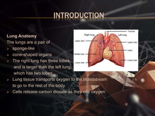
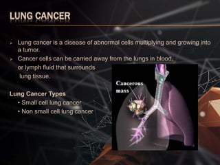
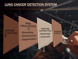
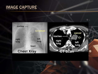
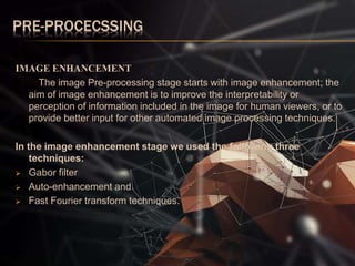
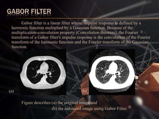
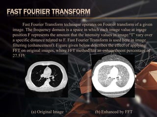
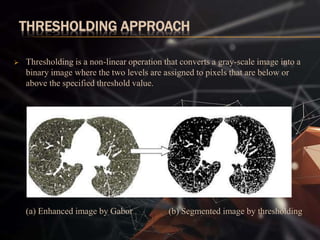
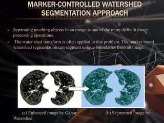
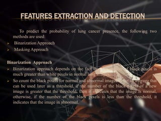
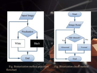
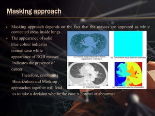
BMS COLLEGE OF ENGINEERING
BANGALORE 56019, INDIA
DEPARTMENTOFELECTRONICSAndCOMMUNICATIONENGINEERING
IMAGE PROCESSING PROJECT PRESENTATION
CANCER CELL DETECTION USING DIGITAL
IMAGE PROCESSING
By l kajikho,manish shah,bikram,adnan,sameep
INTRODUCTION
Lung Anatomy
The lungs are a pair of
 sponge-like
 cone-shaped organs
 The right lung has three lobes,
and is larger than the left lung,
which has two lobes
 Lung tissue transports oxygen to the bloodstream
to go to the rest of the body.
 Cells release carbon dioxide as they use oxygen
LUNG CANCER
 Lung cancer is a disease of abnormal cells multiplying and growing into
a tumor.
 Cancer cells can be carried away from the lungs in blood,
or lymph fluid that surrounds
lung tissue.
Lung Cancer Types
• Small cell lung cancer
• Non small cell lung cancer
LUNG CANCER DETECTION SYSTEM
IMAGE CAPTURE
PRE-PROCECSSING
IMAGE ENHANCEMENT
The image Pre-processing stage starts with image enhancement; the
aim of image enhancement is to improve the interpretability or
perception of information included in the image for human viewers, or to
provide better input for other automated image processing techniques.
In the image enhancement stage we used the following three
techniques:
 Gabor filter
 Auto-enhancement and
 Fast Fourier transform techniques.
GABOR FILTER
Gabor filter is a linear filter whose impulse response is defined by a
harmonic function multiplied by a Gaussian function. Because of the
multiplication-convolution property (Convolution theorem), the Fourier
transform of a Gabor filter's impulse response is the convolution of the Fourier
transform of the harmonic function and the Fourier transform of the Gaussian
function.
(a) (b)
Figure describes (a) the original image and
(b) the enhanced image using Gabor Filter.
FAST FOURIER TRANSFORM
Fast Fourier Transform technique operates on Fourier transform of a given
image. The frequency domain is a space in which each image value at image
position F represents the amount that the intensity values in image “I” vary over
a specific distance related to F. Fast Fourier Transform is used here in image
filtering (enhancement). Figure given below describes the effect of applying
FFT on original images, where FFT method has an enhancement percentage of
27.51%.
(a) Original Image (b) Enhanced by FFT
IMAGE SEGMENTATION
Segmentation divides the image into its constituent regions or
objects.Image segmentation is the process of assigning a label to every pixel in
an image such that pixels with the same label share certain visual
characteristics.
Image segmentation are of two types:
 Thresholding approach
 Marker-Controlled Watershed Segmentation Approach
THRESHOLDING APPROACH
 Thresholding is a non-linear operation that converts a gray-scale image into a
binary image where the two levels are assigned to pixels that are below or
above the specified threshold value.
(a) Enhanced image by Gabor (b) Segmented image by thresholding
MARKER-CONTROLLED WATERSHED
SEGMENTATION APPROACH
 Separating touching objects in an image is one of the more difficult image
processing operations.
 The water shed transform is often applied to this problem. The marker based
watershed segmentation can segment unique boundaries from an image.
(a) Enhanced image by Gabor (b) Segmented image by
Watershed
FEATURES EXTRACTION AND DETECTION
To predict the probability of lung cancer presence, the following two
methods are used:
 Binarization Approach
 Masking Approach
Binarization Approach
 Binarization approach depends on the fact that the number of black pixels is
much greater than white pixels in normal lung images.
 So count the black pixels for normal and abnormal images to get an average that
can be used later as a threshold, if the number of the black pixels of a new
image is greater that the threshold, then it indicates that the image is normal,
otherwise, if the number of the black pixels is less than the threshold, it
indicates that the image in abnormal.
Fig. Binnarization method procedure Fig. Binarization check method
flowchart
Masking approach
 Masking approach depends on the fact that the masses are appeared as white
connected areas inside lungs
 The appearance of solid
blue colour indicates
normal case while
appearance of RGB masses
indicates the presence of
cancer
Therefore, combining
Binarization and Masking
approaches together will lead
us to take a decision whethe the case is normal or abnormal
CONCLUSIONS
 Lung cancer is the most dangerous and widespread in the world according to
stage the discovery of the cancer cells in the lungs.
 An image improvement technique plays a very important and essential role to
avoid serious stages and to reduce its percentage distribution in the world
THE END

CANCER CELL DETECTION USING DIGITAL IMAGE PROCESSING

  • 1.
    BMS COLLEGE OFENGINEERING BANGALORE 56019, INDIA DEPARTMENTOFELECTRONICSAndCOMMUNICATIONENGINEERING IMAGE PROCESSING PROJECT PRESENTATION CANCER CELL DETECTION USING DIGITAL IMAGE PROCESSING By l kajikho,manish shah,bikram,adnan,sameep
  • 2.
    INTRODUCTION Lung Anatomy The lungsare a pair of  sponge-like  cone-shaped organs  The right lung has three lobes, and is larger than the left lung, which has two lobes  Lung tissue transports oxygen to the bloodstream to go to the rest of the body.  Cells release carbon dioxide as they use oxygen
  • 3.
    LUNG CANCER  Lungcancer is a disease of abnormal cells multiplying and growing into a tumor.  Cancer cells can be carried away from the lungs in blood, or lymph fluid that surrounds lung tissue. Lung Cancer Types • Small cell lung cancer • Non small cell lung cancer
  • 4.
  • 5.
  • 6.
    PRE-PROCECSSING IMAGE ENHANCEMENT The imagePre-processing stage starts with image enhancement; the aim of image enhancement is to improve the interpretability or perception of information included in the image for human viewers, or to provide better input for other automated image processing techniques. In the image enhancement stage we used the following three techniques:  Gabor filter  Auto-enhancement and  Fast Fourier transform techniques.
  • 7.
    GABOR FILTER Gabor filteris a linear filter whose impulse response is defined by a harmonic function multiplied by a Gaussian function. Because of the multiplication-convolution property (Convolution theorem), the Fourier transform of a Gabor filter's impulse response is the convolution of the Fourier transform of the harmonic function and the Fourier transform of the Gaussian function. (a) (b) Figure describes (a) the original image and (b) the enhanced image using Gabor Filter.
  • 8.
    FAST FOURIER TRANSFORM FastFourier Transform technique operates on Fourier transform of a given image. The frequency domain is a space in which each image value at image position F represents the amount that the intensity values in image “I” vary over a specific distance related to F. Fast Fourier Transform is used here in image filtering (enhancement). Figure given below describes the effect of applying FFT on original images, where FFT method has an enhancement percentage of 27.51%. (a) Original Image (b) Enhanced by FFT
  • 9.
    IMAGE SEGMENTATION Segmentation dividesthe image into its constituent regions or objects.Image segmentation is the process of assigning a label to every pixel in an image such that pixels with the same label share certain visual characteristics. Image segmentation are of two types:  Thresholding approach  Marker-Controlled Watershed Segmentation Approach
  • 10.
    THRESHOLDING APPROACH  Thresholdingis a non-linear operation that converts a gray-scale image into a binary image where the two levels are assigned to pixels that are below or above the specified threshold value. (a) Enhanced image by Gabor (b) Segmented image by thresholding
  • 11.
    MARKER-CONTROLLED WATERSHED SEGMENTATION APPROACH Separating touching objects in an image is one of the more difficult image processing operations.  The water shed transform is often applied to this problem. The marker based watershed segmentation can segment unique boundaries from an image. (a) Enhanced image by Gabor (b) Segmented image by Watershed
  • 12.
    FEATURES EXTRACTION ANDDETECTION To predict the probability of lung cancer presence, the following two methods are used:  Binarization Approach  Masking Approach Binarization Approach  Binarization approach depends on the fact that the number of black pixels is much greater than white pixels in normal lung images.  So count the black pixels for normal and abnormal images to get an average that can be used later as a threshold, if the number of the black pixels of a new image is greater that the threshold, then it indicates that the image is normal, otherwise, if the number of the black pixels is less than the threshold, it indicates that the image in abnormal.
  • 13.
    Fig. Binnarization methodprocedure Fig. Binarization check method flowchart
  • 14.
    Masking approach  Maskingapproach depends on the fact that the masses are appeared as white connected areas inside lungs  The appearance of solid blue colour indicates normal case while appearance of RGB masses indicates the presence of cancer Therefore, combining Binarization and Masking approaches together will lead us to take a decision whethe the case is normal or abnormal
  • 15.
    CONCLUSIONS  Lung canceris the most dangerous and widespread in the world according to stage the discovery of the cancer cells in the lungs.  An image improvement technique plays a very important and essential role to avoid serious stages and to reduce its percentage distribution in the world
  • 16.