This document evaluates and compares the performance of various segmentation algorithms for detecting brain tumors in MRI images, including hierarchical self-organizing mapping (HSOM), region growing, Otsu, K-means, and fuzzy C-means. It finds that HSOM performs best according to evaluation metrics like segmentation accuracy, Rand index, global consistency error, and variation of information. HSOM is able to segment brain tumor images with higher accuracy and consistency compared to other algorithms like region growing, Otsu, K-means and fuzzy C-means.
Geometric Correction for Braille Document Images csandit
Image processing is an important research area in computer vision. clustering is an unsupervised
study. clustering can also be used for image segmentation. there exist so many methods for image
segmentation. image segmentation plays an important role in image analysis.it is one of the first
and the most important tasks in image analysis and computer vision. this proposed system
presents a variation of fuzzy c-means algorithm that provides image clustering. the kernel fuzzy
c-means clustering algorithm (kfcm) is derived from the fuzzy c-means clustering
algorithm(fcm).the kfcm algorithm that provides image clustering and improves accuracy
significantly compared with classical fuzzy c-means algorithm. the new algorithm is called
gaussian kernel based fuzzy c-means clustering algorithm (gkfcm)the major characteristic of
gkfcm is the use of a fuzzy clustering approach ,aiming to guarantee noise insensitiveness and
image detail preservation.. the objective of the work is to cluster the low intensity in homogeneity
area from the noisy images, using the clustering method, segmenting that portion separately using
content level set approach. the purpose of designing this system is to produce better segmentation
results for images corrupted by noise, so that it can be useful in various fields like medical image
analysis, such as tumor detection, study of anatomical structure, and treatment planning.
Hybrid Approach for Brain Tumour Detection in Image Segmentationijtsrd
In this paper we have considered illustrating a few techniques. But the numbers of techniques are so large they cannot be all addressed. Image segmentation forms the basics of pattern recognition and scene analysis problems. The segmentation techniques are numerous in number but the choice of one technique over the other depends only on the application or requirements of the problem that is being considered. Analysis of cluster is a descriptive assignment that perceive homogenous group of objects and it is also one of the fundamental analytical method in facts mining. The main idea of this is to present facts about brain tumour detection system and various data mining methods used in this system. This is focuses on scalable data systems, which include a set of tools and mechanisms to load, extract, and improve disparate data power to perform complex transformations and analysis will be measured between the way of measuring the Furrier and Wavelet Transform distance. Sandeep | Jyoti Kataria "Hybrid Approach for Brain Tumour Detection in Image Segmentation" Published in International Journal of Trend in Scientific Research and Development (ijtsrd), ISSN: 2456-6470, Volume-4 | Issue-6 , October 2020, URL: https://www.ijtsrd.com/papers/ijtsrd33409.pdf Paper Url: https://www.ijtsrd.com/medicine/other/33409/hybrid-approach-for-brain-tumour-detection-in-image-segmentation/sandeep
Medical Image segmentation using Image Mining conceptsEditor IJMTER
Image differencing is usually done by subtracting the low-level skin texture like strength
in images that are already associated. This paper extracts high-level skin texture in order to find out
an efficient image differencing method for the analysis of Brain Tumor. On the other hand, this
produces sets of skin texture that are both spatial. We demonstrate a technique that avoids arbitrary
spatial constraints and is robust in the presence of sound, outliers, and imaging artifact, while
outperforming even profitable products in the analysis of Brain Tumor images. First, the landmark
are establish, and then the top entrant are sorted into a end set. Second, the top sets of the two
descriptions are then differenced through a cluster judgment. The symmetry of the human body is
utilized to increase the accuracy of the finding. We imitate this technique in an effort to understand
and ultimately capture the judgment of the radiologist. The image differencing with clustered
contrast process determines the being there of Brain Tumor. Using the most favorable features
extracted from normal and tumor regions of MRI by using arithmetical features, classifiers are used
to categorize and segment the tumor portion in irregular images. Both the difficult and preparation
phase gives the proportion of accuracy on each parameter in neural networks, which gives the idea to
decide the best one to be used in supplementary works. The results showed outperformance of
algorithm when compared with classification accuracy which works as shows potential tool for
classification and requires extension in brain tumor analysis.
FUZZY SEGMENTATION OF MRI CEREBRAL TISSUE USING LEVEL SET ALGORITHMAM Publications
The current study investigated a median filter with the fuzzy level set method to propose fuzzy segmentation of magnetic resonance imaging (MRI) cerebral tissue images. An MRI image was used as an input image. A median filter and fuzzy c-means (FCM) clustering were utilized to remove image noise and create image clusters, respectively. The image clusters showed initial and final cluster centers. The level set method was then used for segmentation after separating and extracting white matter from gray matter. Fuzzy c-means was sensitive to the choice of the initial cluster center. Improper center selection caused the method to produce suboptimal solutions. The proposed algorithm was successfully utilized to segment MRI cerebral tissue images. The algorithm efficiently performed segmentation of test MRI cerebral tissue images compared with algorithms proposed in previous studies.
Comparative analysis of multimodal medical image fusion using pca and wavelet...IJLT EMAS
nowadays, there are a lot of medical images and their
numbers are increasing day by day. These medical images are
stored in large database. To minimize the redundancy and
optimize the storage capacity of images, medical image fusion is
used. The main aim of medical image fusion is to combine
complementary information from multiple imaging modalities
(Eg: CT, MRI, PET etc.) of the same scene. After performing
image fusion, the resultant image is more informative and
suitable for patient diagnosis. There are some fusion techniques
which are described in this paper to obtain fused image. This
paper presents two approaches to image fusion, namely Spatial
Fusion and Transform Fusion. This paper describes Techniques
such as Principal Component Analysis which is spatial domain
technique and Discrete Wavelet Transform, Stationary Wavelet
Transform which are Transform domain techniques.
Performance metrics are implemented to evaluate the
performance of image fusion algorithm. An experimental result
shows that image fusion method based on Stationary Wavelet
Transform is better than Principal Component Analysis and
Discrete Wavelet Transform.
MRIIMAGE SEGMENTATION USING LEVEL SET METHOD AND IMPLEMENT AN MEDICAL DIAGNOS...cseij
Image segmentation plays a vital role in image processing over the last few years. The goal of image segmentation is to cluster the pixels into salient image regions i.e., regions corresponding to individual surfaces, objects, or natural parts of objects. In this paper, we propose a medical diagnosis system by using level set method for segmenting the MRI image which investigates a new variational level set algorithm without re- initialization to segment the MRI image and to implement a competent medical diagnosis system by using MATLAB. Here we have used the speed function and the signed distance function of the image in segmentation algorithm. This system consists of thresholding technique, curve evolution technique and an eroding technique. Our proposed system was tested on some MRI Brain images, giving promising results by detecting the normal or abnormal condition specially the existence of tumers. This system will be applied to both simulated and real images with promising results
A Dualistic Sub-Image Histogram Equalization Based Enhancement and Segmentati...inventy
Research Inventy : International Journal of Engineering and Science is published by the group of young academic and industrial researchers with 12 Issues per year. It is an online as well as print version open access journal that provides rapid publication (monthly) of articles in all areas of the subject such as: civil, mechanical, chemical, electronic and computer engineering as well as production and information technology. The Journal welcomes the submission of manuscripts that meet the general criteria of significance and scientific excellence. Papers will be published by rapid process within 20 days after acceptance and peer review process takes only 7 days. All articles published in Research Inventy will be peer-reviewed.
Survey on Brain MRI Segmentation TechniquesEditor IJMTER
Image segmentation is aimed at cutting out, a ROI (Region of Interest) from an image. For
medical images, segmentation is done for: studying the anatomical structure, identifying ROI ie tumor
or any other abnormalities, identifying the increase in tissue volume in a region, treatment planning.
Currently there are many different algorithms available for image segmentation. This paper lists and
compares some of them. Each has their own advantages and limitations.
MIP AND UNSUPERVISED CLUSTERING FOR THE DETECTION OF BRAIN TUMOUR CELLSAM Publications
Image processing is widely used in biomedical applications. Image processing can be used to analyze
different MRI brain images in order to get the abnormality in the image .The objective is to extract meaningful
information from the imaged signals. Image segmentation is a process of partitioning an image in to different parts.
The division in to parts is often based on the characteristics of the pixels in the image. In our paper the segmentation
of the tumour tissues is carried out using k-means and fuzzy c-means clustering.Tumour can be found and faster
detection is achieved with only few seconds for execution. The input image of the brain is taken from the available
database and the presence of tumourin input image can be detected.
SEGMENTATION OF MAGNETIC RESONANCE BRAIN TUMOR USING INTEGRATED FUZZY K-MEANS...ijcsit
Segmentation is a process of partitioning the image into several objects. It plays a vital role in many fields
such as satellite, remote sensing, object identification, face tracking and most importantly in medical field.
In radiology, magnetic resonance imaging (MRI) is used to investigate the human body processes and
functions of organisms. In hospitals, this technique has been using widely for medical diagnosis, to find the
disease stage and follow-up without exposure to ionizing radiation.Here in this paper, we proposed a novel
MR brain image segmentation method for detecting the tumor and finding the tumor area with improved
performance over conventional segmentation techniques such as fuzzy c means (FCM), K-means and even
that of manual segmentation in terms of precision time and accuracy. Simulation performance shows that
the proposed scheme has performed superior to the existing segmentation methods.
IJRET : International Journal of Research in Engineering and Technology is an international peer reviewed, online journal published by eSAT Publishing House for the enhancement of research in various disciplines of Engineering and Technology. The aim and scope of the journal is to provide an academic medium and an important reference for the advancement and dissemination of research results that support high-level learning, teaching and research in the fields of Engineering and Technology. We bring together Scientists, Academician, Field Engineers, Scholars and Students of related fields of Engineering and Technology
Multi fractal analysis of human brain mr imageeSAT Journals
Abstract In computer programming, code smell may origin of latent problems in source code. Detecting and resolving bad smells remain time intense for software engineers despite proposals on bad smell detecting and refactoring tools. Numerous code smells have been recognized yet the sequence in which the detection and resolution of different kinds of code smells are performed because software engineers do not know how to optimize sequence. In this paper, the novel refactoring approach is proposed to improve the performance of programs. In this recommended approach the code smells are automatically detected and refactored. The simulation results propose the reduction of time over the semi-automated refactoring are achieved when code smells are refactored by using multi-step automated refactoring. Keywords: Code smell, multi step refactoring, detection, code resolution, restructuring etc
Image registration is the fundamental task used to
match two or more partially overlapping images taken, for
example, at different times,from different sensors, or from
different viewpoints and stitch these images into one
panoramic image comprising the whole scene. It is
afundamental image processing technique and is very useful
in integrating information from different sensors, finding
changes in images taken at different times, inferring threedimensional
information from stereo images, and recognizing
model-based objects.
This paper overviews the theoretical aspects of an image
registration problem. The purpose of this paper is to present a
survey of image registration techniques. This technique of
image registration aligns two images geometrically. These two
images are reference image and sensed image. The ultimate
purpose of digital image filtering is to support the visual
identification of certain features expressed by characteristic
shapes and patterns. Numerous recipes, algorithms and ready
made programs exist nowadays that predominantly have in
common that users have to set certain parameters.
Particularly if processing is fast and shows results rather
immediately, the choice of parameters may be guided by
making the image ―looking nice‖. However, in practical
situations most users are not in a mood to ―play around‖
with a displayed image, particularly if they are in a stressy
situation as it may encountered in security applications. The
requirements for the application of digital image processing
under such circumstances will be discussed with an example
of automaticfiltering without manual parameter settings that
even entails the advantage of delivering unbiased results
An ensemble classification algorithm for hyperspectral imagessipij
Hyperspectral image analysis has been used for many purposes in environmental monitoring, remote
sensing, vegetation research and also for land cover classification. A hyperspectral image consists of many
layers in which each layer represents a specific wavelength. The layers stack on top of one another making
a cube-like image for entire spectrum. This work aims to classify the hyperspectral images and to produce
a thematic map accurately. Spatial information of hyperspectral images is collected by applying
morphological profile and local binary pattern. Support vector machine is an efficient classification
algorithm for classifying the hyperspectral images. Genetic algorithm is used to obtain the best feature
subjected for classification. Selected features are classified for obtaining the classes and to produce a
thematic map. Experiment is carried out with AVIRIS Indian Pines and ROSIS Pavia University. Proposed
method produces accuracy as 93% for Indian Pines and 92% for Pavia University.
Segmentation of Tumor Region in MRI Images of Brain using Mathematical Morpho...CSCJournals
This paper introduces an efficient detection of brain tumor from cerebral MRI images. The methodology consists of two steps: enhancement and segmentation. To improve the quality of images and limit the risk of distinct regions fusion in the segmentation phase an enhancement process is applied. We applied mathematical morphology to increase the contrast in MRI images and to segment MRI images. Some of experimental results on brain images show the feasibility and the performance of the proposed approach.
PERFORMANCE ANALYSIS OF CLUSTERING BASED IMAGE SEGMENTATION AND OPTIMIZATION ...cscpconf
Partitioning of an image into several constituent components is called image segmentation.
Myriad algorithms using different methods have been proposed for image segmentation. Many
clustering algorithms and optimization techniques are also being used for segmentation of
images. A major challenge in segmentation evaluation comes from the fundamental conflict
between generality and objectivity. As there is a glut of image segmentation techniques
available today, customer who is the real user of these techniques may get obfuscated. In this
paper to address the above described problem some image segmentation techniques are evaluated based on their consistency in different applications. Based on the parameters used quantification of different clustering algorithms is done.
Segmentation of Brain MR Images for Tumor Extraction by Combining Kmeans Clus...CSCJournals
Segmentation of images holds an important position in the area of image processing. It becomes more important while typically dealing with medical images where pre-surgery and post surgery decisions are required for the purpose of initiating and speeding up the recovery process [5] Computer aided detection of abnormal growth of tissues is primarily motivated by the necessity of achieving maximum possible accuracy. Manual segmentation of these abnormal tissues cannot be compared with modern day’s high speed computing machines which enable us to visually observe the volume and location of unwanted tissues. A well known segmentation problem within MRI is the task of labeling voxels according to their tissue type which include White Matter (WM), Grey Matter (GM) , Cerebrospinal Fluid (CSF) and sometimes pathological tissues like tumor etc. This paper describes an efficient method for automatic brain tumor segmentation for the extraction of tumor tissues from MR images. It combines Perona and Malik anisotropic diffusion model for image enhancement and Kmeans clustering technique for grouping tissues belonging to a specific group. The proposed method uses T1, T2 and PD weighted gray level intensity images. The proposed technique produced appreciative results
“Study The Different Parameters of Sewage Treatment With UASB & SBR Technolog...IOSR Journals
Abstract: Every community produces both liquid and solid wastes and air emissions. The liquid wastewastewater-is
essentially the water supply of the community after it has been used in a variety of applications.
From the standpoint of sources of generation, wastewater may be defined as a combination of the liquid or
water-carried wastes removed from residences, institutions, commercial and industrial establishments, together
with such groundwater, surfacewater and stromwater as may be present. This waste water through sewer comes
to the sewage treatment plant so that parameters are reduced and treated wastewater be disposed into water or
land. For treating the sewage UASB( UP FLOW ANAEROBIC SLUDGE BLANKET) and SBR(SEQUENCING
BATCH REACTOR) technologies are mostly used.
All the parameters of these samples were analyzed using standard methods prescribed in “Standard methods for
examination of water and wastewater”. It was observed that pH & temperature values at outlet by both the
processes are almost same. Reading were taking on two consecutive days and value of Biochemical Oxygen
Demand by UASB process was 32, 32mg/l and by SBR process was 11, 16mg/l. Chemical oxygen Demand by
UASB process was 112, 96mg/l and by SBR process was 32, 34mg/l. Total Suspended Solids by UASB process
was 58, 44mg/l and by SBR process was 10, 12mg/l. Both the processes were used for treating the wastewater
and the SBR process showed better results as comparative to UASB.
International Journal of Engineering Research and Applications (IJERA) is an open access online peer reviewed international journal that publishes research and review articles in the fields of Computer Science, Neural Networks, Electrical Engineering, Software Engineering, Information Technology, Mechanical Engineering, Chemical Engineering, Plastic Engineering, Food Technology, Textile Engineering, Nano Technology & science, Power Electronics, Electronics & Communication Engineering, Computational mathematics, Image processing, Civil Engineering, Structural Engineering, Environmental Engineering, VLSI Testing & Low Power VLSI Design etc.
IJRET : International Journal of Research in Engineering and Technology is an international peer reviewed, online journal published by eSAT Publishing House for the enhancement of research in various disciplines of Engineering and Technology. The aim and scope of the journal is to provide an academic medium and an important reference for the advancement and dissemination of research results that support high-level learning, teaching and research in the fields of Engineering and Technology. We bring together Scientists, Academician, Field Engineers, Scholars and Students of related fields of Engineering and Technology.
International Journal of Computational Engineering Research (IJCER) is dedicated to protecting personal information and will make every reasonable effort to handle collected information appropriately. All information collected, as well as related requests, will be handled as carefully and efficiently as possible in accordance with IJCER standards for integrity and objectivity.
Mammogram image segmentation using rough clusteringeSAT Journals
Abstract The mammography is the most effective procedure to diagnosis the breast cancer at an early stage. This paper proposes mammogram image segmentation using Rough K-Means (RKM) clustering algorithm. The median filter is used for pre-processing of image and it is normally used to reduce noise in an image. The 14 Haralick features are extracted from mammogram image using Gray Level Co-occurrence Matrix (GLCM) for different angles. The features are clustered by K-Means, Fuzzy C-Means (FCM) and Rough K-Means algorithms to segment the region of interests for classification. The result of the segmentation algorithms compared and analyzed using Mean Square Error (MSE) and Root Means Square Error (RMSE). It is observed that the proposed method produces better results that the existing methods. Keywords— Mammogram, Data mining, Image Processing, Feature Extraction, Rough K- Means and Image Segmentation
International Journal of Research in Engineering and Science is an open access peer-reviewed international forum for scientists involved in research to publish quality and refereed papers. Papers reporting original research or experimentally proved review work are welcome. Papers for publication are selected through peer review to ensure originality, relevance, and readability.
GAUSSIAN KERNEL BASED FUZZY C-MEANS CLUSTERING ALGORITHM FOR IMAGE SEGMENTATIONcscpconf
Image processing is an important research area in computer vision. clustering is an unsupervised study. clustering can also be used for image segmentation. there exist so many methods for image segmentation. image segmentation plays an important role in image analysis.it is one of the first and the most important tasks in image analysis and computer vision. this proposed system presents a variation of fuzzy c-means algorithm that provides image clustering. the kernel fuzzy
c-means clustering algorithm (kfcm) is derived from the fuzzy c-means clustering algorithm(fcm).the kfcm algorithm that provides image clustering and improves accuracy significantly compared with classical fuzzy c-means algorithm. the new algorithm is called gaussian kernel based fuzzy c-means clustering algorithm (gkfcm)the major characteristic of gkfcm is the use of a fuzzy clustering approach ,aiming to guarantee noise insensitiveness and image detail preservation.. the objective of the work is to cluster the low intensity in homogeneity area from the noisy images, using the clustering method, segmenting that portion separately using content level set approach. the purpose of designing this system is to produce better segmentation results for images corrupted by noise, so that it can be useful in various fields like medical image analysis, such as tumor detection, study of anatomical structure, and treatment planning.
GAUSSIAN KERNEL BASED FUZZY C-MEANS CLUSTERING ALGORITHM FOR IMAGE SEGMENTATIONcsandit
Image processing is an important research area in computer vision. clustering is an unsupervised
study. clustering can also be used for image segmentation. there exist so many methods for image
segmentation. image segmentation plays an important role in image analysis.it is one of the first
and the most important tasks in image analysis and computer vision. this proposed system
presents a variation of fuzzy c-means algorithm that provides image clustering. the kernel fuzzy
c-means clustering algorithm (kfcm) is derived from the fuzzy c-means clustering
algorithm(fcm).the kfcm algorithm that provides image clustering and improves accuracy
significantly compared with classical fuzzy c-means algorithm. the new algorithm is called
gaussian kernel based fuzzy c-means clustering algorithm (gkfcm)the major characteristic of
gkfcm is the use of a fuzzy clustering approach ,aiming to guarantee noise insensitiveness and
image detail preservation.. the objective of the work is to cluster the low intensity in homogeneity
area from the noisy images, using the clustering method, segmenting that portion separately using
content level set approach. the purpose of designing this system is to produce better segmentation
results for images corrupted by noise, so that it can be useful in various fields like medical image
analysis, such as tumor detection, study of anatomical structure, and treatment planning.
A Review on Image Segmentation using Clustering and Swarm Optimization Techni...IJSRD
The process of dividing an image into multiple regions (set of pixels) is known as Image segmentation. It will make an image easy and smooth to evaluate. Image segmentation objective is to generate image more simple and meaningful. In this paper present a survey on image segmentation general segmentation techniques, clustering algorithms and optimization methods. Also a study of different research also been presented. The latest research in each of image segmentation methods is presented in this study. This paper presents the recent research in biologically inspired swarm optimization techniques, including ant colony optimization algorithm, particle swarm optimization algorithm, artificial bee colony algorithm and their hybridizations, which are applied in several fields.
MAGNETIC RESONANCE BRAIN IMAGE SEGMENTATIONVLSICS Design
Segmentation of tissues and structures from medical images is the first step in many image analysis applications developed for medical diagnosis. With the growing research on medical image segmentation, it is essential to categorize the research outcomes and provide researchers with an overview of the existing segmentation techniques in medical images. In this paper, different image segmentation methods applied on magnetic resonance brain images are reviewed. The selection of methods includes sources from image processing journals, conferences, books, dissertations and thesis. The conceptual details of the methods are explained and mathematical details are avoided for simplicity. Both broad and detailed categorizations of reviewed segmentation techniques are provided. The state of art research is provided with emphasis on developed techniques and image properties used by them. The methods defined are not always mutually independent. Hence, their inter relationships are also stated. Finally, conclusions are drawn summarizing commonly used techniques and their complexities in application.
International Journal of Engineering Research and Applications (IJERA) is an open access online peer reviewed international journal that publishes research and review articles in the fields of Computer Science, Neural Networks, Electrical Engineering, Software Engineering, Information Technology, Mechanical Engineering, Chemical Engineering, Plastic Engineering, Food Technology, Textile Engineering, Nano Technology & science, Power Electronics, Electronics & Communication Engineering, Computational mathematics, Image processing, Civil Engineering, Structural Engineering, Environmental Engineering, VLSI Testing & Low Power VLSI Design etc.
An Investigation into Brain Tumor Segmentation Techniques IIRindia
A tumor is an anomalous mass in the brain which can be cancerous. Such anomalous growth within this restricted space or inside the covering skull can cause problems. Detecting brain tumors from images of medical modalities like CT scan or MRI involves segmentation (Division into parts) for analysis and can be a challenging task. Accurate segmentation of brain images is very essential for proper diagnosis of tumor and non-tumor areas for clinical analysis. This paper details on segmentation algorithms for brain images, advantages, disadvantages and a comparison of the algorithms.
Comparitive study of brain tumor detection using morphological operatorseSAT Journals
Abstract
Segmentation divides an image into foreground object and the background object. In our case foreground object is brain tumor and background is CSF, white matter, and grey matter. Aim of our study is to detect the tumor and remove the background completely and compare the morphological operations that can be used for this purpose. Segmentation remains a challenging area for researchers since many segmentation methods results in over segmentation or under segmentation and hence, leads to the false interpretation of the results. The proposed work is the comparative study of the morphological segmentation methods for segmenting brain tumor from MRI images. Before segmentation, filtration process is carried out using two method, Non Local mean filter and median filter and their results are compared using MSE and PSNR. NL mean filter preserves sharp edges and fine details in an image hence, preferred over median filter. Also tumor location is identified, to get an approximate idea about the position of the tumor in the brain i.e. in which part the brain tumor is located. The tumor is identified by using different algorithms which are based on morphology such as watershed segmentation, morphological erosion, and hole filling algorithm and comparison between them is carried out based on parameters like accuracy, sensitivity and elapsed time. Each of the segmentation results are compared with the tumor obtained using interactive tool present in MATLAB R2013b.
Keywords: Brain tumor, MRI images, Image segmentation, Morphology, Erosion, Thresholding, Hole filling, Watershed segmentation
Human brain is the most complex structure where identifying the tumor like diseases are extremely challenging because differentiating the components of a brain is complex. In this paper, pillar k-means algorithm is used for segmentation of brain tumor from magnetic resonance image (MRI).Generally, the brain tumor is detected by radiologist through analysis of MR images which takes longer time. The pillar k-means algorithm’s experimental results clarify the effectiveness of our approach to improve the segmentation quality, accuracy, and computational time. Classify, the tumor from the brain MR images using Bayesian classification.
Explore the innovative world of trenchless pipe repair with our comprehensive guide, "The Benefits and Techniques of Trenchless Pipe Repair." This document delves into the modern methods of repairing underground pipes without the need for extensive excavation, highlighting the numerous advantages and the latest techniques used in the industry.
Learn about the cost savings, reduced environmental impact, and minimal disruption associated with trenchless technology. Discover detailed explanations of popular techniques such as pipe bursting, cured-in-place pipe (CIPP) lining, and directional drilling. Understand how these methods can be applied to various types of infrastructure, from residential plumbing to large-scale municipal systems.
Ideal for homeowners, contractors, engineers, and anyone interested in modern plumbing solutions, this guide provides valuable insights into why trenchless pipe repair is becoming the preferred choice for pipe rehabilitation. Stay informed about the latest advancements and best practices in the field.
Industrial Training at Shahjalal Fertilizer Company Limited (SFCL)MdTanvirMahtab2
This presentation is about the working procedure of Shahjalal Fertilizer Company Limited (SFCL). A Govt. owned Company of Bangladesh Chemical Industries Corporation under Ministry of Industries.
TECHNICAL TRAINING MANUAL GENERAL FAMILIARIZATION COURSEDuvanRamosGarzon1
AIRCRAFT GENERAL
The Single Aisle is the most advanced family aircraft in service today, with fly-by-wire flight controls.
The A318, A319, A320 and A321 are twin-engine subsonic medium range aircraft.
The family offers a choice of engines
About
Indigenized remote control interface card suitable for MAFI system CCR equipment. Compatible for IDM8000 CCR. Backplane mounted serial and TCP/Ethernet communication module for CCR remote access. IDM 8000 CCR remote control on serial and TCP protocol.
• Remote control: Parallel or serial interface.
• Compatible with MAFI CCR system.
• Compatible with IDM8000 CCR.
• Compatible with Backplane mount serial communication.
• Compatible with commercial and Defence aviation CCR system.
• Remote control system for accessing CCR and allied system over serial or TCP.
• Indigenized local Support/presence in India.
• Easy in configuration using DIP switches.
Technical Specifications
Indigenized remote control interface card suitable for MAFI system CCR equipment. Compatible for IDM8000 CCR. Backplane mounted serial and TCP/Ethernet communication module for CCR remote access. IDM 8000 CCR remote control on serial and TCP protocol.
Key Features
Indigenized remote control interface card suitable for MAFI system CCR equipment. Compatible for IDM8000 CCR. Backplane mounted serial and TCP/Ethernet communication module for CCR remote access. IDM 8000 CCR remote control on serial and TCP protocol.
• Remote control: Parallel or serial interface
• Compatible with MAFI CCR system
• Copatiable with IDM8000 CCR
• Compatible with Backplane mount serial communication.
• Compatible with commercial and Defence aviation CCR system.
• Remote control system for accessing CCR and allied system over serial or TCP.
• Indigenized local Support/presence in India.
Application
• Remote control: Parallel or serial interface.
• Compatible with MAFI CCR system.
• Compatible with IDM8000 CCR.
• Compatible with Backplane mount serial communication.
• Compatible with commercial and Defence aviation CCR system.
• Remote control system for accessing CCR and allied system over serial or TCP.
• Indigenized local Support/presence in India.
• Easy in configuration using DIP switches.
Vaccine management system project report documentation..pdfKamal Acharya
The Division of Vaccine and Immunization is facing increasing difficulty monitoring vaccines and other commodities distribution once they have been distributed from the national stores. With the introduction of new vaccines, more challenges have been anticipated with this additions posing serious threat to the already over strained vaccine supply chain system in Kenya.
COLLEGE BUS MANAGEMENT SYSTEM PROJECT REPORT.pdfKamal Acharya
The College Bus Management system is completely developed by Visual Basic .NET Version. The application is connect with most secured database language MS SQL Server. The application is develop by using best combination of front-end and back-end languages. The application is totally design like flat user interface. This flat user interface is more attractive user interface in 2017. The application is gives more important to the system functionality. The application is to manage the student’s details, driver’s details, bus details, bus route details, bus fees details and more. The application has only one unit for admin. The admin can manage the entire application. The admin can login into the application by using username and password of the admin. The application is develop for big and small colleges. It is more user friendly for non-computer person. Even they can easily learn how to manage the application within hours. The application is more secure by the admin. The system will give an effective output for the VB.Net and SQL Server given as input to the system. The compiled java program given as input to the system, after scanning the program will generate different reports. The application generates the report for users. The admin can view and download the report of the data. The application deliver the excel format reports. Because, excel formatted reports is very easy to understand the income and expense of the college bus. This application is mainly develop for windows operating system users. In 2017, 73% of people enterprises are using windows operating system. So the application will easily install for all the windows operating system users. The application-developed size is very low. The application consumes very low space in disk. Therefore, the user can allocate very minimum local disk space for this application.
Hybrid optimization of pumped hydro system and solar- Engr. Abdul-Azeez.pdffxintegritypublishin
Advancements in technology unveil a myriad of electrical and electronic breakthroughs geared towards efficiently harnessing limited resources to meet human energy demands. The optimization of hybrid solar PV panels and pumped hydro energy supply systems plays a pivotal role in utilizing natural resources effectively. This initiative not only benefits humanity but also fosters environmental sustainability. The study investigated the design optimization of these hybrid systems, focusing on understanding solar radiation patterns, identifying geographical influences on solar radiation, formulating a mathematical model for system optimization, and determining the optimal configuration of PV panels and pumped hydro storage. Through a comparative analysis approach and eight weeks of data collection, the study addressed key research questions related to solar radiation patterns and optimal system design. The findings highlighted regions with heightened solar radiation levels, showcasing substantial potential for power generation and emphasizing the system's efficiency. Optimizing system design significantly boosted power generation, promoted renewable energy utilization, and enhanced energy storage capacity. The study underscored the benefits of optimizing hybrid solar PV panels and pumped hydro energy supply systems for sustainable energy usage. Optimizing the design of solar PV panels and pumped hydro energy supply systems as examined across diverse climatic conditions in a developing country, not only enhances power generation but also improves the integration of renewable energy sources and boosts energy storage capacities, particularly beneficial for less economically prosperous regions. Additionally, the study provides valuable insights for advancing energy research in economically viable areas. Recommendations included conducting site-specific assessments, utilizing advanced modeling tools, implementing regular maintenance protocols, and enhancing communication among system components.
Final project report on grocery store management system..pdfKamal Acharya
In today’s fast-changing business environment, it’s extremely important to be able to respond to client needs in the most effective and timely manner. If your customers wish to see your business online and have instant access to your products or services.
Online Grocery Store is an e-commerce website, which retails various grocery products. This project allows viewing various products available enables registered users to purchase desired products instantly using Paytm, UPI payment processor (Instant Pay) and also can place order by using Cash on Delivery (Pay Later) option. This project provides an easy access to Administrators and Managers to view orders placed using Pay Later and Instant Pay options.
In order to develop an e-commerce website, a number of Technologies must be studied and understood. These include multi-tiered architecture, server and client-side scripting techniques, implementation technologies, programming language (such as PHP, HTML, CSS, JavaScript) and MySQL relational databases. This is a project with the objective to develop a basic website where a consumer is provided with a shopping cart website and also to know about the technologies used to develop such a website.
This document will discuss each of the underlying technologies to create and implement an e- commerce website.
Immunizing Image Classifiers Against Localized Adversary Attacksgerogepatton
This paper addresses the vulnerability of deep learning models, particularly convolutional neural networks
(CNN)s, to adversarial attacks and presents a proactive training technique designed to counter them. We
introduce a novel volumization algorithm, which transforms 2D images into 3D volumetric representations.
When combined with 3D convolution and deep curriculum learning optimization (CLO), itsignificantly improves
the immunity of models against localized universal attacks by up to 40%. We evaluate our proposed approach
using contemporary CNN architectures and the modified Canadian Institute for Advanced Research (CIFAR-10
and CIFAR-100) and ImageNet Large Scale Visual Recognition Challenge (ILSVRC12) datasets, showcasing
accuracy improvements over previous techniques. The results indicate that the combination of the volumetric
input and curriculum learning holds significant promise for mitigating adversarial attacks without necessitating
adversary training.
Saudi Arabia stands as a titan in the global energy landscape, renowned for its abundant oil and gas resources. It's the largest exporter of petroleum and holds some of the world's most significant reserves. Let's delve into the top 10 oil and gas projects shaping Saudi Arabia's energy future in 2024.
Courier management system project report.pdfKamal Acharya
It is now-a-days very important for the people to send or receive articles like imported furniture, electronic items, gifts, business goods and the like. People depend vastly on different transport systems which mostly use the manual way of receiving and delivering the articles. There is no way to track the articles till they are received and there is no way to let the customer know what happened in transit, once he booked some articles. In such a situation, we need a system which completely computerizes the cargo activities including time to time tracking of the articles sent. This need is fulfilled by Courier Management System software which is online software for the cargo management people that enables them to receive the goods from a source and send them to a required destination and track their status from time to time.
Democratizing Fuzzing at Scale by Abhishek Aryaabh.arya
Presented at NUS: Fuzzing and Software Security Summer School 2024
This keynote talks about the democratization of fuzzing at scale, highlighting the collaboration between open source communities, academia, and industry to advance the field of fuzzing. It delves into the history of fuzzing, the development of scalable fuzzing platforms, and the empowerment of community-driven research. The talk will further discuss recent advancements leveraging AI/ML and offer insights into the future evolution of the fuzzing landscape.
Performance Evaluation of Basic Segmented Algorithms for Brain Tumor Detection
1. IOSR Journal of Electronics and Communication Engineering (IOSR-JECE)
e-ISSN: 2278-2834,p- ISSN: 2278-8735.Volume 5, Issue 6 (Mar. - Apr. 2013), PP 08-13
www.iosrjournals.org
www.iosrjournals.org 8 | Page
Performance Evaluation of Basic Segmented Algorithms for
Brain Tumor Detection
1
Suchita Yadav , 2
Sachin Meshram
1
(Department of ETC / Chouksey Engineering College, Bilaspur India)
2
(Department of ETC / Chouksey Engineering College, Bilaspur India)
Abstract: In the field of computers segmentation of image plays a very important role. By this method the re-
quired portion of object is traced from the image. In medical image segmentation, clustering is very famous
method . By clustering, an image is divided into a number of various groups or can also be called as clusters.
There are various methods of clustering and thresholding which have been proposed in this paper such as otsu
, region growing , K Means , fuzzy c means and Hierarchical self organizing mapping algorithm. Fuzzy c-means
(FCM) is a method of clustering which allows one piece of data to belong to two or more clusters. This method
(developed by Dunn in 1973 and improved by Bezdek in 1981) is frequently used in pattern recognition. As
process of fuzzy c mean is too slow, this drawback is then removed. In this paper by experimental analysis and
performance parameters the segmentation of hierarchical self organizing mapping method is done in a better
way as compared to other algorithms. The various parameters used for the evaluation of the performance are as
follows: segmentation accuracy (Sa) , area (A), rand index (Ri),and global consistency error (Gce) .
Keywords - area (A), Fuzzy C means, global consistency error (Gce) , HSOM, K means , Otsu , rand index
(Ri), Region Growing , segmentation accuracy (Sa) , and variation of information (Vi).
I. Introduction
The brain is a very important part of the body and is one of the special organ . The brain consists of
large number of cells which grow on increasing from third month upto the seven years of age. Each cell has its
own special function. When some of the cells in the body grow in an orderly way to generate new cells then the
body is healthier. But if cells grow irregularly then various cells which are in excess form a cluster or mass of
tissue known as tumor. Brain tumor is one of the most common and deadly diseases in the world. Detection of
the brain tumor in its early stage is the key of its cure [1]. Brain tumors may be benign or malignant[2].
The term gray level is often used to refer to the intensity of the monochrome images. In the field of
medical, segmentation has wide application . Color images are formed by a combination of individual 2-D im-
ages. An image may be continuous with respect to the x and y coordinates and also in amplitude. A continuous
image is converted to digital image which requires that the coordinates as well as the amplitude be digitized. By
using image segmentation, the image can be divided into various parts such as mutually exclusive and exhausted
regions. Image segmentation is an important and challenging factor in the medical image segmentation [3]. The
ultimate aim in a large number of image processing applications is to extract important features from the image
data, from which a description, interpretation, or understanding of the scene can be provided by the machine
.Image segmentation is too complex to be used in image processing and is very sensitive and important com-
ponent when image is being analyzed. In this paper various methods of image segmentation are used, men-
tioned as follows : Thresholding such as Region growing, Otsu,Clustering such as k means, Fuzzy cmeans and
Hierarchical Self Organizing mapping [4].It utilized the HSOM, thresholding and clustering methods to identify
which type of brain tumor suffered by patient regarding to the image of brain tumor from the Magnetic Reson-
ance Imaging (MRI) and scan as inputs for the network and other methods.
II. Problem Description
The required area of an image is exploited by using the undesired component, atmospheric interfe-
rence. So rather than analyzing the original image, the image segmentation technique is used.Various experi-
ments with published benchmarks are required for this research field to progress [4] .The drawback which oc-
curs in this paper is that image is divided into number of segmentation [5] .This drawback is overcome by se-
lecting an appropriate model for segmentation and then modified with reduced computational time and output is
of high quality.
III. Segmentation Of Image By Thresholding And Clustering Methods
Clustering can be taken as a process of partitioning or grouping a given portion which is an unlabeled
pattern into a large number of clusters such that a group is assigned to similar patterns. Clustering is used for
pattern recognition in image processing, and usually requires a high volume of computation. Thresholding is
2. Performance Evaluation Of Basic Segmented Algorithms For Brain Tumor Detection
www.iosrjournals.org 9 | Page
the simplest method of image segmentation. From a grayscale image, thresholding can be used to create binary
images. During the thresholding process, individual pixels in an image are marked as "object" pixels if their
value is greater than some threshold value (assuming an object to be brighter than the background) and as
"background" pixels otherwise. This convention is known as threshold above. Variants include threshold below,
which is opposite of threshold above; threshold inside, where a pixel is labeled "object" if its value is between
two thresholds; and threshold outside, which is the opposite of threshold inside. Typically, an object pixel is
given a value of “1” while a background pixel is given a value of “0.” Finally, a binary image is created by co-
loring each pixel white or black, depending on a pixel's labels. The major drawback to threshold-based ap-
proaches is that they often lack the sensitivity and specificity needed for accurate classification.
1 Fuzzy C Means Algorithm
The goal of a clustering analysis is to divide a given set of data or objects into a cluster, which
represents subsets or a group. The partition should have two properties: 1. Homogeneity inside clusters: the data,
which belongs to one cluster, should be as similar as possible. 2. Heterogeneity between the clusters: the data,
which belongs to different clusters, should be as different as possible. Clustering is a process to obtain a parti-
tion P of a set E of N objects Xi (i=1, 2,…, N), using the resemblance or dissemblance measure, such as a dis-
tance measure d. A partition P is a set of disjoint subsets of E and the element Ps of P is called cluster and the
centers of the clusters are called centroid or prototypes. Many techniques have been developed for clustering
data. In this report c-means clustering is used. It’s a simple unsupervised learning method which can be used for
data grouping or classification when the number of the clusters is known[4]. It consists of the following steps:
Step 1: Choose the number of clusters - K
Step 2: Set initial centers of clusters c1, c2… ck
Step 3: Classify each vector x [x , x ,....x ] T into the closest centre ci by Euclidean distance measure ||xi - ci ||
= min || xi -ci||
Step 4: Recomputed the estimates for the cluster centers ci. Let ci = [ci1 ,ci2 ,....cin ] T
cim be computed by, cim = Σxli ∈ Clter(Ixlim) Ni .Where, Ni is the number of vectors in the ith cluster. Step
5:If none of the cluster centers changes in step 4 stop; otherwise go to step 3.
2 K Means
It is also one of the clustering method and is very famous because it is simpler and easier in computa-
tion. It is the simplest unsupervised learning algorithms that solve the well known clustering problem. It classi-
fies the input data points into multiple classes based on their intrinsic distance from each other. The algorithm
assumes that the data features form a vector space and tries to find natural clustering in them[4]. The algorithm
which follows for the k-means clustering is given below: The cluster centers are obtained by minimizing the
objective function :
xi ∈ si ............ (1)
1.Initialize the centroids with k random values. 2. Repeat the following steps until the cluster labels of the image
do not change anymore. 3. For each data point, we calculate the Euclidean distance from the data point to the
mean of each cluster :
C (i) = arg min || x (i) - μj ||^2 …………………….......(2)
If the data point is not closest to its own cluster, it will have to be shifted into the closest cluster. If the data
point is already closest to its own cluster, we will not shift it. 4. Compute the new centroid for each of the clus-
ters. Where k is a parameter of the algorithm (the number of clusters to be found), i iterates over the all the in-
tensities, j iterates over all the centroids and μi are the centroid intensities.
3 Region Growing
Region growing comes under region-based segmentation method [5]. In this an image is partition into
regions. There is predefined criteria where there are k clusters Si , i= 1,2,….,k and μi is the centroid or mean
point of all the points, according to which groups of pixels are formed or groups of sub-regions are formed into
larger regions as a result region growing takes place If suppose there is no priori information, then the procedure
is to compute at every pixel the same set of properties that ultimately will be used to compute at every pixel the
same set of properties that ultimately will be used to assign pixels to regions during the growing process.
4 Otsu Method
In image processing, Otsu’s method is used for thresholding [6] of image by histogram method which
is done automatically. The gray level image is converted to a binary image. In this method, the image on which
this thresholding process has to be done is done in such a way that there are two models of histogram, known as
bimodal histogram of the image. In other words, there are two classes of pixels one is background and other is
3. Performance Evaluation Of Basic Segmented Algorithms For Brain Tumor Detection
www.iosrjournals.org 10 | Page
foreground. In image segmentation, the sub-division of image is done by using intensity of background and ob-
ject. So both the different regions are then distinguished by a suitable value of threshold value. It consists of two
classes within class variance and between class variance. In Otsu’s method the value the threshold is chosen
such that it minimizes the intra-class variance, defined a weighted sum of variances of the two classes :
σ2
ω(t) = ω1 (t) σ1
2
(t) + ω2(t) σ2
2
(t) ……………………………….(3)
ω1 & ω2 are weights, probabilities of the two classes separated by a threshold and variances of these
classes.In this, threshold operation is done in such a way that an image is sub-divided into two classes R0 and
R1 at gray level t. Let partitioning of the pixels is done between objects and background. Therefore, R0 = {0, 1,
2, ….t} and R1 = {t+1, t+2 … L-1}. The within-class variance, between class variance, and the total variance are
denoted by σ2w, σ2b, and σ2T respectively. So an optimal value of threshold is obtained by minimizing one of the
following (equivalent) criterion functions with respect to :
t = Arg Min η …………………………….(4)
The value of η lies between 0 & 1. There are two limits in the value of η. Where when η= upper limit =
1, then only two-valued images are given and when η= lower limit = 0, then only single constant gray-level is
given.
5 Hierarchical Self Organizing Mapping (HSOM)
A self organizing map (SOM) comes under unsupervised learning of feedback networks. SOM is
another type of neural networks also known as SOFM (Self Organizing Feature Maps).As in the brain, SOM
also has self organization property. There is direct connection between input and output devices, and output
nodes are also interconnected (different from general feed forward NN).The weights of the output nodes will be
adjusted based on the input connected to them, and also the weights of the neighborhood output nodes. There-
fore, output nodes will be ordered in a natural manner. Similar nodes will be close to each other. There are two
modes in which SOMs operate, such as : training and mapping. Training is a competitive process, also called
vector quantization. Mapping automatically classifies a new input vector. The HSOM is the extension of the
conventional self organizing map used to classify the image row by row. In this lowest level of weight vector, a
higher value of tumor pixels, computation speed is achieved by the HSOM with vector quantization. In the field
of medical, segmentation has wide application. The hierarchical self organizing map has been used for multi
scale image segmentation. The combination of self organization and graphic mapping technique is known as
HSOM. Here hybrid technique is used which has the advantages of HSOM, so as to implement for the MRI im-
age segmentation. MR brain image is loaded into MATLAB 7.0. in the form of matrix. Next initialize the va-
riables sigma, weight vector and winning neuron .In that Calculate the neighborhood function, weight vector
and winning neuron .Here neuron is the input and winning neuron is the output [3].
Fig 3.1 : Flow chart of HSOM Method
4. Performance Evaluation Of Basic Segmented Algorithms For Brain Tumor Detection
www.iosrjournals.org 11 | Page
IV. Results And Discussion
The proposed algorithms have been implemented using MATLAB. Here various methods of image
segmentation are analyzed and discussed. In this system, based on the power of Artificial Neural Network
(ANN), a computer-aided brain tumor diagnosis based on hierarchical Self Organizing Mapping is done. It is too
much complex to measure the segmentation of image because of no common algorithm. So in order to measure
the quality of segmentation of image the statistical measurements are used. The experiment is conducted over the
five MRI images using the algorithms Hierarchical Self Organizing mapping, Region Growing, Otsu,K Means
and FCM. To evaluate the performance various parameter values are found such as rand index (Ri), global con-
sistency error (Gce), segmentation accuracy (Sa), variations of information (Vi), and area (a).
Rand Index : The Rand index (RI) counts the fraction of pairs of pixels and pixels are those whose labeling are
consistent between the segmentation which was computed and the ground truth averaging across multiple ground
truth segmentations.
Global Consistency Error : The Global consistency error (Gce) measures the extent by which one segmentation
can be viewed as a refinement of the other.
Variation of Information : The variation of information (Vi) represents the distance between two segmentations
as the average conditional entropy of one segmentation given the other, and as a result measures the amount of
randomness in one segmentation which cannot be explained by the other.
In this paper the comparison of methods i.e. cluster based algorithms, thresholding algorithms
and hierarchal self organizing mapping algorithms was done and HSOM was found as the best method for image
segmentation .Fig 4.1(a) shows the original MRI image, Fig 4.1(b) shows the ground truth image, Fig 4.1(c)
shows the segmented image of HSOM method, Fig4.1(d), (e),(f) and (g) shows the segmented images of Region
Growing, Otsu,K Means and FCM respectivly.Table4.1 shows the Segmentation Accuracy of different MRI
Images which is best for HSOM method as compared to other methods. Table 4.2 shows Areas of Ground Truth
Images and Segmented Images.Table 4.3 shows Performance Parameters for different methods.Table 4.4 shows
Average values of Ri, Gce and Vi for various methods. The Graph 4.1 shows the comparison of performance
parameters for various methods. The Graph shows the comparison of average values of rand index, average
values of global consistency error,and average values of variation of information for various methods. For better
performance Ri should be higher comparatively, Gce and Vi should be lower which is clear from the Graph 4.1.
The performance parameters chart reveals that the rand index of hierarchical self organizing mapping is higher
than others while the global consistency error and variation of information error of hierarchical self organizing
mapping is lower than others.
Fig 4.1: Results of Different MRI Images by applying various methods shown in the fig (a) Original Image, fig
(b) Ground Truth image fig (c) to fig (g) Segmented Images of HSOM, Region Growing , Otsu , K Means , FCM
For image-1
Fig (a) (b) (c) (d) (e) (f) (g)
For image-2
For image-3
For image-4
5. Performance Evaluation Of Basic Segmented Algorithms For Brain Tumor Detection
www.iosrjournals.org 12 | Page
For image-5
Table-4.1 : Segmentation Accuracy of different MRI Images
Images Methods
Hierarchical
Self Organiz-
ing Mapping
Region
Growing
Otsu K means
Fuzzy C
means
1
Segmentation Accura-
cy
94.6622 91.7153 89.8599 167.5377 107.9257
2 96.2918 96.0347 91.2184 93.5783 97.9332
3 99.0744 93.5562 89.6722 70.1631 103.721
4 93.6552 89.3195 67.9729 169.8229 135.7683
5 99.4895 100.0756 96.8309 70.7531 116.6366
Table-4.2 : Areas of Ground Truth and Segmented Images
Images Methods
Hierarchical Self
Organizing Map-
ping
Region
Growing
Otsu
K
means
Fuzzy C
means
1
Area 1 (Ground Truth Image) 8.9322 8.9415 8.9608 8.948 8.944
Area 2 8.4554 8.2007 8.0522 14.9913 9.6529
2
Area 1 (Ground Truth Image) 34.2238 34.1763 34.1872 34.2281 34.2142
Area 2 32.9548 32.8242 31.185 32.0301 33.5071
3
Area 1 (Ground Truth Image) 82.8334 83.0213 83.3506 83.3506 83.3621
Area 2 82.0667 77.6715 74.7423 58.4813 86.464
4
Area 1 (Ground Truth Image) 2.9107 2.9229 2.913 2.9107 2.9129
Area 2 2.726 2.6107 1.98 4.93 3.9548
5
Area 1 (Ground Truth Image) 54.7087 54.7087 54.6859 54.6933 54.6814
Area 2 54.4274 54.75 52.93 38.6972 63.7785
Table 4.3 : Performance Parameters for different methods
Im
age
s
Methods/ Parameters
Hierarchical Self Or-
ganizing Mapping
Region Growing Otsu
K
means
Fuzzy C
means
1
Rand Index 0.9037 0.7866 0.7118 0.7438 0.7499
Global Consistency Error 0.2713 0.4426 0.5639 0.5305 0.5458
Variation of Information 1.7587 3.3427 4.4336 4.3895 4.5025
2
Rand Index 0.956 0.8808 0.7845 0.7767 0.834
Global Consistency Error 0.1365 0.2957 0.3957 0.4105 0.3457
Variation of Information 0.9222 2.2362 3.2923 3.7277 2.8733
3
Rand Index 0.9764 0.8009 0.7687 0.818 0.8347
Global Consistency Error 0.1134 0.3446 0.453 0.3919 0.3738
Variation of Information 0.7604 3.1271 3.8279 3.3293 3.0942
4
Rand Index 0.9228 0.7184 0.6667 0.6793 0.7108
Global Consistency Error 0.1543 0.3534 0.4765 0.6056 0.5579
Variation of Information 1.1188 3.0827 3.9671 4.9737 4.1204
5
Rand Index 0.9794 0.823 0.7865 0.7735 0.776
Global Consistency Error 0.1102 0.3734 0.3951 0.3691 0.3576
Variation of Information 0.6893 3.1773 3.4585 3.3129 3.2637
6. Performance Evaluation Of Basic Segmented Algorithms For Brain Tumor Detection
www.iosrjournals.org 13 | Page
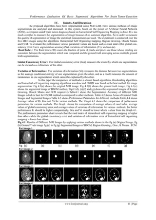
Table 4.4: Average values of Parameters for different methods
Methods Rand Index
Global Consistency
Error
Variation of
Information
Hierarchical Self Organizing Mapping 0.94766 0.15714 1.0498
Region Growing 0.80194 0.36194 2.9932
OTSU 0.74364 0.45684 3.79714
K MEANS 0.75826 0.46152 3.94662
FUZZY CMEANS 0.78108 0.43616 4.5025
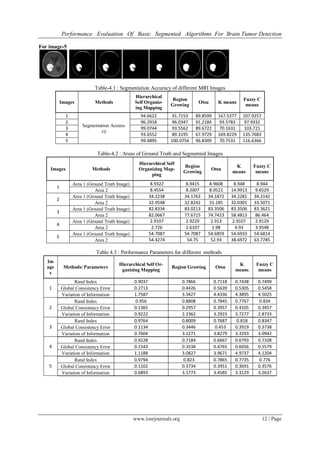
Graph 4.1 : Comparison of Average Values of Ri, Gce & Vi from Various methods
In this graph 4.1 axis of various methods showing 1, 2 ,3 ,4 and 5 which represents HSOM, Region Growing ,
Otsu, K Means and Fuzzy CMeans methods respectively.
References
[1] Arjan Simonetti , ”Investigation of brain tumor classification and its reliability using chemometrics on MR spectroscopy and MR
imaging data”,2004.
[2] Noworolski , S.M.; Nelson, S.J.; Henry, R.G.; Day, M.R.; Wald, L.L.; Star-Lack, J.; Vigneron, D.B. Magnetic resonance in medi-
cine 1999, 41, 21-29.
[3] T.Logeswari1 and M.Karnan2,”An improved implementation of brain tumor detection using segmentation based on soft compu-
ting”,Journal of Cancer Research and Experimental Oncology Vol. 2 pp. 06-014, March, 2010.
[4] B.Sathya and R. Manavalan ,”Image Segmentation by Clustering Methods: Performance Analysis”, IJCA vol 29-No.11,September
2011.
[5] Rafael C.Gonzalez and Richard E. Woods,”Digital Image Processing”,Second Edition,Prentice Hall of India Private Limited ,New
Delhi – 2007.
[6] OtSu NA. “Threshold selection method from aray level histogram”. IEEE Trans. On system, man, and cybernetics. Vol. 9. No. 1.
Pp. 62-66, 1979.
[7] Krishna Kant Singh and Akansha Singh, “A study of Image Segmentation Algorithms for Different Types Of Images”, IJCSI Inter-
national Journal of Computer Science Issues 2010.
[8] Mrs. Bharati R. JipKate et al., A comparative Analysis of Fuzzy C-Means Clustering and K-Means Clustering Algorithm”, IJCER /
May-June, 2012 / Vol. 21 / Issue No. 3 / 737-739.
[9] M. Sezgin and B. Sankur (2004). “Survey over image thresholding techniques and quantitative performance evaluation”. Journal of
Electronic Imaging 13(1) : 146-165.
0
1
2
3
4
5
1 2 3 4 5
AverageValues
VariousMethods
Comparison of Average Values of Ri,Gce & Vi from Various
methods
Ri
Gce
Vi