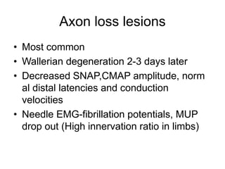
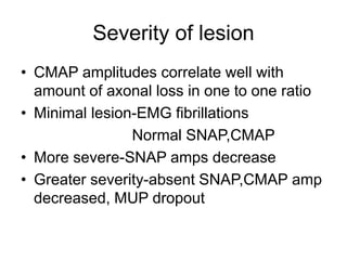
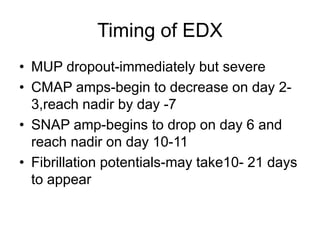
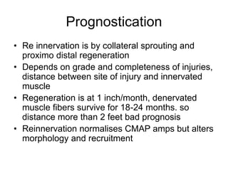
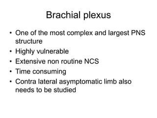
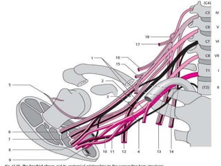
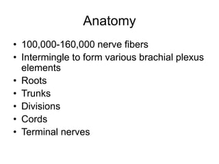
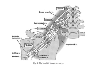
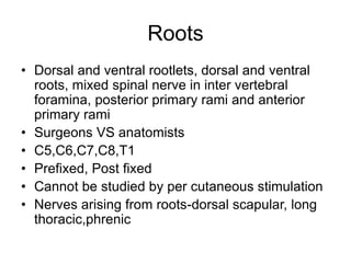
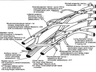
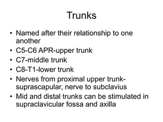
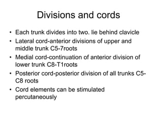
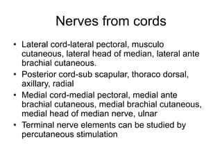
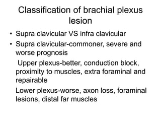
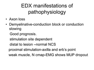
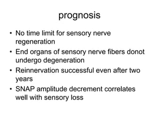
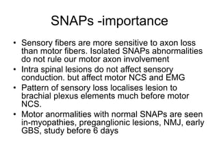
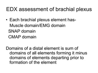
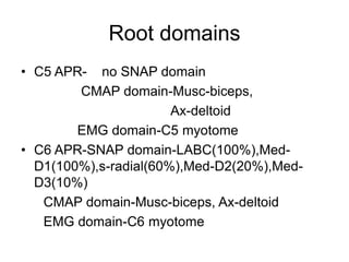
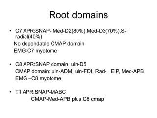
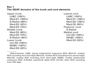
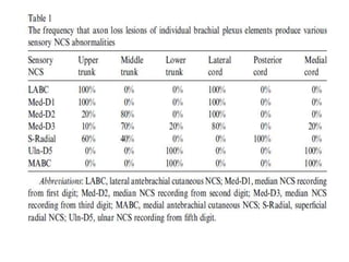
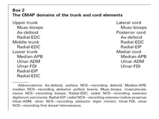
This document discusses the electrophysiological evaluation of the brachial plexus using nerve conduction studies and electromyography. It describes the anatomy of the brachial plexus including its roots, trunks, divisions, cords, and terminal nerves. It explains how to assess each element of the brachial plexus using sensory nerve action potentials, compound muscle action potentials, and electromyography findings. It also discusses the patterns seen with different types of brachial plexus lesions and provides guidance on timing of electrodiagnostic testing and prognostication.
Transcranial Motor Evoked Potentials Monitoring per aACNS guidelinesAnurag Tewari MD
Motor evoked potentials (MEPs) are electrical signals recorded from neural tissue or
muscle following activation of central motor pathways. They complement other clinical
neurophysiology techniques, such as somatosensory evoked potentials (SEPs), in the assessment
of the nervous system, especially during intraoperative neurophysiologic monitoring (IONM).
what is RNS and what the techniques to perform this test in the lab. Its significance in the evaluation and diagnosis of NMJ disorders like MG, LEMBS etc..
This presentation describes the common conditions, anatomy and the ideal ways to do and perform nerve conduction studies in lower limbs. It is nicely depicted with self explanatory pictures.
This presentation is an introduction to the principles of Nerve Conduction Study and is entirely sourced from the book by David C Preston and Barbara E Shapiro: Electromyography and Neuromuscular disorders, 3rd Edition
Delivery of electrical current to a specific subcortical grey matter target to stimulate a desired group of nerve cells which results in specific modulation the output of the involved neurocirciut.
Spinal cord injury is a low incidence, high cost disability requiring tremendous changes in an individual’s lifestyle
Tetraplegia - lesion
Paraplegia - lesion
Title: Sense of Smell
Presenter: Dr. Faiza, Assistant Professor of Physiology
Qualifications:
MBBS (Best Graduate, AIMC Lahore)
FCPS Physiology
ICMT, CHPE, DHPE (STMU)
MPH (GC University, Faisalabad)
MBA (Virtual University of Pakistan)
Learning Objectives:
Describe the primary categories of smells and the concept of odor blindness.
Explain the structure and location of the olfactory membrane and mucosa, including the types and roles of cells involved in olfaction.
Describe the pathway and mechanisms of olfactory signal transmission from the olfactory receptors to the brain.
Illustrate the biochemical cascade triggered by odorant binding to olfactory receptors, including the role of G-proteins and second messengers in generating an action potential.
Identify different types of olfactory disorders such as anosmia, hyposmia, hyperosmia, and dysosmia, including their potential causes.
Key Topics:
Olfactory Genes:
3% of the human genome accounts for olfactory genes.
400 genes for odorant receptors.
Olfactory Membrane:
Located in the superior part of the nasal cavity.
Medially: Folds downward along the superior septum.
Laterally: Folds over the superior turbinate and upper surface of the middle turbinate.
Total surface area: 5-10 square centimeters.
Olfactory Mucosa:
Olfactory Cells: Bipolar nerve cells derived from the CNS (100 million), with 4-25 olfactory cilia per cell.
Sustentacular Cells: Produce mucus and maintain ionic and molecular environment.
Basal Cells: Replace worn-out olfactory cells with an average lifespan of 1-2 months.
Bowman’s Gland: Secretes mucus.
Stimulation of Olfactory Cells:
Odorant dissolves in mucus and attaches to receptors on olfactory cilia.
Involves a cascade effect through G-proteins and second messengers, leading to depolarization and action potential generation in the olfactory nerve.
Quality of a Good Odorant:
Small (3-20 Carbon atoms), volatile, water-soluble, and lipid-soluble.
Facilitated by odorant-binding proteins in mucus.
Membrane Potential and Action Potential:
Resting membrane potential: -55mV.
Action potential frequency in the olfactory nerve increases with odorant strength.
Adaptation Towards the Sense of Smell:
Rapid adaptation within the first second, with further slow adaptation.
Psychological adaptation greater than receptor adaptation, involving feedback inhibition from the central nervous system.
Primary Sensations of Smell:
Camphoraceous, Musky, Floral, Pepperminty, Ethereal, Pungent, Putrid.
Odor Detection Threshold:
Examples: Hydrogen sulfide (0.0005 ppm), Methyl-mercaptan (0.002 ppm).
Some toxic substances are odorless at lethal concentrations.
Characteristics of Smell:
Odor blindness for single substances due to lack of appropriate receptor protein.
Behavioral and emotional influences of smell.
Transmission of Olfactory Signals:
From olfactory cells to glomeruli in the olfactory bulb, involving lateral inhibition.
Primitive, less old, and new olfactory systems with different path
Knee anatomy and clinical tests 2024.pdfvimalpl1234
This includes all relevant anatomy and clinical tests compiled from standard textbooks, Campbell,netter etc..It is comprehensive and best suited for orthopaedicians and orthopaedic residents.
Adv. biopharm. APPLICATION OF PHARMACOKINETICS : TARGETED DRUG DELIVERY SYSTEMSAkankshaAshtankar
MIP 201T & MPH 202T
ADVANCED BIOPHARMACEUTICS & PHARMACOKINETICS : UNIT 5
APPLICATION OF PHARMACOKINETICS : TARGETED DRUG DELIVERY SYSTEMS By - AKANKSHA ASHTANKAR
share - Lions, tigers, AI and health misinformation, oh my!.pptxTina Purnat
• Pitfalls and pivots needed to use AI effectively in public health
• Evidence-based strategies to address health misinformation effectively
• Building trust with communities online and offline
• Equipping health professionals to address questions, concerns and health misinformation
• Assessing risk and mitigating harm from adverse health narratives in communities, health workforce and health system
ABDOMINAL TRAUMA in pediatrics part one.drhasanrajab
Abdominal trauma in pediatrics refers to injuries or damage to the abdominal organs in children. It can occur due to various causes such as falls, motor vehicle accidents, sports-related injuries, and physical abuse. Children are more vulnerable to abdominal trauma due to their unique anatomical and physiological characteristics. Signs and symptoms include abdominal pain, tenderness, distension, vomiting, and signs of shock. Diagnosis involves physical examination, imaging studies, and laboratory tests. Management depends on the severity and may involve conservative treatment or surgical intervention. Prevention is crucial in reducing the incidence of abdominal trauma in children.
Local Advanced Lung Cancer: Artificial Intelligence, Synergetics, Complex Sys...Oleg Kshivets
Overall life span (LS) was 1671.7±1721.6 days and cumulative 5YS reached 62.4%, 10 years – 50.4%, 20 years – 44.6%. 94 LCP lived more than 5 years without cancer (LS=2958.6±1723.6 days), 22 – more than 10 years (LS=5571±1841.8 days). 67 LCP died because of LC (LS=471.9±344 days). AT significantly improved 5YS (68% vs. 53.7%) (P=0.028 by log-rank test). Cox modeling displayed that 5YS of LCP significantly depended on: N0-N12, T3-4, blood cell circuit, cell ratio factors (ratio between cancer cells-CC and blood cells subpopulations), LC cell dynamics, recalcification time, heparin tolerance, prothrombin index, protein, AT, procedure type (P=0.000-0.031). Neural networks, genetic algorithm selection and bootstrap simulation revealed relationships between 5YS and N0-12 (rank=1), thrombocytes/CC (rank=2), segmented neutrophils/CC (3), eosinophils/CC (4), erythrocytes/CC (5), healthy cells/CC (6), lymphocytes/CC (7), stick neutrophils/CC (8), leucocytes/CC (9), monocytes/CC (10). Correct prediction of 5YS was 100% by neural networks computing (error=0.000; area under ROC curve=1.0).
Muktapishti is a traditional Ayurvedic preparation made from Shoditha Mukta (Purified Pearl), is believed to help regulate thyroid function and reduce symptoms of hyperthyroidism due to its cooling and balancing properties. Clinical evidence on its efficacy remains limited, necessitating further research to validate its therapeutic benefits.
These simplified slides by Dr. Sidra Arshad present an overview of the non-respiratory functions of the respiratory tract.
Learning objectives:
1. Enlist the non-respiratory functions of the respiratory tract
2. Briefly explain how these functions are carried out
3. Discuss the significance of dead space
4. Differentiate between minute ventilation and alveolar ventilation
5. Describe the cough and sneeze reflexes
Study Resources:
1. Chapter 39, Guyton and Hall Textbook of Medical Physiology, 14th edition
2. Chapter 34, Ganong’s Review of Medical Physiology, 26th edition
3. Chapter 17, Human Physiology by Lauralee Sherwood, 9th edition
4. Non-respiratory functions of the lungs https://academic.oup.com/bjaed/article/13/3/98/278874
Rasamanikya is a excellent preparation in the field of Rasashastra, it is used in various Kushtha Roga, Shwasa, Vicharchika, Bhagandara, Vatarakta, and Phiranga Roga. In this article Preparation& Comparative analytical profile for both Formulationon i.e Rasamanikya prepared by Kushmanda swarasa & Churnodhaka Shodita Haratala. The study aims to provide insights into the comparative efficacy and analytical aspects of these formulations for enhanced therapeutic outcomes.
- Video recording of this lecture in English language: https://youtu.be/kqbnxVAZs-0
- Video recording of this lecture in Arabic language: https://youtu.be/SINlygW1Mpc
- Link to download the book free: https://nephrotube.blogspot.com/p/nephrotube-nephrology-books.html
- Link to NephroTube website: www.NephroTube.com
- Link to NephroTube social media accounts: https://nephrotube.blogspot.com/p/join-nephrotube-on-social-media.html
2. Brachial plexus
• One of the most complex and largest PNS
structure
• Highly vulnerable
• Extensive non routine NCS
• Time consuming
• Contra lateral asymptomatic limb also
needs to be studied
3.
4. Anatomy
• 100,000-160,000 nerve fibers
• Intermingle to form various brachial plexus
elements
• Roots
• Trunks
• Divisions
• Cords
• Terminal nerves
5.
6. Roots
• Dorsal and ventral rootlets, dorsal and ventral
roots, mixed spinal nerve in inter vertebral
foramina, posterior primary rami and anterior
primary rami
• Surgeons VS anatomists
• C5,C6,C7,C8,T1
• Prefixed, Post fixed
• Cannot be studied by per cutaneous stimulation
• Nerves arising from roots-dorsal scapular, long
thoracic,phrenic
7.
8. Trunks
• Named after their relationship to one
another
• C5-C6 APR-upper trunk
• C7-middle trunk
• C8-T1-lower trunk
• Nerves from proximal upper trunk-
suprascapular, nerve to subclavius
• Mid and distal trunks can be stimulated in
supraclavicular fossa and axilla
9.
10. Divisions and cords
• Each trunk divides into two. lie behind clavicle
• Lateral cord-anterior divisions of upper and
middle trunk C5-7roots
• Medial cord-continuation of anterior division of
lower trunk C8-T1roots
• Posterior cord-posterior division of all trunks C5-
C8 roots
• Cord elements can be stimulated
percutaneously
11.
12. Nerves from cords
• Lateral cord-lateral pectoral, musculo
cutaneous, lateral head of median, lateral ante
brachial cutaneous.
• Posterior cord-sub scapular, thoraco dorsal,
axillary, radial
• Medial cord-medial pectoral, medial ante
brachial cutaneous, medial brachial cutaneous,
medial head of median nerve, ulnar
• Terminal nerve elements can be studied by
percutaneous stimulation
13.
14. Classification of brachial plexus
lesion
• Supra clavicular VS infra clavicular
• Supra clavicular-commoner, severe and
worse prognosis
Upper plexus-better, conduction block,
proximity to muscles, extra foraminal and
repairable
Lower plexus-worse, axon loss, foraminal
lesions, distal far muscles
15. EDX manifestations of
pathophysiology
• Axon loss
• Demyelinative-conduction block or conduction
slowing
Good prognosis.
stimulation site dependent
distal to lesion –normal NCS
proximal stimulation-axilla and erb’s point
weak muscle, N cmap-EMG shows MUP dropout
16. Axon loss lesions
• Most common
• Wallerian degeneration 2-3 days later
• Decreased SNAP,CMAP amplitude, norm
al distal latencies and conduction
velocities
• Needle EMG-fibrillation potentials, MUP
drop out (High innervation ratio in limbs)
17. Severity of lesion
• CMAP amplitudes correlate well with
amount of axonal loss in one to one ratio
• Minimal lesion-EMG fibrillations
Normal SNAP,CMAP
• More severe-SNAP amps decrease
• Greater severity-absent SNAP,CMAP amp
decreased, MUP dropout
18. Timing of EDX
• MUP dropout-immediately but severe
• CMAP amps-begin to decrease on day 2-
3,reach nadir by day -7
• SNAP amp-begins to drop on day 6 and
reach nadir on day 10-11
• Fibrillation potentials-may take10- 21 days
to appear
19. Prognostication
• Re innervation is by collateral sprouting and
proximo distal regeneration
• Depends on grade and completeness of injuries,
distance between site of injury and innervated
muscle
• Regeneration is at 1 inch/month, denervated
muscle fibers survive for 18-24 months. so
distance more than 2 feet bad prognosis
• Reinnervation normalises CMAP amps but alters
morphology and recruitment
20. prognosis
• No time limit for sensory nerve
regeneration
• End organs of sensory nerve fibers donot
undergo degeneration
• Reinnervation successful even after two
years
• SNAP amplitude decrement correlates
well with sensory loss
21. SNAPs -importance
• Sensory fibers are more sensitive to axon loss
than motor fibers. Isolated SNAPs abnormalities
do not rule our motor axon involvement
• Intra spinal lesions do not affect sensory
conduction. but affect motor NCS and EMG
• Pattern of sensory loss localises lesion to
brachial plexus elements much before motor
NCS.
• Motor anormalities with normal SNAPs are seen
in-myopathies, preganglionic lesions, NMJ, early
GBS, study before 6 days
22. EDX assessment of brachial plexus
• Each brachial plexus element has-
Muscle domain/EMG domain
SNAP domain
CMAP domain
Domains of a distal element is sum of
domains of all elements forming it minus
domains of elements departing prior to
formation of the element
28. EMG domains
• Upper trunk-(C5 plus C6) minus dorsal scapular,
long thoracic nerve.
• Middle trunk-C7 domain minus serratus anterior
• Lower trunk-C8 plus T1 APR
• Lateral cord-upper and middle trunks minus
supra scapular, subscapular, thoraco dorsal,
radial, axillary nerve
• Posterior cord-sum of sub scapular, thoraco
dorsal ,axillary and radial
• Medial cord-lower trunk minus posterior division
elements