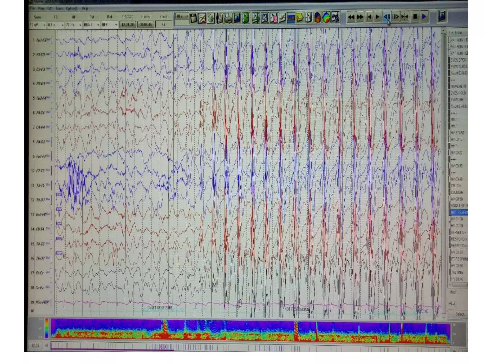
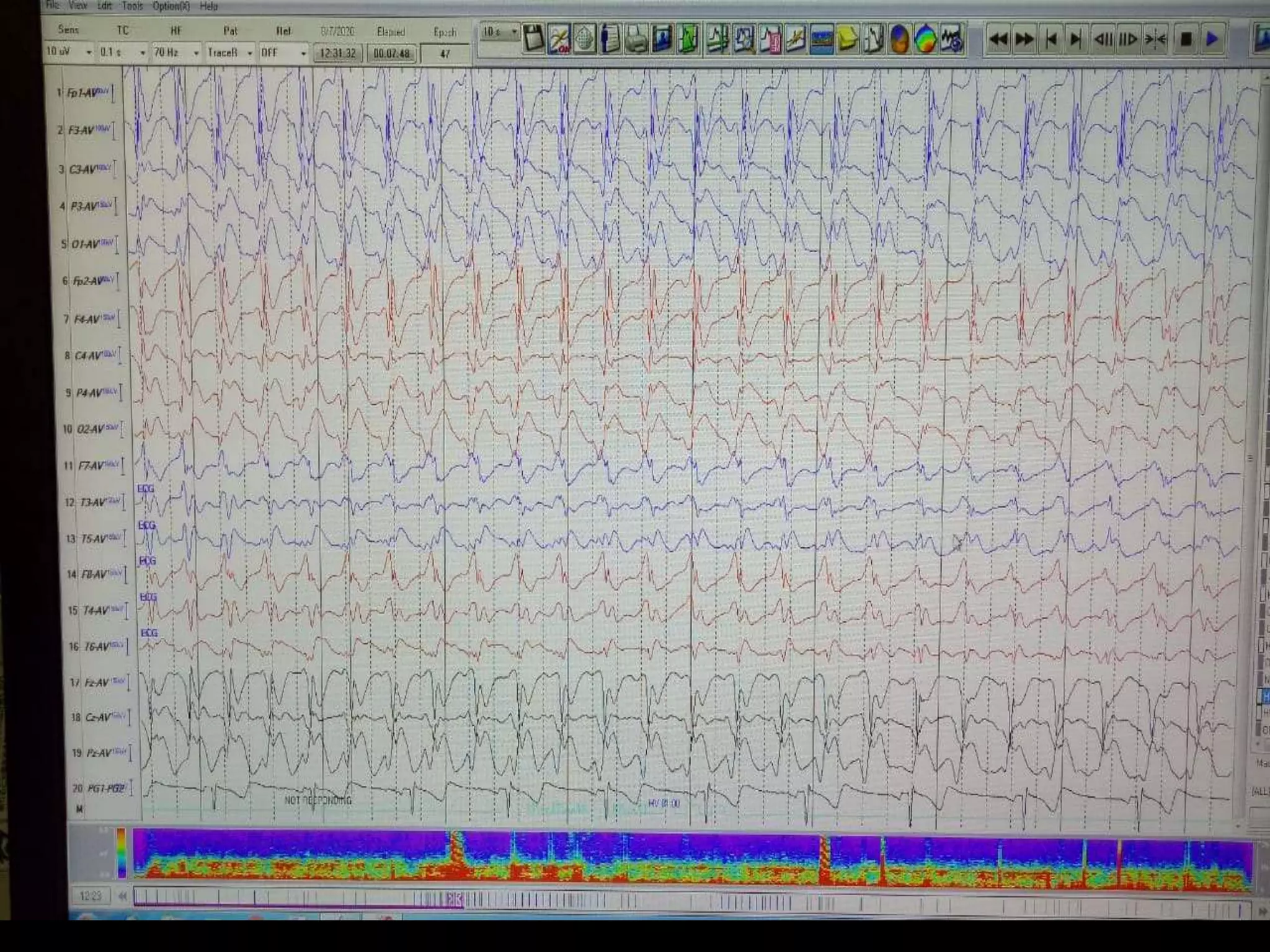
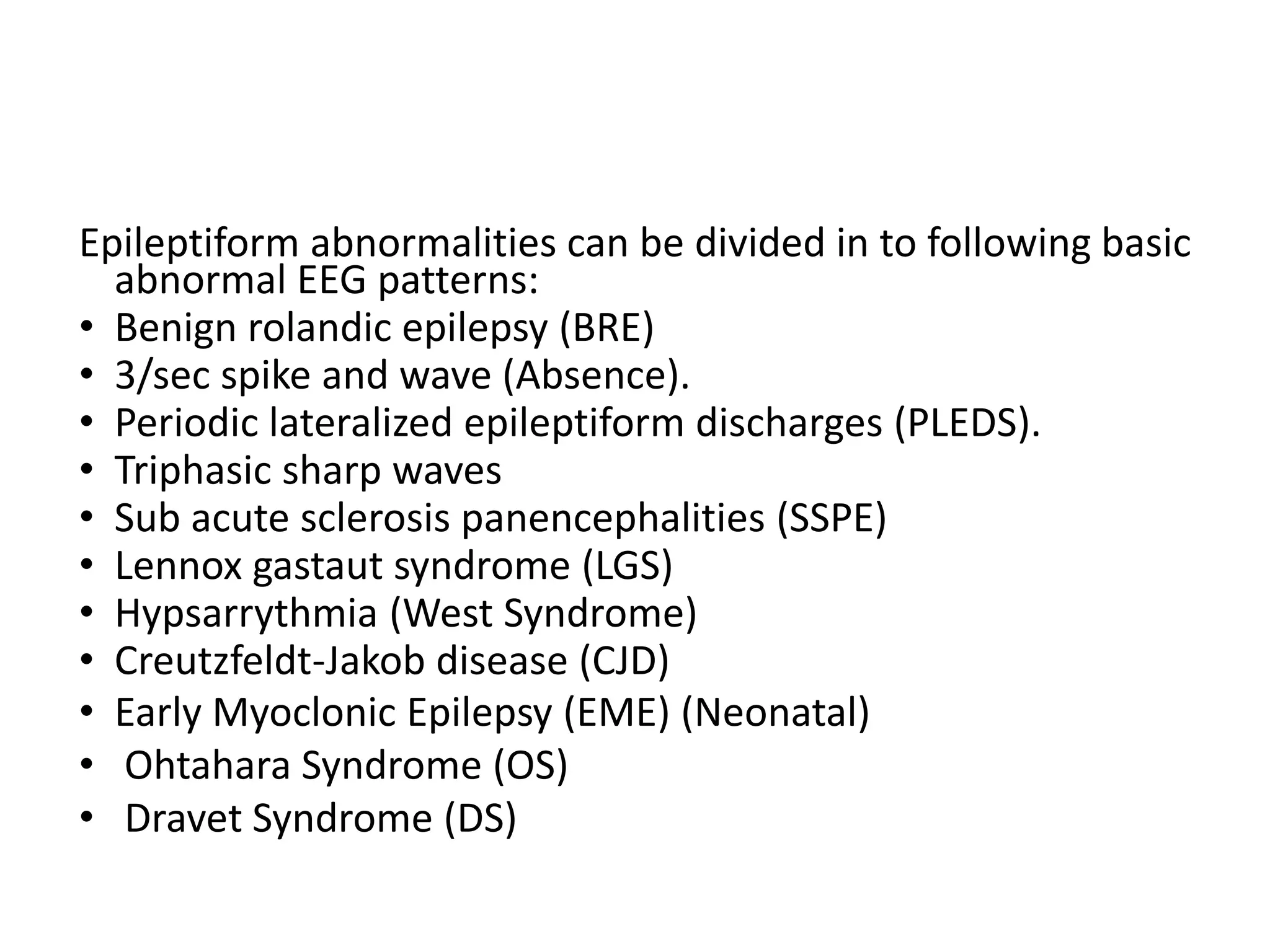
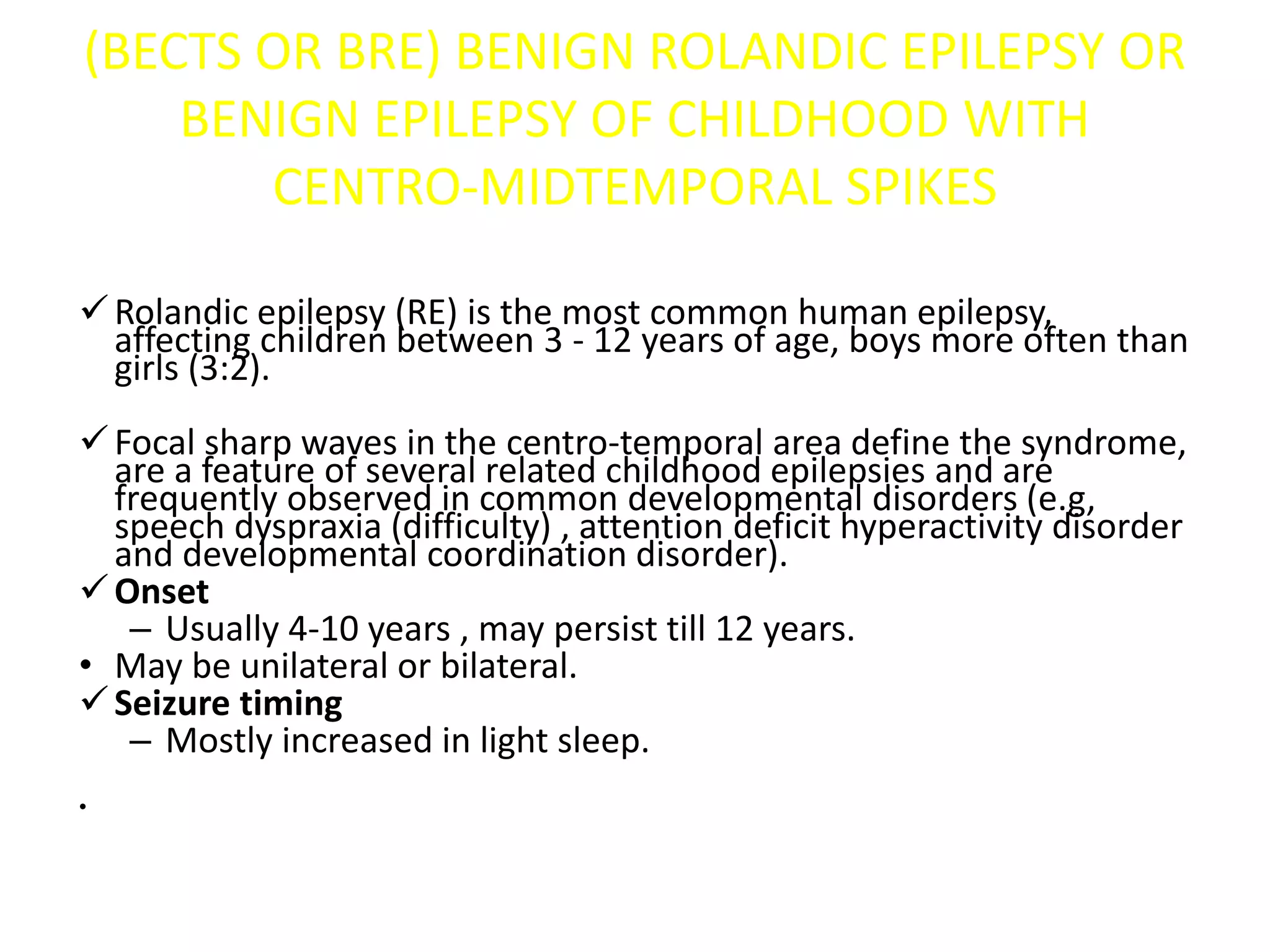
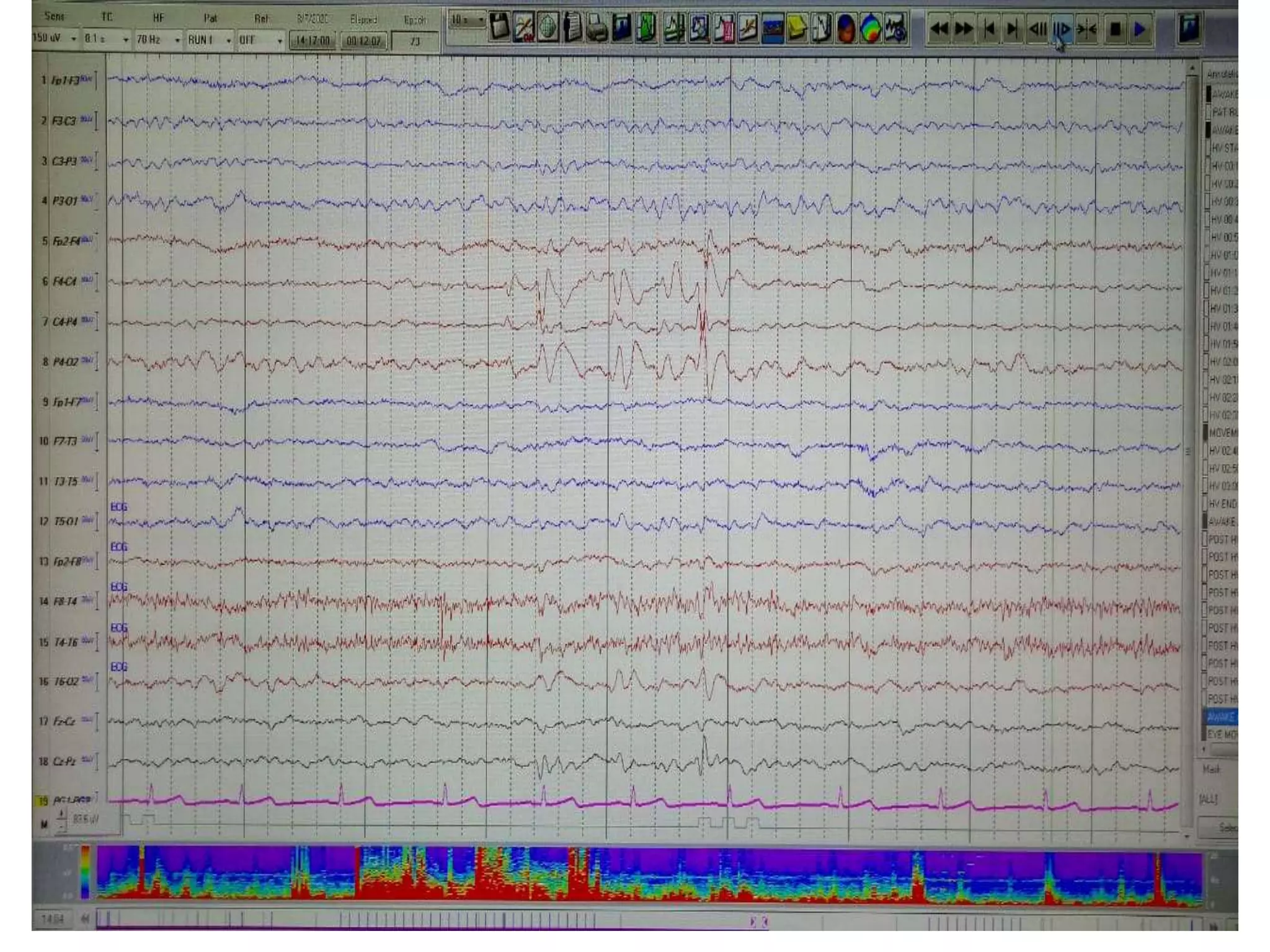
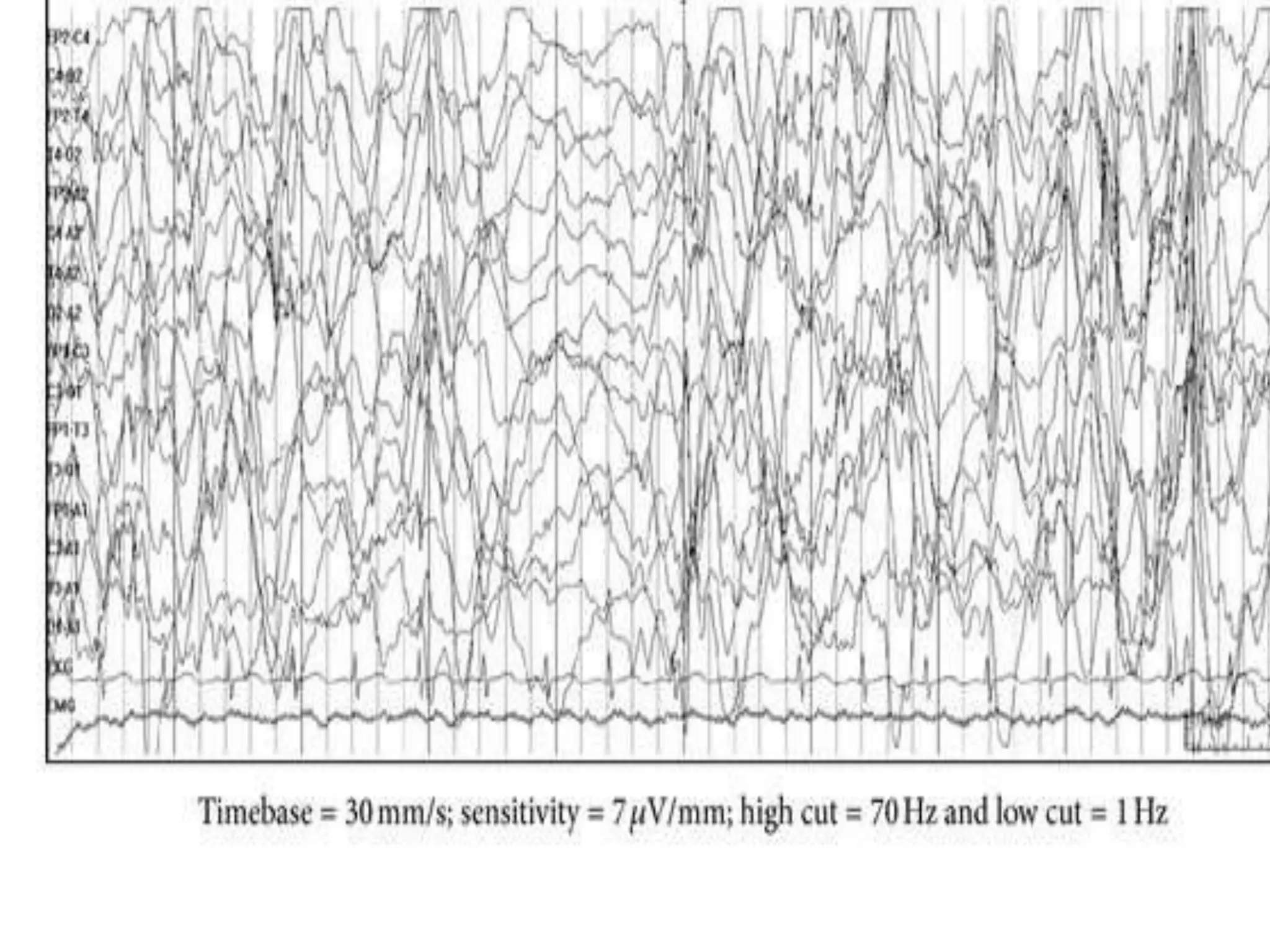
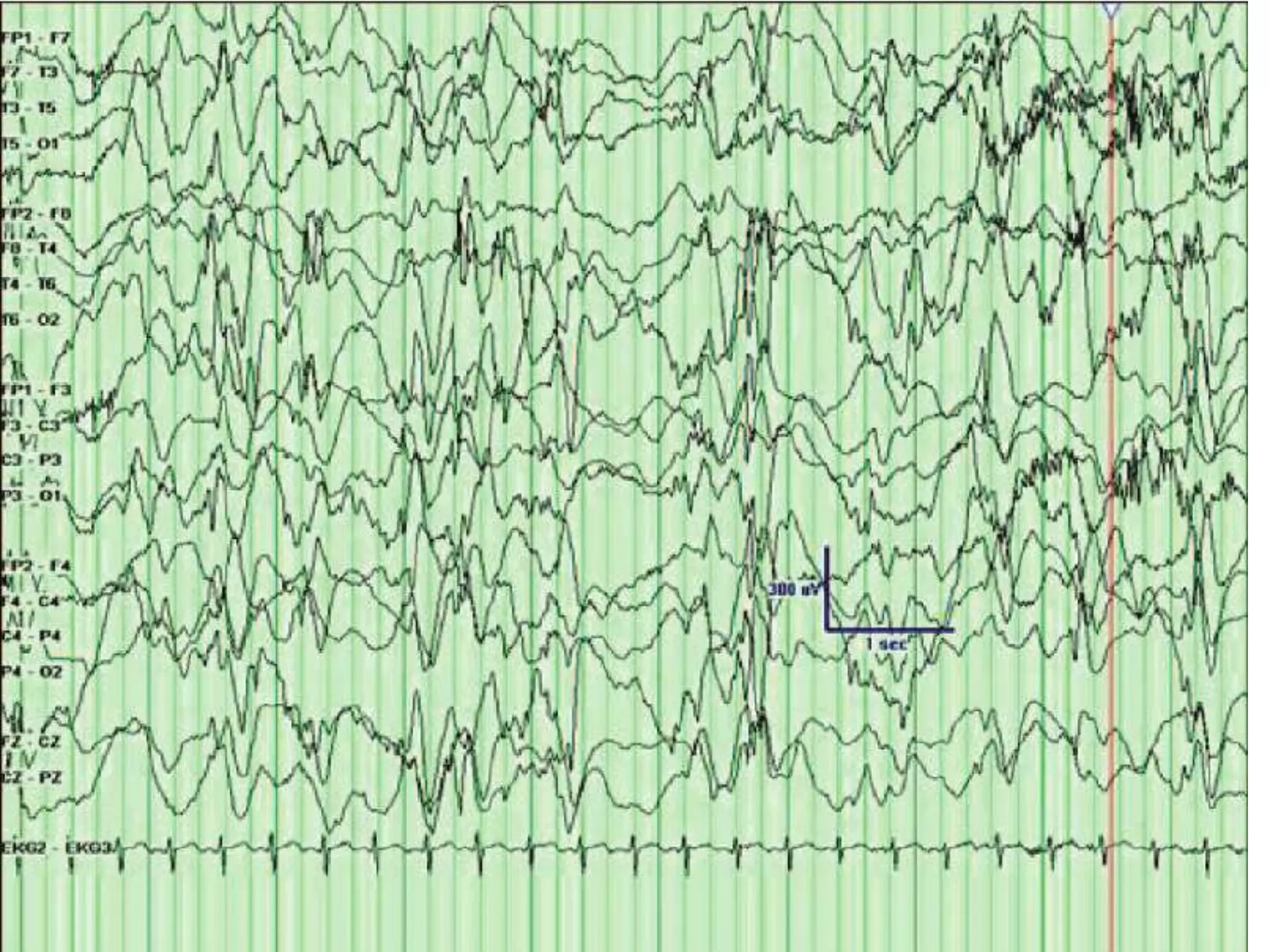
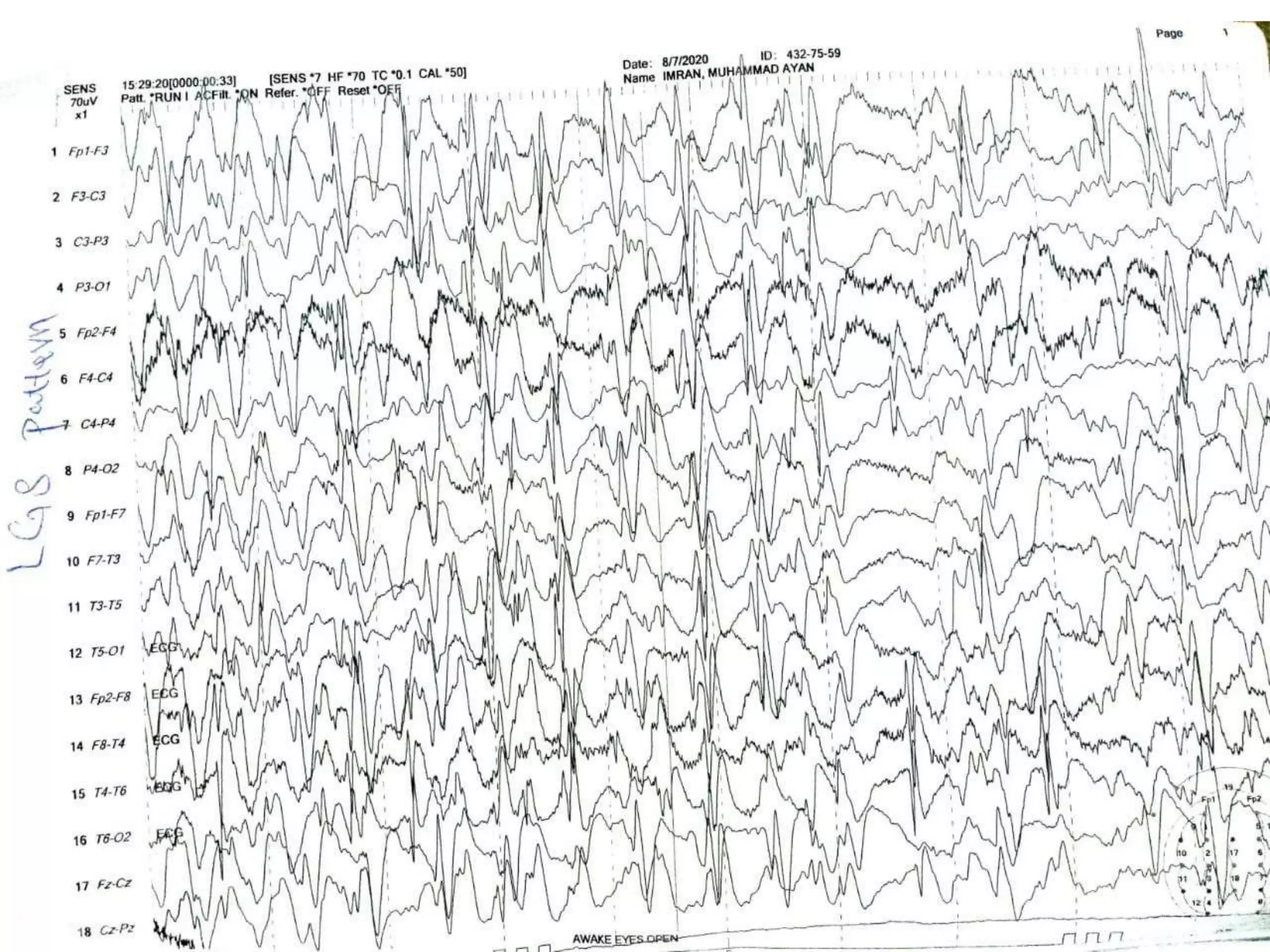
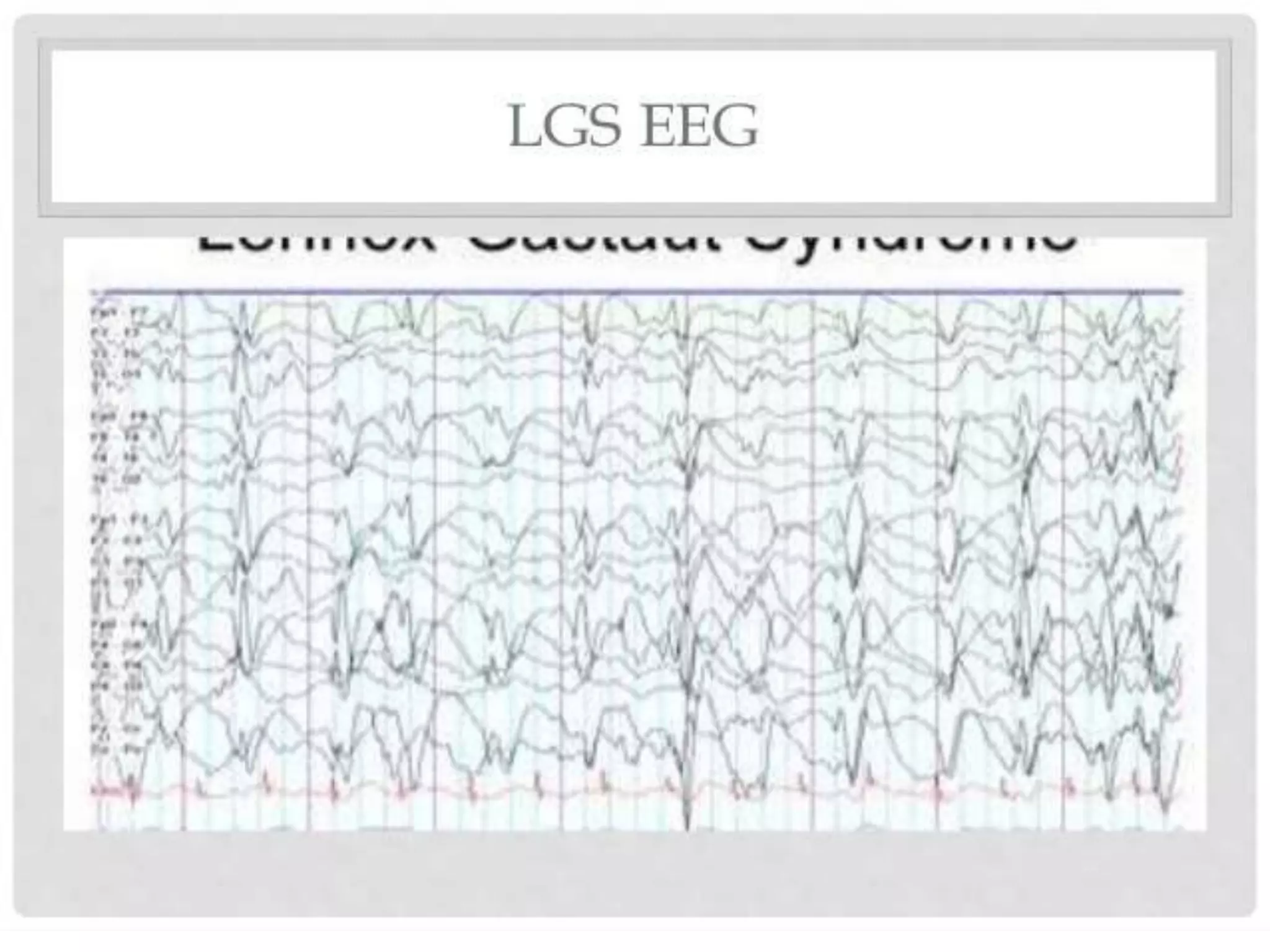
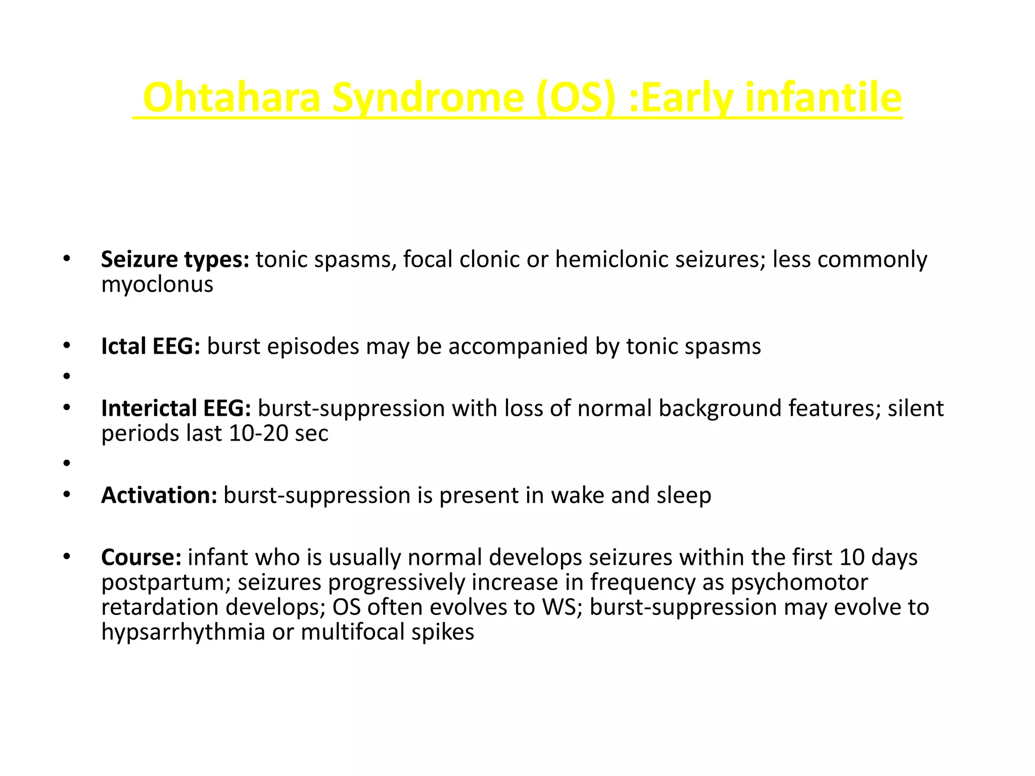
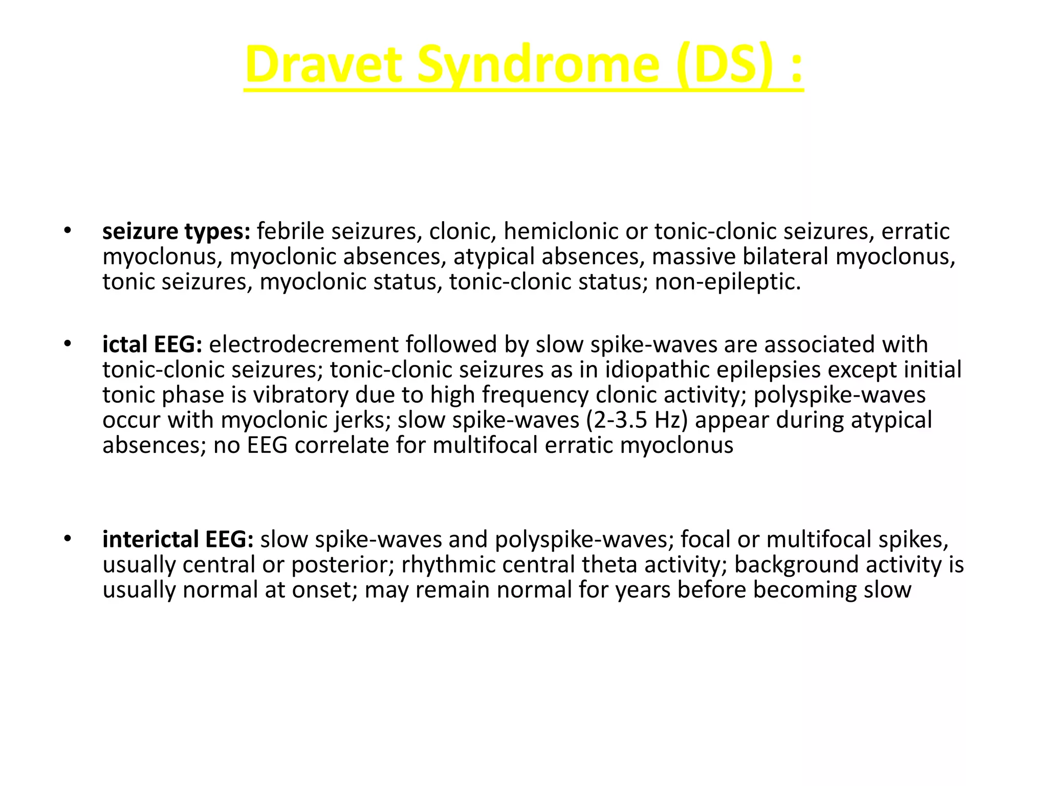
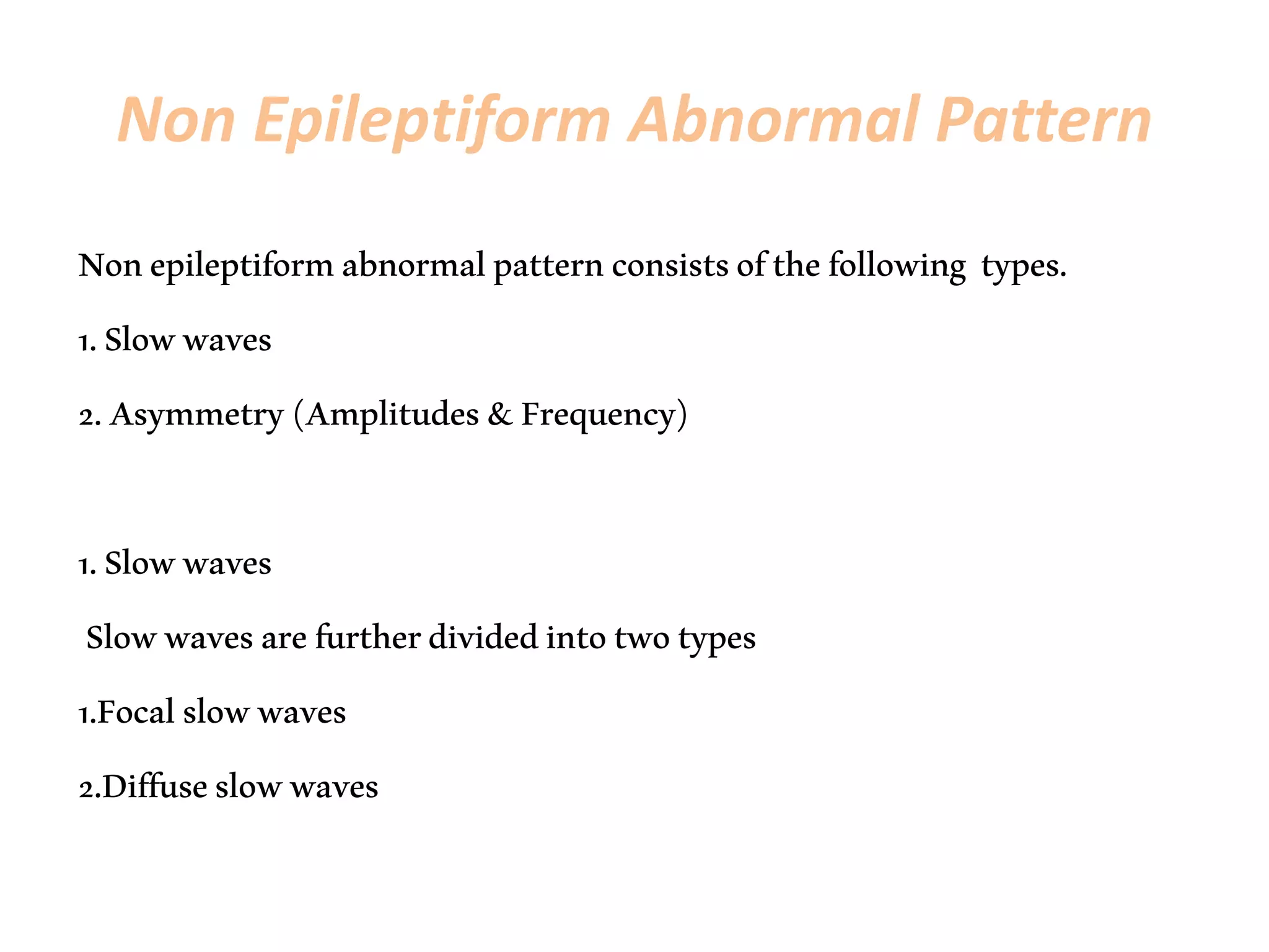
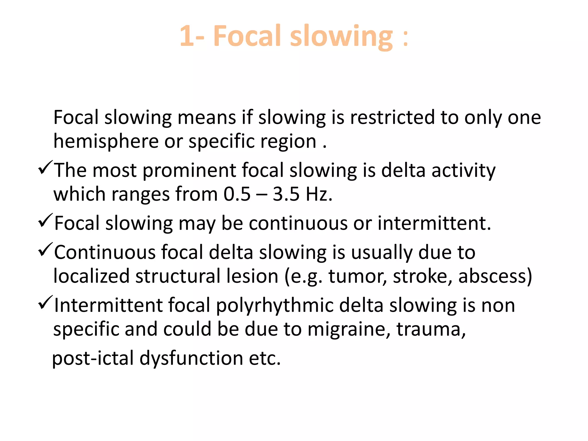
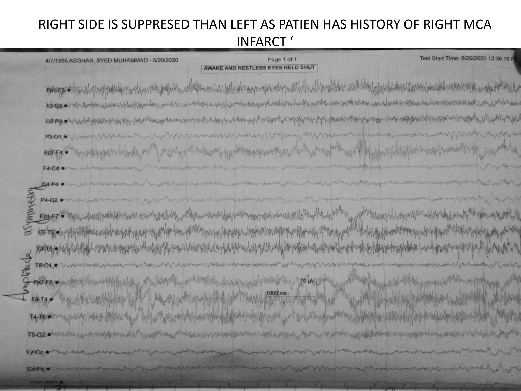
CLINICAL NEUROPHYSIOLOGY AGA
KHAN UNIVERSITY HOSPITAL KARACHI
PAKISTAN
Assignment:AbnormalEEGpatterns
Name:FaizanAbdullah
Designation:TraineeTechnologistNeurophysiology
Date:27/08/20
Content
 Introduction
 Abnormal EEG types
 Epileptic EEG patterns
– Benign rolandic epilepsy (BRE)
– 3/sec spike and wave
(Absence).
– Periodic lateralized
epileptiform discharges
(PLEDS).
– Triphasic sharp waves
– Sub acute sclerosis
panencephalities (SSPE)
– Lennox gastaut syndrome
(LGS)
– Hypsarrythmia (West
Syndrome)
– Creutzfeldt-Jakob disease
(CJD)
– Early Myoclonic Epilepsy (EME)
(Neonatal)
– Ohtahara Syndrome (OS)
– Dravet Syndrome (DS)
 Non epileptic EEG
abnormalities
 References
EEG STANDS FOR
• An electroencephalogram (EEG) is a test used
to find problems related to electrical activity
of the brain. An EEG tracks and records brain
wave patterns. Small metal discs with thin
wires (electrodes) are placed on the scalp, and
then send signals to a computer to record the
results.
• EEG depends upon age state of the patient
because EEG varies according to age and state
EEG ABNORMALITY
Any activity which does not correlate with the
age and state of the patient.
Abnormality could be of the following two
main types of activity.
1. Abnormal Non-epileptiform pattern
2. Epileptiform Pattern
Epileptiform abnormal pattern
Epileptiform activity mainly consists of spike and sharp waves.
• Spike :
A transient clearly distinguished from the background activity
with pointed peak, duration from 20-70ms mostly followed by slow
wave.
• Sharp wave :
A transient which is clearly distinguished from the background
activity with blunted peak, duration from 70-200ms.
• Polyspikes :
It is complex clearly distinguished from the background
activity with multiple spikes ,mostly followed by slow wave. With
more tghen 3spikes.
MAINLY TWO TYPES OF EPILEPTIFORM
ACTIVITY
Focal epileptiform activity:
Focal epileptiform activity (spike and sharp
waves) consists of spike and sharp waves that
appear on one or a few neighboring electrodes.
Generalized epileptiform activity:
Epileptiform discharges (spike and sharp
waves)
that appear over most of all parts of both
hemisphere
and usually have symmetrical shape, amplitude
and
timing in corresponding areas.
Epileptiform abnormalities can be divided in to following basic
abnormal EEG patterns:
• Benign rolandic epilepsy (BRE)
• 3/sec spike and wave (Absence).
• Periodic lateralized epileptiform discharges (PLEDS).
• Triphasic sharp waves
• Sub acute sclerosis panencephalities (SSPE)
• Lennox gastaut syndrome (LGS)
• Hypsarrythmia (West Syndrome)
• Creutzfeldt-Jakob disease (CJD)
• Early Myoclonic Epilepsy (EME) (Neonatal)
• Ohtahara Syndrome (OS)
• Dravet Syndrome (DS)
(BECTS OR BRE) BENIGN ROLANDIC EPILEPSY OR
BENIGN EPILEPSY OF CHILDHOOD WITH
CENTRO-MIDTEMPORAL SPIKES
Rolandic epilepsy (RE) is the most common human epilepsy,
affecting children between 3 - 12 years of age, boys more often than
girls (3:2).
Focal sharp waves in the centro-temporal area define the syndrome,
are a feature of several related childhood epilepsies and are
frequently observed in common developmental disorders (e.g,
speech dyspraxia (difficulty) , attention deficit hyperactivity disorder
and developmental coordination disorder).
Onset
– Usually 4-10 years , may persist till 12 years.
• May be unilateral or bilateral.
Seizure timing
– Mostly increased in light sleep.
•
• Inter-ictal shows frontal +ve centro-temporal
–ve spikes
• Ictal initially shows decremental response
• Common in girls as compaire to boys 3:2
Clinically:
.Patient complaint of paresthesias unilateral face
and tongue numbness.
.Could be GTCS but unilateral
ABSENCE SEIZURES
The three Hz spike and wave pattern is suggestive of
idiopathic Generalized epilepsy.
• Characteristics:
It is classically described with typical absence epilepsies when bursts
of 3Hz spike and wave is Gen, regular, symmetrical, synchronous and
maximal in the anterior head region
Clinical symptoms: fluttering of eyes and automatism.
It usually occurs in patients aged b/w 5-12 years.
Onset : At 2 years and can be appeared till 14 years.
• Often faster at onset and slows down toward end.
• Accentuated (more prominent) by HV (50-80% pt) and photic
stimulation (about 20% of pt).
CHILDHOOD ABSENCE SEIZURES
• ONSET 3-12YERAS
• Eeg shows 3HZ OEDA have a smaller risk of
developing tonic clonic seizures
• Absence seuzures without myoclonus are
higher chances of remission.
JUVENILE ABSENCE EPILEPSY
• The onset of this type is around 10-12 years
even later.
• Spike wave is faster than 3hz may have
polyspikes.
• This group of patients likely to develop GTCS
PERIODIC LATERIZED EPILEPTIC
DISCHARGES
 PLEDs are basically triphasic with sharply contoured wave followed by a slow wave mostly occurring
unilaterally with duration 100-300 msec and amplitude 100-300 often present with fast rhythm b/w
discharges.
 The term "periodic" was first used by Cobb , in 1950.
 Periodic EEG patterns consist of various forms discharges, usually epileptiform in appearance, and
apply to waves or complexes occurring in sequence at an approximately regular rate or intermittently
regular intervals.
 They are commonly classified as periodic lateralized epileptiform discharges (PLEDs),
Characteristics:
• Periodic recurrence every 0.5-4.0 sec.
• With frequency of 1-2 /sec.
• Etiology (Cause): acute infarction, CVA, tumor, anoxia and herpes simplex encephalitis, abscess etc.
 This term was first used by Chatrian in 1964
 A pattern consists of lateralized complexes, usually recurring every 1.0 to 2.0 seconds, and often (but not always)
appear as sharp waves or spikes which may be followed by a slow wave at periodic intervals.
 The discharges may repeat as fast as 3/s or as slow as 10/min
 PLEDs may be subclassified into PLEDs-proper and PLEDs-plus.
 PLEDs-proper, in which the periodicity of the discharges is relatively stable, uniform, and there are no associated
rhythmic discharges
 PLEDs-plus, in which the periodicity of the discharges is variable and there is associated low amplitude rhythmic
activity with the discharges and its more associated with seizures.
 PLEDs tend to be transient and resolve spontaneously within 2 to 3 weeks,
ETIOLOGY: Acute brain injury, cerebral
infarction, glioblastoma, brain abscesses, viral
encephalitis (especially related to the Herpes
simplex virus), Creutzfeldt-Jakob disease (CJD),
hematomas and, less frequently, in demyelinating
diseases, anoxia, primary epilepsia, migraine and
fat embolism syndrome.
TRIPHASIC SHARP WAVES
These are typical and sharply contoured wave discharges.
Characteristics
Having three phase -ve, +ve and then -ve.
• Initial is sharp component.
• FORMS LAG ANTERIOR TO POTERIOR
• Appeared with rhythmic train of 1.5-2.5 /sec.
• Having medium to high voltage (>70uv) .
• Bilaterally synchronous and symmetrical
• These waves are having anterior posterior lag of 25-140 sec.
• Best seen in referential montage.
• Appeared in hypoxic state, subdural hematoma, intoxication (excite
by the action of a chemical substance ), mainly in hepatic
encephalopathy.
• Seen in deep impairment of consciousness but occasionally in awake
state.
SUBACUTE SCLEROSING
PANENCEPHALITIS
• Repetitive bursts of high voltage spike, sharp and slow wave complexes,
recurring after every 4-15 seconds.
• More prominent in awaking state.
• Gen. but maximum over fronto-central areas.
• Frequency ranges 4-15/sec
• All the waves are Stereotyped (morphologically similar)
• Bilaterally synchronous
• Periodic high voltage 300-1500uv of sharp and slow waves complex with an
irregular delta wave superimposed.
• CLINICALLY:
• PATIENT WILL BE MENTIALLY RETARDED .
• It relates with measles and myoclonic jerks
• JERKS WILL BE TIME LOCKED
• It is an inflammatory disease that occurs in children and adolescents and is
believed to be caused by measles virus
HYHPSARRYTHMIA
• Hypsarrhythmia is an abnormal pattern, consisting of
high amplitude and irregular paroxysmal sharp wave,
spike, poly spikes, independent, multifocal and focal
spike on all cortical region with a background of chaotic
(disorderly) and disorganized activity
• It is a pattern seen in patient with infantile spasm
• Hypsarrhythmia is frequently found in patients with
West syndrome .
• Appeared at the age of 4-6 or 13 months.
• Clinically patient is mentally and physically retarded.
• Also diffuse slowing is present.
LENOX GASTAUT SYNDROME
• It is the disease of the later phase of hypsarrythmia
which usually shows slow spike and wave discharges
and may show discharges of gen spikes.
• Duration 2.5/sec or less.
• The discharges may be Gen and synchronous
• Highest amplitudes in midline frontal region (Fz).
• The discharges may become continuous during sleep.
• Patient gradually becomes mentally retarded.
• Within the age of 11-12 years most of the patients die,
but some may live upto 20 years, depending on the
affecting of syndrome.
Creutzfeldt-Jakob disease (CJD)
• CJD is at times called a human form of mad cow
disease in which the brain tissue develops holes
and takes on a sponge-like texture. This is due to a
type of infectious protein called a prion.
• Prions are mis-folded proteins which replicate by
converting their properly folded counterparts.
• CLINICAL SYMTOMS:
• Symptoms of CJD are rapidly progressive
dementia, leading to memory loss, personality
changes and hallucinations.
• This is accompanied by physical problems such as speech
impairment, jerky movements (myoclonus), balance and
coordination dysfunction (ataxia), changes in gait, rigid
posture, and seizures.
EEG FINDINGS:
• Periodic discharges with sharp wave or a sharp triphasic
complex.
• 100 -300msec duration
• Repetition rate of 0.5-2/sec.
• Shows severely disorganized & generalized synchrony
background.
• Some cases show unilateral discharges over one
hemispheres.
Early Myoclonic Epilepsy (EME) in
infancy
• Seizure types:
Erratic focal myoclonus, focal clonic or tonic seizures,
tonic spasms; less commonly: massive bilateral myoclonus,
epileptic spasms
• Ictal EEG: onset of focal seizures are similar to neonatal
seizures; generalized discharges are seen during massive
myoclonus; no EEG correlate for erratic myoclonus
• Interictal EEG: burst-suppression with loss of normal
background features; silent periods last 3-10 sec
• Activation: burst-suppression, almost limited to sleep
Ohtahara Syndrome (OS) :Early infantile
• Seizure types: tonic spasms, focal clonic or hemiclonic seizures; less commonly
myoclonus
• Ictal EEG: burst episodes may be accompanied by tonic spasms
•
• Interictal EEG: burst-suppression with loss of normal background features; silent
periods last 10-20 sec
•
• Activation: burst-suppression is present in wake and sleep
• Course: infant who is usually normal develops seizures within the first 10 days
postpartum; seizures progressively increase in frequency as psychomotor
retardation develops; OS often evolves to WS; burst-suppression may evolve to
hypsarrhythmia or multifocal spikes
Dravet Syndrome (DS) :
• seizure types: febrile seizures, clonic, hemiclonic or tonic-clonic seizures, erratic
myoclonus, myoclonic absences, atypical absences, massive bilateral myoclonus,
tonic seizures, myoclonic status, tonic-clonic status; non-epileptic.
• ictal EEG: electrodecrement followed by slow spike-waves are associated with
tonic-clonic seizures; tonic-clonic seizures as in idiopathic epilepsies except initial
tonic phase is vibratory due to high frequency clonic activity; polyspike-waves
occur with myoclonic jerks; slow spike-waves (2-3.5 Hz) appear during atypical
absences; no EEG correlate for multifocal erratic myoclonus
• interictal EEG: slow spike-waves and polyspike-waves; focal or multifocal spikes,
usually central or posterior; rhythmic central theta activity; background activity is
usually normal at onset; may remain normal for years before becoming slow
Non Epileptiform Abnormal Pattern
Nonepileptiformabnormalpatternconsistsofthefollowing types.
1.Slowwaves
2.Asymmetry(Amplitudes&Frequency)
1.Slowwaves
Slowwavesarefurtherdividedintotwotypes
1.Focalslowwaves
2.Diffuseslowwaves
1- Focal slowing :
Focal slowing means if slowing is restricted to only one
hemisphere or specific region .
The most prominent focal slowing is delta activity
which ranges from 0.5 – 3.5 Hz.
Focal slowing may be continuous or intermittent.
Continuous focal delta slowing is usually due to
localized structural lesion (e.g. tumor, stroke, abscess)
Intermittent focal polyrhythmic delta slowing is non
specific and could be due to migraine, trauma,
post-ictal dysfunction etc.
Diffuse slowing
Diffuse slowing/generalized slowing means
synchronous and symmetrical slowing that
appears in the whole background throughout the
recording which indicates the involvement of both
hemispheres of the brain or cerebral dysfunction.
While reporting diffuse slowing, age, state
(alertness) and medications of the patient must
be known.
•
2. Asymmetry (Amplitudes &
Frequency)
 Asymmetric background could be amplitude-
wise or frequency-wise.
More than 50% amplitude wise asymmetry
and 1 or more than 1% frequency wise
asymmetry throughout the recording is
considerable and it shows hemispheric
abnormality, considering the age and state of
the patient.
RIGHT SIDE IS SUPPRESED THAN LEFT AS PATIEN HAS HISTORY OF RIGHT MCA
INFARCT ‘
References
• PracticalforclinicalneurophysiologyEEGbyyamadathoruandElizabethmeng
• AtlasEEG
• Peerlectures

Abnormal EEG patterns

  • 1.
    CLINICAL NEUROPHYSIOLOGY AGA KHANUNIVERSITY HOSPITAL KARACHI PAKISTAN Assignment:AbnormalEEGpatterns Name:FaizanAbdullah Designation:TraineeTechnologistNeurophysiology Date:27/08/20
  • 2.
    Content  Introduction  AbnormalEEG types  Epileptic EEG patterns – Benign rolandic epilepsy (BRE) – 3/sec spike and wave (Absence). – Periodic lateralized epileptiform discharges (PLEDS). – Triphasic sharp waves – Sub acute sclerosis panencephalities (SSPE) – Lennox gastaut syndrome (LGS) – Hypsarrythmia (West Syndrome) – Creutzfeldt-Jakob disease (CJD) – Early Myoclonic Epilepsy (EME) (Neonatal) – Ohtahara Syndrome (OS) – Dravet Syndrome (DS)  Non epileptic EEG abnormalities  References
  • 3.
    EEG STANDS FOR •An electroencephalogram (EEG) is a test used to find problems related to electrical activity of the brain. An EEG tracks and records brain wave patterns. Small metal discs with thin wires (electrodes) are placed on the scalp, and then send signals to a computer to record the results. • EEG depends upon age state of the patient because EEG varies according to age and state
  • 4.
    EEG ABNORMALITY Any activitywhich does not correlate with the age and state of the patient. Abnormality could be of the following two main types of activity. 1. Abnormal Non-epileptiform pattern 2. Epileptiform Pattern
  • 5.
    Epileptiform abnormal pattern Epileptiformactivity mainly consists of spike and sharp waves. • Spike : A transient clearly distinguished from the background activity with pointed peak, duration from 20-70ms mostly followed by slow wave. • Sharp wave : A transient which is clearly distinguished from the background activity with blunted peak, duration from 70-200ms. • Polyspikes : It is complex clearly distinguished from the background activity with multiple spikes ,mostly followed by slow wave. With more tghen 3spikes.
  • 7.
    MAINLY TWO TYPESOF EPILEPTIFORM ACTIVITY Focal epileptiform activity: Focal epileptiform activity (spike and sharp waves) consists of spike and sharp waves that appear on one or a few neighboring electrodes.
  • 9.
    Generalized epileptiform activity: Epileptiformdischarges (spike and sharp waves) that appear over most of all parts of both hemisphere and usually have symmetrical shape, amplitude and timing in corresponding areas.
  • 12.
    Epileptiform abnormalities canbe divided in to following basic abnormal EEG patterns: • Benign rolandic epilepsy (BRE) • 3/sec spike and wave (Absence). • Periodic lateralized epileptiform discharges (PLEDS). • Triphasic sharp waves • Sub acute sclerosis panencephalities (SSPE) • Lennox gastaut syndrome (LGS) • Hypsarrythmia (West Syndrome) • Creutzfeldt-Jakob disease (CJD) • Early Myoclonic Epilepsy (EME) (Neonatal) • Ohtahara Syndrome (OS) • Dravet Syndrome (DS)
  • 13.
    (BECTS OR BRE)BENIGN ROLANDIC EPILEPSY OR BENIGN EPILEPSY OF CHILDHOOD WITH CENTRO-MIDTEMPORAL SPIKES Rolandic epilepsy (RE) is the most common human epilepsy, affecting children between 3 - 12 years of age, boys more often than girls (3:2). Focal sharp waves in the centro-temporal area define the syndrome, are a feature of several related childhood epilepsies and are frequently observed in common developmental disorders (e.g, speech dyspraxia (difficulty) , attention deficit hyperactivity disorder and developmental coordination disorder). Onset – Usually 4-10 years , may persist till 12 years. • May be unilateral or bilateral. Seizure timing – Mostly increased in light sleep. •
  • 14.
    • Inter-ictal showsfrontal +ve centro-temporal –ve spikes • Ictal initially shows decremental response • Common in girls as compaire to boys 3:2 Clinically: .Patient complaint of paresthesias unilateral face and tongue numbness. .Could be GTCS but unilateral
  • 19.
    ABSENCE SEIZURES The threeHz spike and wave pattern is suggestive of idiopathic Generalized epilepsy. • Characteristics: It is classically described with typical absence epilepsies when bursts of 3Hz spike and wave is Gen, regular, symmetrical, synchronous and maximal in the anterior head region Clinical symptoms: fluttering of eyes and automatism. It usually occurs in patients aged b/w 5-12 years. Onset : At 2 years and can be appeared till 14 years. • Often faster at onset and slows down toward end. • Accentuated (more prominent) by HV (50-80% pt) and photic stimulation (about 20% of pt).
  • 20.
    CHILDHOOD ABSENCE SEIZURES •ONSET 3-12YERAS • Eeg shows 3HZ OEDA have a smaller risk of developing tonic clonic seizures • Absence seuzures without myoclonus are higher chances of remission.
  • 21.
    JUVENILE ABSENCE EPILEPSY •The onset of this type is around 10-12 years even later. • Spike wave is faster than 3hz may have polyspikes. • This group of patients likely to develop GTCS
  • 26.
    PERIODIC LATERIZED EPILEPTIC DISCHARGES PLEDs are basically triphasic with sharply contoured wave followed by a slow wave mostly occurring unilaterally with duration 100-300 msec and amplitude 100-300 often present with fast rhythm b/w discharges.  The term "periodic" was first used by Cobb , in 1950.  Periodic EEG patterns consist of various forms discharges, usually epileptiform in appearance, and apply to waves or complexes occurring in sequence at an approximately regular rate or intermittently regular intervals.  They are commonly classified as periodic lateralized epileptiform discharges (PLEDs), Characteristics: • Periodic recurrence every 0.5-4.0 sec. • With frequency of 1-2 /sec. • Etiology (Cause): acute infarction, CVA, tumor, anoxia and herpes simplex encephalitis, abscess etc.
  • 27.
     This termwas first used by Chatrian in 1964  A pattern consists of lateralized complexes, usually recurring every 1.0 to 2.0 seconds, and often (but not always) appear as sharp waves or spikes which may be followed by a slow wave at periodic intervals.  The discharges may repeat as fast as 3/s or as slow as 10/min  PLEDs may be subclassified into PLEDs-proper and PLEDs-plus.  PLEDs-proper, in which the periodicity of the discharges is relatively stable, uniform, and there are no associated rhythmic discharges  PLEDs-plus, in which the periodicity of the discharges is variable and there is associated low amplitude rhythmic activity with the discharges and its more associated with seizures.  PLEDs tend to be transient and resolve spontaneously within 2 to 3 weeks,
  • 28.
    ETIOLOGY: Acute braininjury, cerebral infarction, glioblastoma, brain abscesses, viral encephalitis (especially related to the Herpes simplex virus), Creutzfeldt-Jakob disease (CJD), hematomas and, less frequently, in demyelinating diseases, anoxia, primary epilepsia, migraine and fat embolism syndrome.
  • 30.
    TRIPHASIC SHARP WAVES Theseare typical and sharply contoured wave discharges. Characteristics Having three phase -ve, +ve and then -ve. • Initial is sharp component. • FORMS LAG ANTERIOR TO POTERIOR • Appeared with rhythmic train of 1.5-2.5 /sec. • Having medium to high voltage (>70uv) . • Bilaterally synchronous and symmetrical • These waves are having anterior posterior lag of 25-140 sec. • Best seen in referential montage. • Appeared in hypoxic state, subdural hematoma, intoxication (excite by the action of a chemical substance ), mainly in hepatic encephalopathy. • Seen in deep impairment of consciousness but occasionally in awake state.
  • 35.
    SUBACUTE SCLEROSING PANENCEPHALITIS • Repetitivebursts of high voltage spike, sharp and slow wave complexes, recurring after every 4-15 seconds. • More prominent in awaking state. • Gen. but maximum over fronto-central areas. • Frequency ranges 4-15/sec • All the waves are Stereotyped (morphologically similar) • Bilaterally synchronous • Periodic high voltage 300-1500uv of sharp and slow waves complex with an irregular delta wave superimposed. • CLINICALLY: • PATIENT WILL BE MENTIALLY RETARDED . • It relates with measles and myoclonic jerks • JERKS WILL BE TIME LOCKED • It is an inflammatory disease that occurs in children and adolescents and is believed to be caused by measles virus
  • 40.
    HYHPSARRYTHMIA • Hypsarrhythmia isan abnormal pattern, consisting of high amplitude and irregular paroxysmal sharp wave, spike, poly spikes, independent, multifocal and focal spike on all cortical region with a background of chaotic (disorderly) and disorganized activity • It is a pattern seen in patient with infantile spasm • Hypsarrhythmia is frequently found in patients with West syndrome . • Appeared at the age of 4-6 or 13 months. • Clinically patient is mentally and physically retarded. • Also diffuse slowing is present.
  • 45.
    LENOX GASTAUT SYNDROME •It is the disease of the later phase of hypsarrythmia which usually shows slow spike and wave discharges and may show discharges of gen spikes. • Duration 2.5/sec or less. • The discharges may be Gen and synchronous • Highest amplitudes in midline frontal region (Fz). • The discharges may become continuous during sleep. • Patient gradually becomes mentally retarded. • Within the age of 11-12 years most of the patients die, but some may live upto 20 years, depending on the affecting of syndrome.
  • 48.
    Creutzfeldt-Jakob disease (CJD) •CJD is at times called a human form of mad cow disease in which the brain tissue develops holes and takes on a sponge-like texture. This is due to a type of infectious protein called a prion. • Prions are mis-folded proteins which replicate by converting their properly folded counterparts. • CLINICAL SYMTOMS: • Symptoms of CJD are rapidly progressive dementia, leading to memory loss, personality changes and hallucinations.
  • 49.
    • This isaccompanied by physical problems such as speech impairment, jerky movements (myoclonus), balance and coordination dysfunction (ataxia), changes in gait, rigid posture, and seizures. EEG FINDINGS: • Periodic discharges with sharp wave or a sharp triphasic complex. • 100 -300msec duration • Repetition rate of 0.5-2/sec. • Shows severely disorganized & generalized synchrony background. • Some cases show unilateral discharges over one hemispheres.
  • 51.
    Early Myoclonic Epilepsy(EME) in infancy • Seizure types: Erratic focal myoclonus, focal clonic or tonic seizures, tonic spasms; less commonly: massive bilateral myoclonus, epileptic spasms • Ictal EEG: onset of focal seizures are similar to neonatal seizures; generalized discharges are seen during massive myoclonus; no EEG correlate for erratic myoclonus • Interictal EEG: burst-suppression with loss of normal background features; silent periods last 3-10 sec • Activation: burst-suppression, almost limited to sleep
  • 52.
    Ohtahara Syndrome (OS):Early infantile • Seizure types: tonic spasms, focal clonic or hemiclonic seizures; less commonly myoclonus • Ictal EEG: burst episodes may be accompanied by tonic spasms • • Interictal EEG: burst-suppression with loss of normal background features; silent periods last 10-20 sec • • Activation: burst-suppression is present in wake and sleep • Course: infant who is usually normal develops seizures within the first 10 days postpartum; seizures progressively increase in frequency as psychomotor retardation develops; OS often evolves to WS; burst-suppression may evolve to hypsarrhythmia or multifocal spikes
  • 53.
    Dravet Syndrome (DS): • seizure types: febrile seizures, clonic, hemiclonic or tonic-clonic seizures, erratic myoclonus, myoclonic absences, atypical absences, massive bilateral myoclonus, tonic seizures, myoclonic status, tonic-clonic status; non-epileptic. • ictal EEG: electrodecrement followed by slow spike-waves are associated with tonic-clonic seizures; tonic-clonic seizures as in idiopathic epilepsies except initial tonic phase is vibratory due to high frequency clonic activity; polyspike-waves occur with myoclonic jerks; slow spike-waves (2-3.5 Hz) appear during atypical absences; no EEG correlate for multifocal erratic myoclonus • interictal EEG: slow spike-waves and polyspike-waves; focal or multifocal spikes, usually central or posterior; rhythmic central theta activity; background activity is usually normal at onset; may remain normal for years before becoming slow
  • 55.
    Non Epileptiform AbnormalPattern Nonepileptiformabnormalpatternconsistsofthefollowing types. 1.Slowwaves 2.Asymmetry(Amplitudes&Frequency) 1.Slowwaves Slowwavesarefurtherdividedintotwotypes 1.Focalslowwaves 2.Diffuseslowwaves
  • 56.
    1- Focal slowing: Focal slowing means if slowing is restricted to only one hemisphere or specific region . The most prominent focal slowing is delta activity which ranges from 0.5 – 3.5 Hz. Focal slowing may be continuous or intermittent. Continuous focal delta slowing is usually due to localized structural lesion (e.g. tumor, stroke, abscess) Intermittent focal polyrhythmic delta slowing is non specific and could be due to migraine, trauma, post-ictal dysfunction etc.
  • 58.
    Diffuse slowing Diffuse slowing/generalizedslowing means synchronous and symmetrical slowing that appears in the whole background throughout the recording which indicates the involvement of both hemispheres of the brain or cerebral dysfunction. While reporting diffuse slowing, age, state (alertness) and medications of the patient must be known. •
  • 60.
    2. Asymmetry (Amplitudes& Frequency)  Asymmetric background could be amplitude- wise or frequency-wise. More than 50% amplitude wise asymmetry and 1 or more than 1% frequency wise asymmetry throughout the recording is considerable and it shows hemispheric abnormality, considering the age and state of the patient.
  • 61.
    RIGHT SIDE ISSUPPRESED THAN LEFT AS PATIEN HAS HISTORY OF RIGHT MCA INFARCT ‘
  • 62.