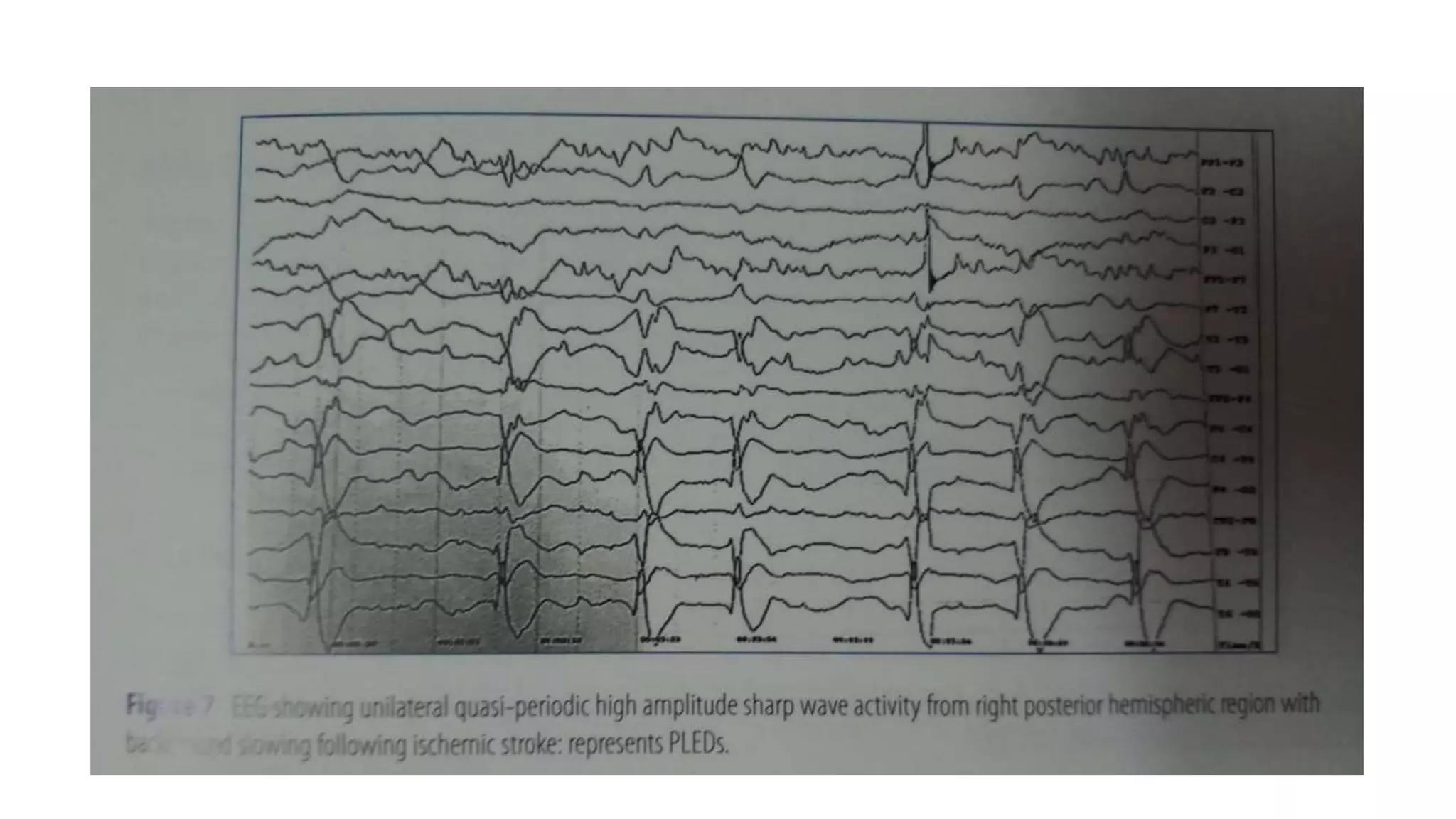
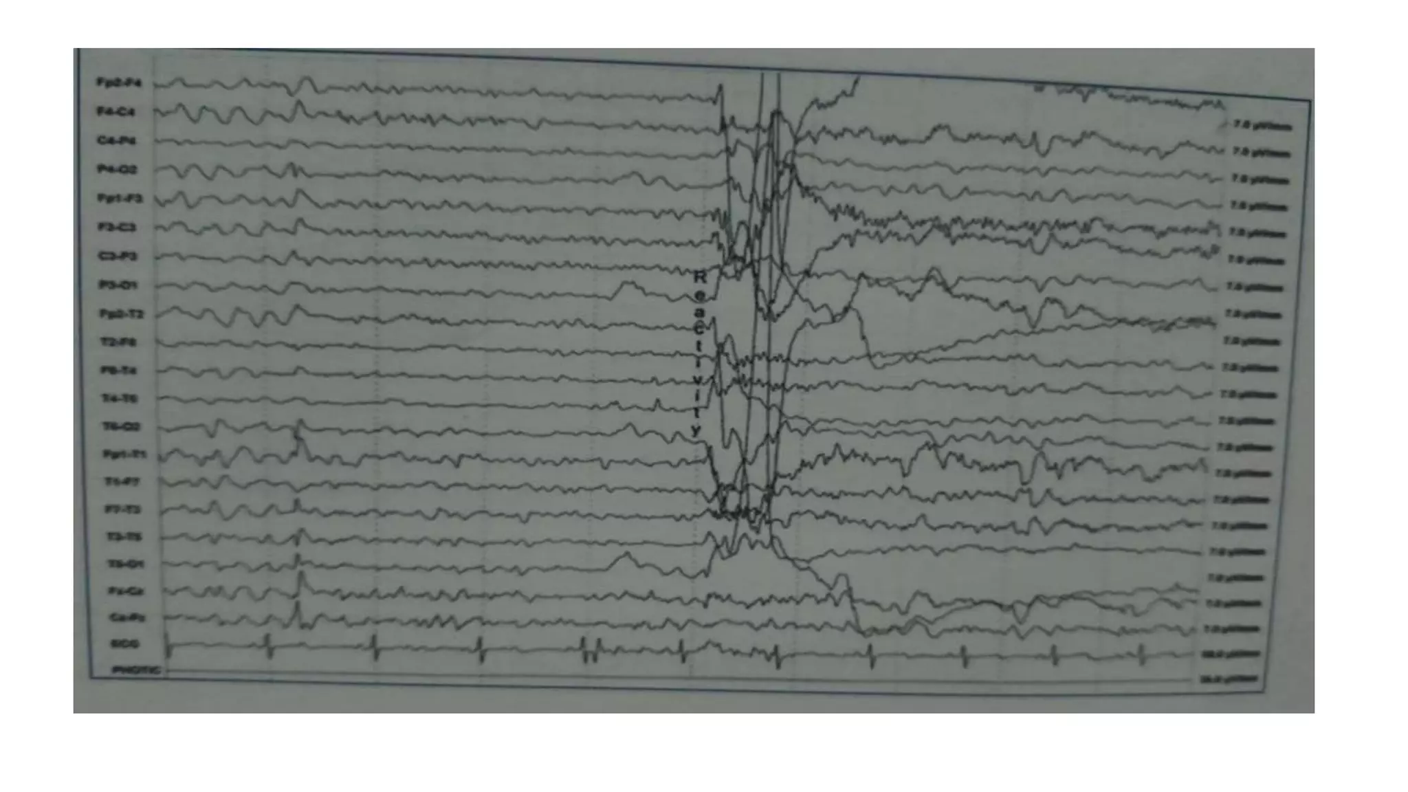
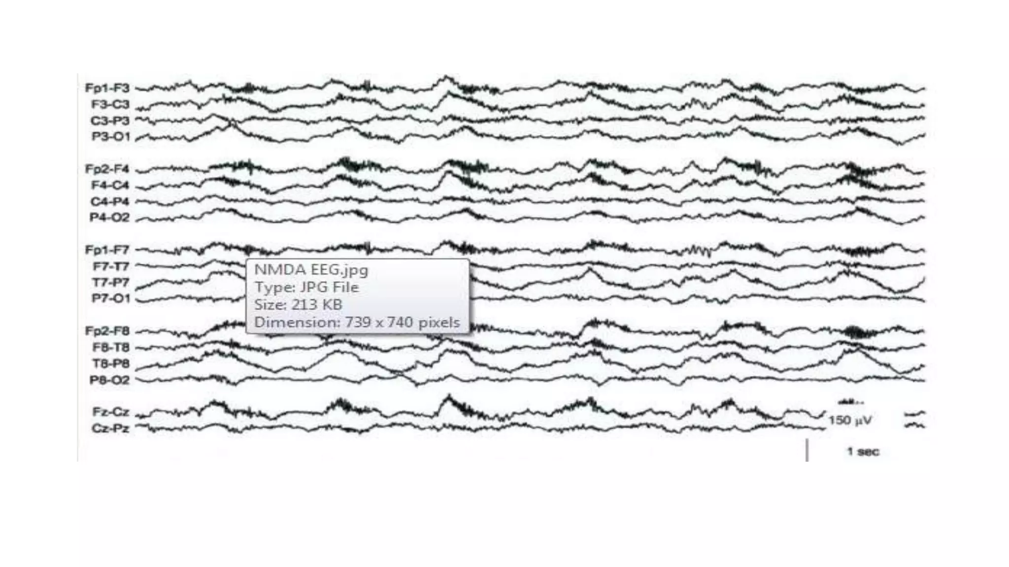
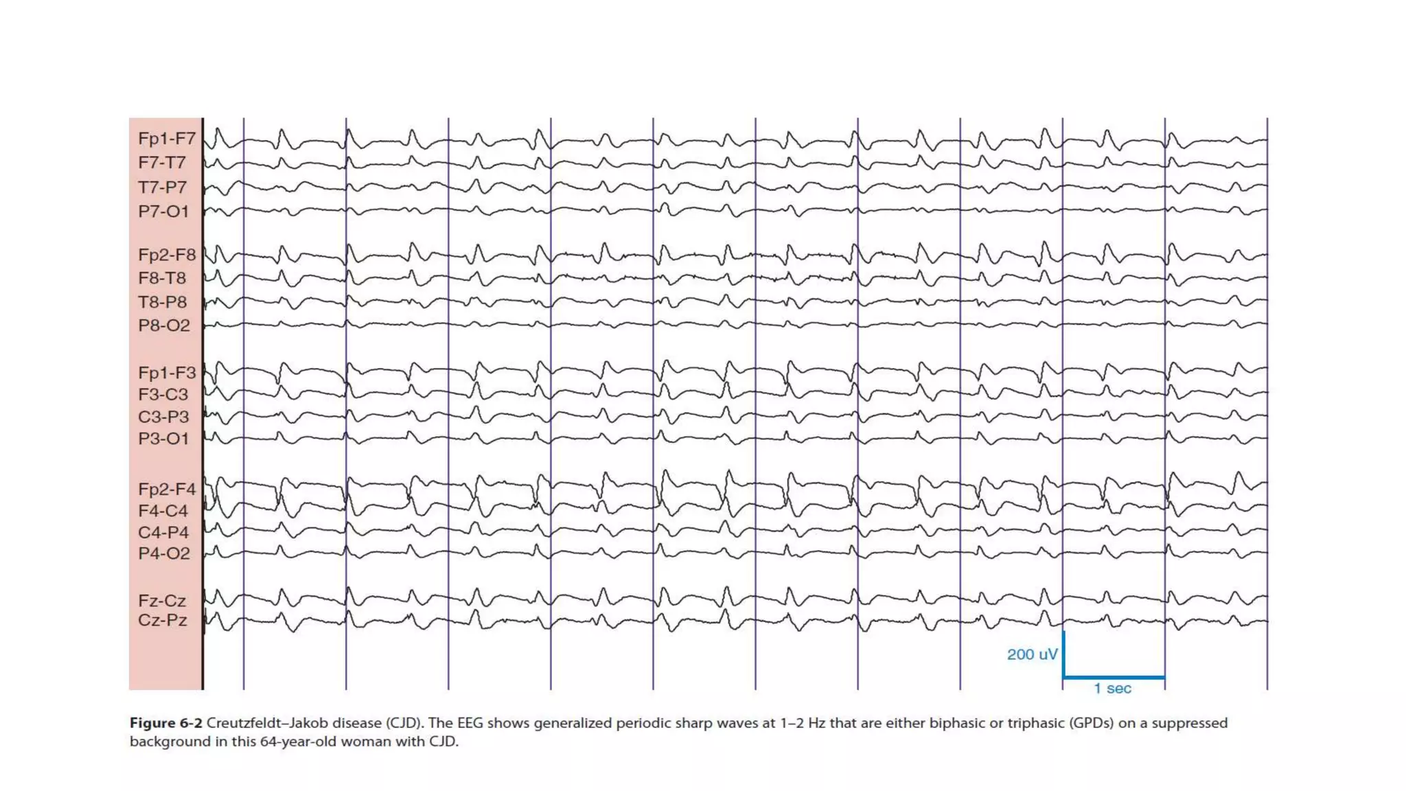
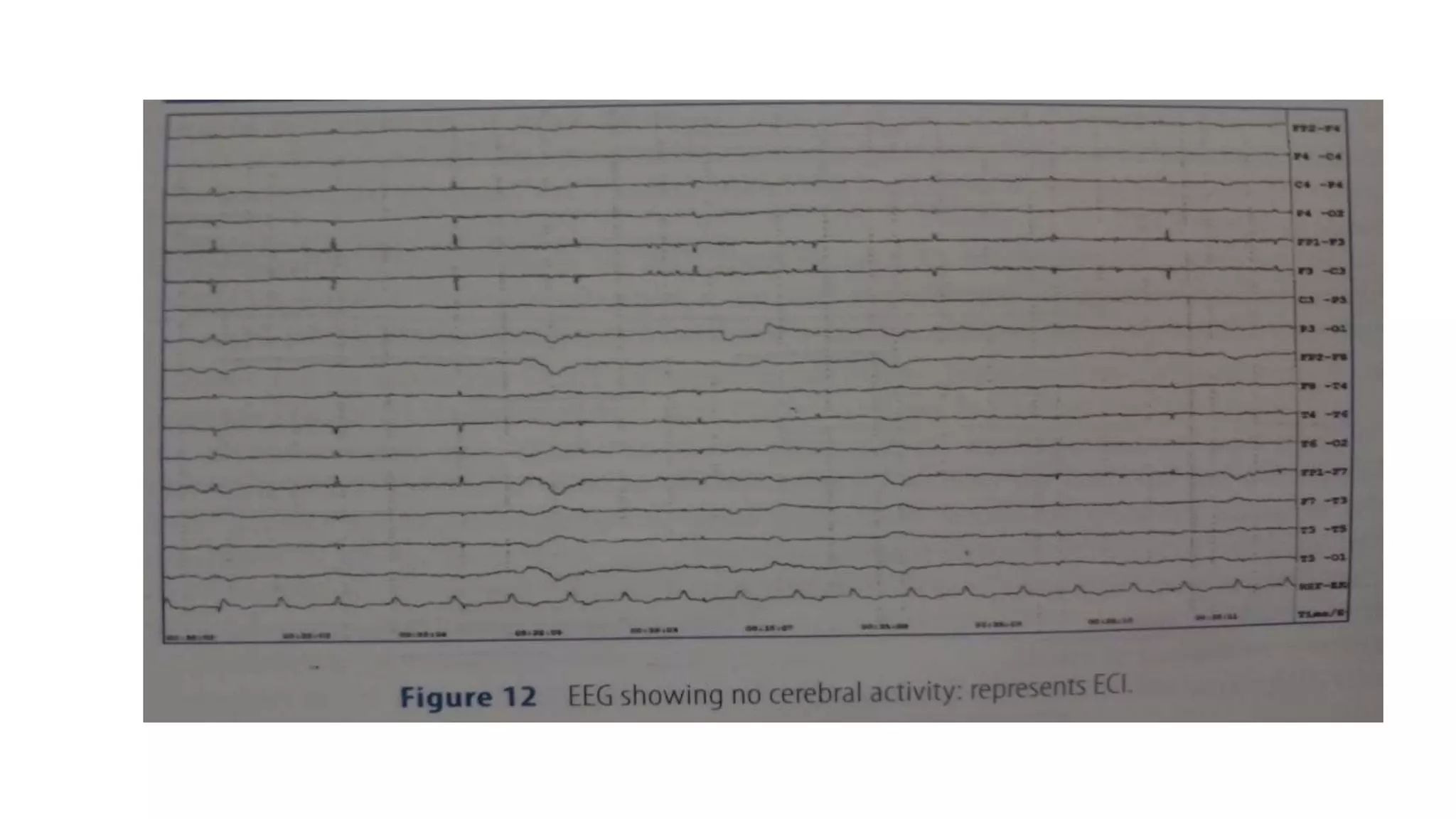
The document summarizes EEG patterns seen in various encephalopathies. It describes diffuse slowing, triphasic waves, burst suppression, periodic epileptiform discharges (PLEDs, BIPLEDs, GPEDs), alpha coma, spindle coma and beta coma patterns. Specific patterns are seen in hepatic encephalopathy, uremia, Hashimoto's encephalopathy, NMDAR encephalitis, Creutzfeldt-Jakob disease, subacute sclerosing panencephalitis. Criteria for periodic discharges and electrocerebral inactivity seen in brain death are also outlined.
2. Usefulness
• To identify seizures or NCSE
• To detect functional cerebral dysfunction in patients with normal
imaging
• To demonstrate focal or lateralized abnormalities
4. Triphasic waves
• Medium to high amplitude (usually >70µV), diffuse, frontally
predominant waves.
• Most prominent component is a positive sharp wave that is preceded
by a short-duration negative sharp wave and followed by a long-
duration negative slow wave.
• Duration-0.15 to 0.25 seconds and recur singly or in serial trains
almost periodically
• GPDs with triphasic morphology- hepatic encephalopathy, uremia,
sepsis, and electrolyte disturbances.
5.
6. BURST-SUPPRESSION
• Burst suppression (BS) is an EEG pattern consisting of alternative
period of slow/spike waves of high amplitude (the burst) and periods
of so called flat EEG (the suppression).
• The amplitude of bursts is between 20-100 µV.
• Suppressions also vary in amplitude in the range of electro-cerebral
inactivity (ECI; <20 µV to 50 µV).
7. • As abrupt change in amplitude describes this pattern, the amplitude
difference must be clearly visible and must be >50%.
• Bursts are usually bilateral synchronous, generalized
• Duration- 1-3 seconds
8. Pathogenesis -Results from suppression of cortical activity via GABA-
ergic mechanisms with breakthrough EEG activity due to intact
glutaminergic transmission.
• General anesthesia.
• Medically induced-in patients with refractory status epilepticus.
• Cardiopulmonary arrest who suffer from cerebral anoxia.
• Encountered in neonates(Severe cerebral damage)
9.
10. Periodic epileptiform discharges
• PLEDs consist of sharp waves and/ or spikes, associated with slow
waves, occurring at periodic intervals.
• Duration-100-300 msec, amplitude(100-300µV) and they recur every
0.5-4 sec.
• PLEDs stand out from the background activity due to their higher
amplitudes.
• The periodicity of the discharges is the result of a disconnection of
the cortex from the subcortical structures
• Significance-indicative of an acute focal brain dysfunction or unilateral
brain lesion
11.
12. • BIPLEDs are PLEDs seen in both hemispheres, in an independent and
asynchronous manner.
• BIPLEDs may present as asynchronous complexe.
• Differing in morphology, amplitude, rate of repetition, and site of
maximal involvement.
• Acute multifocal structural lesions, with or without metabolic
disturbances.
• Most common-anoxic encepahalopathy,CNS infections
13.
14. • GPEDs
• Periodic complexes occupying at least 50% of a standard 30-minutes
EEG, over both hemispheres in a symmetric, diffuse, and synchronous
manner.
• Characterized by spikes/polyspike or sharp wave of high amplitude
• Toxic-metabolic encephalopathies, anoxic brain injury, CJD, and
nonconvulsive status epilepticus.
15.
16. COMA
REACTIVE EEG
• The EEG is said to be reactive when there is a change in cerebral
rhythm to stimulation, which includes change in amplitude or
frequency.
• Eye blink artifacts or muscle artifacts do not count.
• It is important for the EEG technician to stimulate comatose patients
(noxious stimulation/passive eye opening) and note the time of
stimulation on EEG.
17.
18. Alpha coma
• Rhythmic alpha frequency in unconscious patient
• Monomorphic,symmetrical,prominently in the frontal derivations but
may be diffusely represented.
• There is no response to external stimuli or passive opening of the
eyes.
• Sleep wake cycles are absent.
• Widespread cerebral damage (as from anoxia)
• Prognosis poor
19.
20. Spindle coma
• Mono-rhythmic, non-reactive, 11-14 Hz waveforms, occurring
paroxysmally on a delta background.
• Head injury, midbrain strokes, drug toxicity, and cerebral anoxia.
• Spindle coma is associated with preserved brain stem reflexes and
sparing of normal thalamo-cortical pathways
• Carries a better prognosis.
21.
22. Beta coma
• Beta coma is characterized by high-amplitude beta activity,
sometimes frontally predominant.
• Intoxication with barbiturates or benzodiazepines
• favorable outcome.
23.
24. • Hepatic- Slowing of PDR which is followed by frequency in theta and
delta rhythm. Triphasic waves(25%)
• Uremic-Slowing of background activity. Triphasic waves(15-20%).
Epileptiform activities(b/l spike and wave) more common in uremic
• HSE- PLED,BIPLEDS or focal temporal slowing
25. • Hashomoto encephalopathy
• Mild to severe generalized slowing or frontal intermittent rhythmic
delta activity, triphasic waves, photomyogenic response, and
photoparoxysmal response.
• With clinical improvement, the EEG background activity also improves
and returns to normal,
• Rate of resolution of the EEG abnormalities is usually slower than the
rate of resolution of clinical abnormalities
• The EEG is useful for diagnosis.
26.
27.
28. NMDAR encepahlitis
• Nearly continuous combination of delta activity (1–3 Hz) with
superimposed fast activity (20–30 Hz) usually in the beta range
• Seen in NMDA-R encephalitis
• It is most often symmetric and synchronous
• Detected broadly across all head regions with predominance in
frontal regions
29.
30. Creutzfeldt–Jakob disease (CJD)
• Background rhythms become fragmented and are destroyed.
• Diffuse slowing appears and increases.
• Later, the distinctive periodic sharp wave discharges, often at 1 Hz, are recorded
• At first, the discharges may be more irregular and even focal, only later becoming
generalized and synchronous.
• Background activity decreases in amplitude.
• Eventually the EEG is dominated by the periodic discharges with no discernible
background.
31. Criteria
• Strictly periodic activity- variability of intercomplex interval is <500ms
Periodic activity is continuous for atleast 1 seconds
• Bi/triphasic morphology of periodic complexes
• Duration- 100-600 ms
• Periodic complex may be generalized or lateralized,but not regional or
asynchronous
32.
33. SSPE
• Periodic, stereotyped, generalized high voltage discharges
• I- periodic discharges seen in sleep
• II-In wakefulness. Bilateral synchrounus symmetrical bursts,
Amplitude- 200-500mv.
• Each bursts consists of polyphasic, momomhorphic delta waves.
Burst every 4-10 seconds(Time locked with myoclonus)
34. • III- slow delta rhythm
• IV- voltage of recording gets smaller and becomes isoelectric
35.
36. BRAIN DEATH
• Electrocerebral inactivity (ECI) is defined as the absence of any waves
of cerebral origin.
• The record should not have activity that exceeds 2 μV, unless that
activity is clear environmental artifact (e.g., an IV drip or cardiac
artifact).
• Low-frequency filters should be set between 0.5 Hz and 1.5 Hz, and
the high-frequency filter should be set at 70 Hz.
37. • Interelectrode impedance should be between 1000 and 10,000
Ohms.
• The EEG should be reviewed at a sensitivity of 2 μV/mm for at least 30
minutes
• Double-distance bipolar montage should be available to maximize the
chances of detecting cerebral activity.
• Reversible disturbances must be excluded (toxic–metabolic
perturbations, hypothermia, or sedating medication).
38.
39.
40. • D/D-non-convulsive status epilepticus (NCSE).
• Usually no anterior-posterior delay in NCSE.
• GPDs that are faster in frequency (>3 Hz) or have evolution meet the
criteria for electrographic seizures.
Editor's Notes
There is a tendency for progressive time lag (25 to 140 ms) of the positive component of the triphasic wave from the anterior to the posterior region (most specific for hepatic encephalopathy).