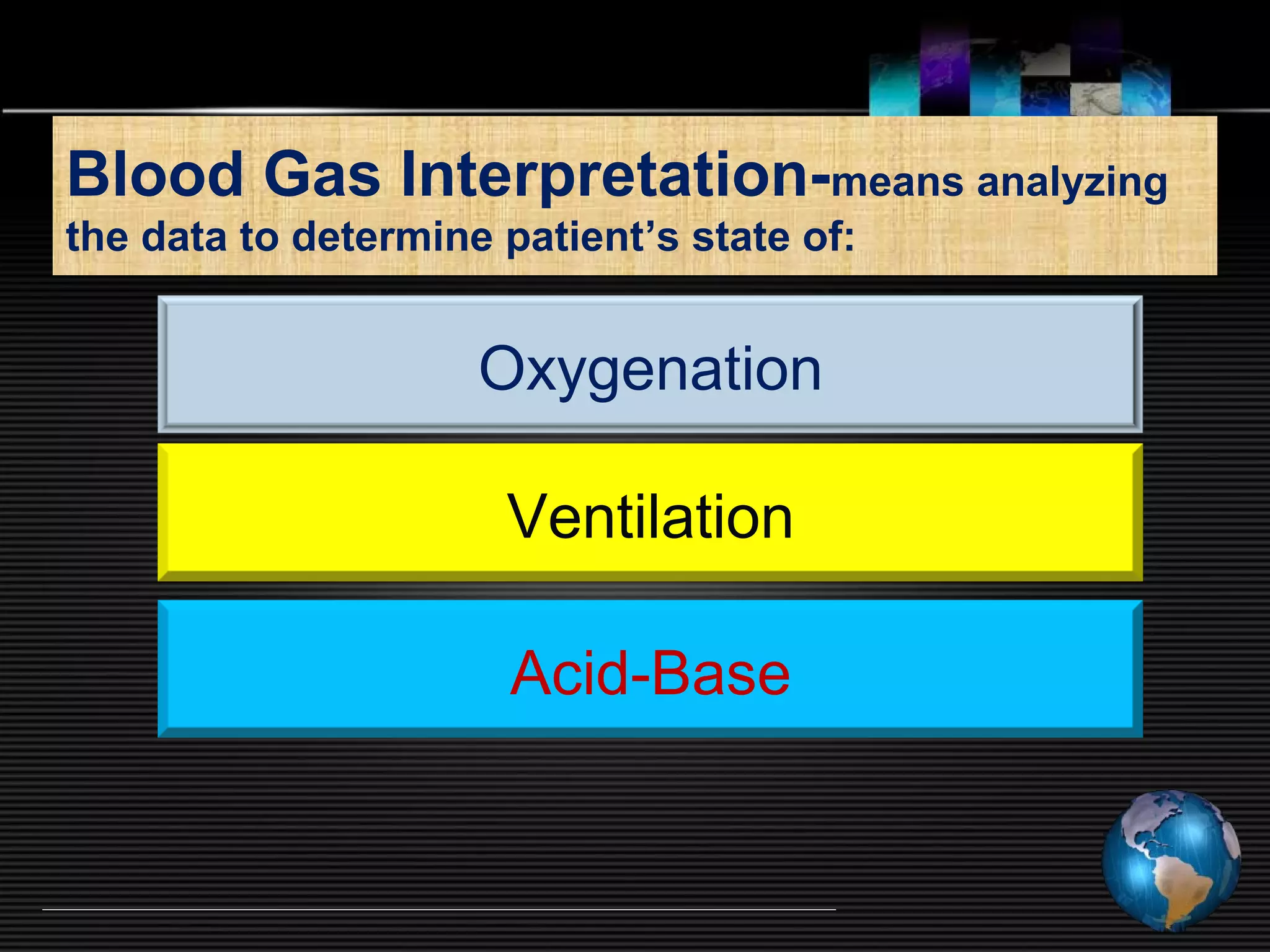
Dr. T.R.Chandrashekar
Intensivist, Liver transplantation.
BMC & RI super-specialty Hospital.
Bangalore.
Arterial Blood Gas Analysis
Why Order an ABG?
 Aids in establishing a diagnosis
 Helps guide treatment plan
 Aids in ventilator management
 Improvement in acid/base management allows
for optimal function of medications
 Acid/base status may alter electrolyte levels
critical to patient status/care
Blood Gas Interpretation-means analyzing
the data to determine patient’s state of:
Ventilation
Oxygenation
Acid-Base
Approach to ABG Interpretation
Assessment
of Acid-Base
Status
Assessment of
Oxygenation &
Ventilation Status
There is an interrelationship, but less
confusing if considered separately…..
Volume –
Osmolality
Electrolytes
-----XXXX Diagnostics----
Blood Gas Report
Measured 37.0 0
C
pH 7.452
pCO2 45.1 mm Hg
pO2 112.3 mm Hg
Calculated Data
HCO3 act 31.2 mmol / L
O2 Sat 98.4 %
O2 ct 15.8
pO2 (A -a) 30.2 mm Hg 
pO2 (a/A) 0.78
Entered Data
FiO2 %
Ct Hb gm/dl
-----XXXX Diagnostics-----
Blood Gas Report
328 03:44 Feb 5 2006
Pt ID 3245 / 00
Measured 37.0 0
C
pH 7.452
pCO2 45.1 mm Hg
pO2 112.3 mm Hg
Corrected 38.6 0
C
pH 7.436
pCO2 47.6 mm Hg
pO2 122.4 mm Hg
Calculated Data
HCO3 act 31.2 mmol / L
HCO3 std 30.5 mmol / L
B E 6.6 mmol / L
O2 ct 15.8 mL / dl
O2 Sat 98.4 %
ct CO2 32.5 mmol / L
pO2 (A -a) 30.2 mm Hg 
pO2 (a/A) 0.78
Entered Data
Temp 38.6 0C
FiO2 30.0 %
ct Hb 10.5 gm/dl
Calculated parameters
Measured parameters
FIO2 X 5=PaO2
Why would I require a ABG ?
 Oxygenation status?- Pulse oximeter SPO2✔
 Ventilation status ? – ETCO2✔
 Acid base status?
 Stable patient
 Normal hemodynamics
 Good UO Surgical stress + Sick patient
Hepatic/ Renal dysfunction
Massive blood/ fluid usage
Specific surgeries- liver
transplantation/ Hypotensive
anaesthesia/ aortic surgeries
IHD/COPD/ Asthma ……
Assessment of Oxygenation
 PaO2=Not informative
 CaO2= DO2- Better
 DO2~VO2=ScVO2 and Lactate- Ideal
O2 delivery is a Cardio-Respiratory function
TIME =TISSUE
Oxygen DonOxygen Don’t Go’t Go
Where the Blood Won’t FlowWhere the Blood Won’t Flow
Oxygen delivery DO2
Oxygen requirement VO2
Oxygen Cascade
 Atmospheric Air- 150 mmHg ( 21%)
PAO2-Alveolar Oxygen-100 mmHg ( CO2 / Water Vapour)
PaO2- 90mm Hg ( A-a difference)
SaO2 ( can be measured if Co-oximeter / calculated ODC)- Limitations
CaO2- Oxygen content (1.34 x Hb x Sao2)
DaO2 = Oxygen delivery = (CaO2 x Cardiac output)
If A-a difference is
more -does it tell us
anything ?
OO22
COCO22
Alveoli
PAO2
Atmospheric air /FIO2
Water vapour is added-
Nose/ upper airway
Alveolar Oxygen
PaO2 (2% dissolved O2)
Measured in ABG
SaO2
O.
D.
C.
98% of O2 is Hb bound-
1.34 x Hb% x Sao2
CaO2-oxygen content +PaO2 x 0.003ml
Oxygen Delivery=CaO2 x Cardiac output
Cardiac output - SV x HR
Preload / Afterload/ Contractility
Oxygen delivery DO2 is a
Cardio- Respiratory Function
=
Intra-operative bleeding Hb
Aspiration/ collapse
Cardiac depressants drugs/ MI
Stress response- Afterload
Causes for DO2
The power of hemoglobin
Normal Hypoxemia Anemia
PaO2 90 mm Hg 45 mm Hg 90 mm Hg
SaO2 98% 80% 98%
Hb 15 g/dL 15 g/dL 7.5 g/dL
CaO2 200 ml/L 163 ml/L 101 ml/L
% change - 18.6% - 49.5%
Restrictive transfusion strategy is the order of the day
PaCO2=60 mmHg
PAO2 = FIO2 (BP-47) – 1.2 (PaCO2)
=.21 (760- 47) – 1.2 (60)
= 150 – 72 = 78
An elevated PaCO2 will lower the PAO2
and as a result will lower the PaO2
We always correlate PaO2 with
FiO2
BUT………………………….
never forget to correlate with
PaCO2
PAO2=FIO2(Barometric Pressure-H2O)-1.2(PCO2)
PAO2 = FIO2 (760– 47 mm Hg)- 1.2 (PaCO2)
PAO2 = 0.21(713)-1.2(40)=100 mmHg
“1.2” is dropped when FIO2 is above 60%.
PAO2 is affected by PCO2
A-aDo2
A-aDo2 = PAO2-PaO2(from ABG)= 10-15 mmHg / Increases with age
Increased P(A-a)O2 -lungs are not transferring oxygen properly from
alveoli into the pulmonary capillaries.
OO22
COCO22
PaO2
Alveoli
PAO2
P(A-
a)O2
Diffusion defect
Interstitial edema ILD/ARDS
V/Q Mismatch-Dead Space
Amendable to increased FIO2
Shunt ( >30%) not amendable to
increased FIO2 / May require PEEP
P(A-a)O2 signifies some sort of
problem within the lungs
If DO2 is normal is everything OK?
 Next question is
 Is DO2~VO2 ?
 Lactate levels
 ScVO2/SVO2
A central venous ABG may
be more informative than
arterial ABG
ScVO2
DO2
Consumption O2
75%
Factors that influence mixed and central venousFactors that influence mixed and central venous
SOSO22
↑VO2 ↓DO2 ↑ DO2 ↓VO2
Stress
Pain
Hyperthermia
Shivering
↓ PaO2
↓ Hb
↓ Cardiac output
↑ PaO2
↑ Hb
↑ Cardiac output
Hypothermia
Anesthesia
_ +
DO2 Oxygen delivery VO2 Oxygen requirement
Summary –Oxygenation assessment
 CaO2 x CO =Delivery
 ScVO2 & Lactate levels = Surrogates for DO2~VO2
 Lacti-Time- prognostic indicator
 Is lactate bad? May not due to anaerobic metabolism
 Body’s response to stress
 Alternate energy shuttle ScVO2 Range
60-80% Normal
60-50% More extraction
warning sign
50-30% Lactic acidosis
Demand > Supply
30-20% Severe lactic acidosis
Cell death
ScVO2
SVO2
> 5-7
65 yr old male with DM IHD –in septic
shock on ventilator
ABG-PaO2-90 PH 7.42, PCO2 43
Hb-12 gm%, Spo2 98%
CaO2-17 Vol%
BP 90/40 mmHg ,Temp 103F
What is the problem ?
ScVO2 58%, Lactate 8 mMoles/L
Fluid resuscitation
Noradrenaline / Dobutamine
Fever control
After 2hrs
ScVO2 68%, Lactate 2 mMoles/L
Case …. PaO2 and DO2 are normal
65 yr old male with DM IHD –in septic
shock on ventilator
ABG-PaO2-90 PH 7.42, PCO2 43
Hb-12 gm%, Spo2 98%
CaO2-17 Vol%
BP 70/40 mmHg ,Temp 102F
What is the problem ?
ScVO2 45% Lactate 10 mMoles/L
Microcirculatory Mitochondrial
Dysfunction (MMDS)
Fluid resuscitation
Noradrenaline / Dobutamine
Fever control
After 2hrs
ScVO2 38%, Lactate 14 mMoles/L
PaO2 and DO2 are normal
Assessment of Ventilatory Status….
Oxygenation Acid-Base
HCO3
PAO2 = FIO2 (BP-47) – 1.2 (PCO2) pH ~ ------------
PaCO2
PaO2
» VCO2 x .863
» PaCO2 = --------------------
» VA
» VA=Minute ventilation-Dead space volume
» f(VT) – f(VD)
PaCO2 is key to the blood gas universe; without understanding
PaCO2 you can’t understand oxygenation or acid-base.
 The ONLY clinical parameter in PaCO2 equation is RR
VCO2=CO2 production
Difficulties in sampling and accurate measurement limits the usefulness
Of dead space in clinical practice =2ml/kg
Breathing pattern’s effect on PaCO2
Patient Vt f MV Description
A (400)(20) = 8.0L/min (slow/deep)
B (200)(40) = 8.0L/min (fast/shallow)
Patient Vt-Vd f Alveolar ventilation
A (400-150) (20) = 5.0L/min
B (200-150) (40) = 2.0L/min
PaCO2 = alveolar ventilation
Not on Minute ventilation which is measured
Condition State of
PaCO2 in blood alveolar ventilation
> 45 mm Hg Hypercapnia Hypoventilation
35 - 45 mm Hg Eucapnia Normal ventilation
< 35 mm Hg Hypocapnia Hyperventilation
PaCO2 abnormalities…
PCO2 / RR- 65 / 7 in Drug overdosage
True hypoventilation
PCO2 / RR - 65 / 37 in bilateral consolidation
Reduced alveolar ventilation/ dead space ventilation
PCO2 / RR - 22 / 37 in post operative patient with pain and fever-
Increased alveolar ventilation
Acid-Base DisturbancesAcid-Base Disturbances
Perioperative acid base disturbances
Sick patients +
Stress of surgery
Decreased organ perfusion
Reduced immunity
Activity of coagulation factors and
complement system impaired
Cardiac depression
Vasoplegia
Arrhythmias
Inotropes or vasopressors do not work
Reduced O2 delivery
O2 delivery is a Cardio-Respiratory function
Hypo perfusion— Anaerobic metabolism-Lactic acidosis
- More Cardiac depression and vasoplegia-Viscous cycle
There is always a underlying cause- treatment of which is
the most important step in acid base problems
Deodorant
Flush
Problem
.Patient has
Peritonitis/ Sepsis
BP 80/60 mmHg
pH of 7.12, HCO3
14, Lactate of 6
PCO2 23
Metabolic acidosis
NAHCO3
Infusion to
correct pH
Fluid
resuscitation
Inotropes/
vasopressors
Surviving
Sepsis bundle
•Fever , pain, shivering-Increased metabolic demands
Limited cardio pulmonary reserves -more problems
•Narcotics , Sedatives, NMB –hypoventilation
•Nasogastric suctioning-Chloride loss- Metabolic alkalosis
•Hypotension may lead to lactic acidosis, renal dysfunction
Septic patients / Elderly / diabetic HT Nephropathy / IHD
Commonly encountered acid base
disturbances seen in Perioperative period
Basics
•[H+]= 40 nEq/L at pH-7.4
•For every 0.3 pH change = [H+] double
160nEq/L
40 nEq/L
16nEq/L
[ H+
] in nEq/L = 10
(9-pH)
CO2 + H2O H2CO3 H+
+ HCO3
-
CO2
H+
HCO3
-
Acid-Base physiology
Respiratory
Metabolic
Bicarbonate is the transport from of CO2 hence both should
move in the same direction
Ventilation controls PCO2
Kidney losses H+ and reabsorbs bicarbonate (HCO3
-
)
PCO2-Respiratory acidosis
(Hypoventilation)
PCO2-Respiratory alkalosis
(Hyperventilation)
HCO3
-
- Metabolic acidosis
HCO3
-
- Metabolic Alkalosis
Very fast 80% in ECF
Starts within minutes
good response by 2hrs,
complete by 12-24 hrs
Starts after few hrs
complete by 5-7 days
Acid-base Balance
Henderson-Hasselbalch Equation
[HCO3
-
]
pH = pK + log -------------
.03 [PaCO2]
For teaching purposes, the H-H equation can be
shortened to its basic relationships:
HCO3
-
( KIDNEY)
pH ~ --------------------
PaCO2 (LUNG)
Maximum compensation
HCO3-= 40/10
CO2=60/10
Stewart approach
• Stewart (physical chemistry principles) suggested that the
traditional Henderson-Hasselbalch explanation of the
underlying physiology and pathophysiology is wrong.
• “Traditional” approach merely looks at a mirror image of that
proposed by Stewart. In fact HH equation is a component of
Stewart approach
• The ‘‘modern’’ approach is clinically difficult, more CPU based
• Requires knowledge of protein and phosphate concentrations
and more electrolytes than may be routinely measured
Characteristics of 1° acid-base disorders
DISORDER PRIMARY
RESPONSES
COMPENSATORY
RESPONSE
Metabolic
acidosis
↓ PH ↓ HCO3
-
↓ pCO2
Metabolic
alkalosis
↑ PH ↑ HCO3
-
↑ pCO2
Respiratory
acidosis
↓ PH ↑ pCO2 ↑ HCO3
-
Respiratory
alkalosis
↑ PH ↓ pCO2 ↓ HCO3
-
pH HCO3 CO2
7.20 15 40
7.25 15 30
7.38 15 20
Un Compensated
Partially Compensated
Fully Compensated
(pH abnormal)
(pH in normal range)
This amount of compensation rarely occurs in Acute situations
Metabolic acidosis with compensatory Respiratory alkalosis
Body’s physiologic response to Primary disorder
in order to bring pH towards NORMAL limit
Full/Partial compensation/uncompensated
BUT never overshoots,
If overshoot pH is present -Take it for granted it is
a MIXED disorder
Normal functioning
• Clinical history
• pH normal, abnormal PCO2 and HCO3
• PCO2 and HCO3 moving in opposite directions
• Degree of anticipated compensation for primary disorder is
inappropriate??
Primary Respiratory disorders
Metabolic compensations
RESPIRATORY disorders CO2 Change lead to change in HCO3
Acidosis-expected change in HCO3 = 1
Alkalosis-expected change in HCO3 = 2 x
Acidosis-expected change in HCO3 =3 x
Alkalosis-expected change in HCO3 = 4 x
Acute respiratory
Chronic respiratory
For every 10 mmHg CO2 change Expected HCO3 Change is given below
Step 5 continued…CompensationStep 5 continued…Compensation
Primary Metabolic disorders
Respiratory compensations
Metabolic disorders compensation
by changing CO2
Metabolic Acidosis: Compensation CO2
Winters’ formula
pCO2 = 1.5 x [HCO3-] + 8 ± 2
 Last two digits of pH = PaCO2
 pH being 7.23 = PaCO2should fall to 23mmHg
Metabolic Alkalosis: Compensation CO2
pCO2 = 0.7x [HCO3-] + 20 ± 5
Unpredictable because increasing CO2 causes increased RR
More anions are unmeasured than are
cations
 Major unmeasured anions
• albumin
• phosphates
• sulfates
• organic anions- ketones and
lactate
Anion gap-AG = [Na+
] - [Cl-
+HCO3
-
]
Anion gap is thus an artifact of
measurement, and not a physiologic reality
1 gm/dl decrease in serum albumin causes a 2.5 drop in the AG.
• Elevated anion gap represents metabolic acidosis
• Normal value: 12 ± 4mmol/L (More than 20 is usually significant)
chandrakavi@gmail.com
• Practice gentle mechanical ventilation
and do not try to bring ABG to perfect
normal.
• Treat the patient not the ABG report
2. Look at pH? (Acidosis /Alkalosis)
3. HCO3
-
// PCO2 (respiratory or metabolic or mixed )
4. Match either pCO2 at the HCO3with pH
(which is primary & which is compensation)
5. Fix the level of compensation.
6.If metabolic acidosis, calculate-Anion gap
7. Treat the Patient not the ABG
1. Consider the clinical settings! Anticipate the disorder
7 steps to analyze ABG
1st
Step-Clinical History
COPD- Chronic
 Respiratory Acidosis-Compensatory Met alkalosis
Post O.P patient -residual NMB, drowsy
 Respiratory Acidosis not well compensated
Cardiac arrest
 Metabolic/Respiratory acidosis
Septic shock
 Metabolic acidosis
2nd
step
Look at the pH - Label it.
70 yr old man operated for # neck femur under
GA/ extubated drowsy shallow breathing on 2l
of O2 Spo2 88%
ABG shows
pH of 7.28- ACIDOSIS
PaCO2 of 80 mm Hg,
HCO3- of 25 mEq/L.
Na+ 143, CL-104
 Label it. (Respiratory or metabolic)
 pH of 7.28,
 PaCO2 of 80 mm Hg-Respiratory acidosis
 HCO3- of 25 mEq/L- Alkalosis
Normal pCONormal pCO22 levels are 35-45mmHg.levels are 35-45mmHg.
Below 35 is alkalosis,Below 35 is alkalosis,
Above 45 is acidosis.Above 45 is acidosis.
3RD
step-Look at -pCO2/ HCO3 which
has moved in acidotic direction
Next match either -pCO2 or HCO3 with the pH
pH of 7.28-Acidosis
PaCO2 of 80 mm Hg-Respiratory acidosis
HCO3- of 25 mEq/L- Compensatory alkalosis
pH and PCO2have moved in same direction
So it is Primary respiratory acidosis
4TH
Step- Find the primary problem and what
is compensatory
HCO3- of 25 mEq/L is going in opposite direction of pH.
Metabolic compensation
Is it full/partial/uncompensated ???
Another disorder- mixed disorder ????
RESPIRATORY disorders- CO2 Change lead to change in HCO3
Acidosis-expected change in HCO3 = 1
Alkalosis-expected change in HCO3 = 2 x
Acidosis-expected change in HCO3 =3 x
Alkalosis-expected change in HCO3 = 4 x
Acute respiratory
Chronic respiratory
For every 10 mmHg CO2 change expected HCO3 Change is given below
Step 5 continued…CompensationStep 5 continued…Compensation
5TH
fix compensation- full or partial??
Do the calculations….
pH of 7.28,
PaCO2 of 80 mm Hg, (80-40=40 mmHg increased)
HCO3- of 25 mEq/L
 PCO2 is increased by =40 (for every 10 rise in CO2 /HCO3
should increase by 1)
 HCO3=should be increased by 4
i.e. 24+4=28( for full compensation)
Respiratory acidosis with partially compensated
metabolic alkalosis
• Calculate the anion gap if it is more
there is Metabolic acidosis
AG = [Na+] - [Cl- +HCO3-AG = [Na+] - [Cl- +HCO3-]]
Sixth Step- AG in case of metabolic acidosis
pH of 7.30, PaCO2 of 80 mm Hg, and
HCO3- of 27 mEq/L. Na+ 143, CL-104
AG+143- (104+27)=140-131=12
7Th
Step
Treat the Patient not the ABG
PCO2-60mmHg,
PO2-54mmHg
RR-40/min,
55 yr old patient with
chronic neurological
weakness conscious,
comfortable
PCO2-60mmHg,
PO2-54mmHg
RR-40/min,
Spinal surgery post OP
extubated with residual NMB
effect and drowsy
Severe respiratory distress
Reversal
/vent support
chandrakavi@gmail.com
• Practice gentle mechanical ventilation and do not try to
bring ABG to perfect normal.
• Treat the patient not the ABG report
 Too much normal saline hyperchloremic acidosis
 Can we explain why increased Cl-
causes Acidosis??
Normal saline infusion leads to
hyperchloremic acidosis
Dilution
of HCO3
-
CO2
CO2 increases
HCO3
-
unchanged
pH falls
Metabolic
CO2
production
pH falls
Water H2O
dissociates
and adds H+
Explanation of acidosis by HH method Explanation of acidosis by Stewart
In NS- Na/Cl is 150 mEq/L
In ECF -Na 145/ Cl 102
Two approaches used in Acid
base evaluation
HCO3
-
( KIDNEY) 20
PaCO2 (LUNG) 1
pH ~
Metabolic’ component of
acid-base physiology is
bicarbonate
Stewart approachTraditional approach
Henderson-Hasselbalch
Water is an important source of H +
Ionic Strength: weak and strong
Electrical Neutrality is maintained at
all times
Metabolic’ component of acid-base
physiology is Strong Ion Difference (SID)
SID=(Na+
+K+
+Ca2+
+Mg2+
)-(Cl-
+Lactate)
is 40 to 42 mEq/L
Two commonly used approaches
pH rely on three independent
Factors
1. SID- Strong ion difference
2. (ATOT)- total concentration of weak
acids (albumin and phosphate)
3. PCO2
‘Metabolic’ component of acid-base
physiology is not dependent on
bicarbonate but instead, predominately
on SID
Henderson-Hasselbalch Stewart approach
HCO3
-
( KIDNEY)
PaCO2 (LUNG)
pH --------- ------~
Modified Stewart approach
= ([Na+] – [Cl-]) – 38 (1)
= 0.25x [4.2–albumin] (2)
Thus true BE = BE – [1 + 2]
At bedside- it works well!
{where 38 is normal average difference in strong ions – Na and Cl}
NaCl effect
Albumin effect
Story, Belmo, Balasubramanyam
where 4.2 is normal serum albumin
Base excess
 The base excess is a calculated value that quantifies metabolic
derangements.
 It hypothetically ‘‘corrects’’ pH to 7.4 by ‘‘adjusting’’ measured
PaCO2 to 40 mmHg, allowing a comparison of the ‘‘corrected’’
HCO3
 with the normal HCO3.(i.e.,24mEq/L)at pH 7.4.
 A negative value means that HCO3 stores are depleted.
Base excess=HCO3+10(pH-7.4)-24
Uncompensated
Respiratory Acidosis
pH = 7.4
PaCO2 = 40
HCO3 = 24
Post op pt –drowsy
Uncompensated
Respiratory Alkalosis
pH = 7.4
PaCO2 = 40
HCO3 = 24
Pt on vent pressure
support has pain
Acute asthmatic
Normal A.B.G.
pH = 7.4
PaCO2 = 40
HCO3 = 24
Partially compensated
Metabolic Acidosis
pH = 7.4
PaCO2 = 40
HCO3 = 24
20 yr old male with Acute Gastroenteritis…..
Case
A 46-year-old man has been in the hospital for two
days with pneumonia. He was recovering but has
just become diaphoretic, dyspneic, and
hypotensive. He is breathing oxygen through a
nasal cannula at 3 l/min.
pH 7.41
PaCO2 20 mm Hg
HCO3- 12 mEq/L
CaO2 17.2 ml O2/dl
PaO2 80 mm Hg
SaO2 95%
Hb 13.3 gm%
How would you characterize his state of oxygenation,
ventilation, and acid-base balance?
Normal pH
Respiratory alkalosis and
Metabolic acidosis.
Winters formula
pCo2=1.5 x 12 +8=26
Case
 Mrs. H is found pulseless and not breathing this
morning. After a couple minutes of CPR she
responds with a pulse and starts breathing on
her own. A blood gas is obtained:
pH----------- 6.89
pCO2 -------70
pO2 ---------42
HCO3------- 13
SaO2-------- 50%
 What is your interpretation?
 What interventions would be appropriate for
Mrs. H?
Mrs. H has a severe metabolic and
respiratory acidosis with hypoxemia
Case …..
 A 44 year old moderately dehydrated man was
admitted with a two day history of acute severe
diarrhea. Electrolyte results: BP 90/60 mmHg
 Na+ 134, K+ 2.9, Cl- 108,
 BUN 31, Cr 1.5.
ABG: pH 7.31
 PCO2 33 mmHg
HCO3 16
 PaO2 93 mmHg
 What is the acid base disorder?
History
Acidosis from diarrhea or
lactic acidosis as a result of
hypovolemia and poor
perfusion.
Normal anion gap acidosis with adequate compensation
 Look at the pH- acidemic.
 What is the process? Look at the PCO2, HCO3- .
PCO2 and HCO3- are abnormal in the same direction,
therefore less likely a mixed acid base disorder.
 Calculate the anion gap
The anion gap is Na - (Cl + HCO3-) = 134 -(108 + 16) = 10
Is compensation adequate? Calculate the estimated PCO2.
Winter's formula;
PCO2 = 1.5 × [HCO3-]) + 8 ± 2 = 1.5 ×16 + 8 ± 2 = 30-34.
Case....
 A 50 year old insulin dependent diabetic woman
was brought to the ED by ambulance. She was
semi-comatose and had been ill for several days.
Current medication was digoxin and a thiazide
diuretic for CHF.
Lab results
Serum chemistry:
 Na 132, K 2.7, Cl 79, Glu 815,
Lactate 0.9 urine ketones 3+
ABG: pH 7.41 PCO2 32
 HCO3- 19 pO2 82
History:
Elevated anion gap acidosis secondary to DKA
Metabolic alkalosis in the setting of thiazide
diuretics use.
Case......
 2. Look at the pH. - Note that the pH is normal which would suggest no
acid base disorder. But remember, pH may be normal in the presence of a
mixed acid base disorder.
 3. What is the process? Look at the PCO2, HCO3- .
PCO2 is low indicating a possible respiratory alkalosis. The HCO3- is also
low indicating a possible metabolic acidosis. Because the pH is normal, we
are unable to distinguish the initial, primary change from the compensatory
response.
 We suspect however that the patient has DKA, and therefore should have a
metabolic acidosis with an anion gap that should be elevated. We can
confirm this by calculating the anion gap.
 4. Calculate the anion gap
The anion gap is Na - (Cl + HCO3-) = 132 -(79 + 19) = 34
Since gap is greater than 16, it is therefore abnormal and confirms the
presence of metabolic acidosis.
 Why is the pH normal? If the patient has metabolic acidosis, we suspect a
low ph unless there is another process acting to counteract the acidosis, i.e
alkalosis.
Delta Gap 34-12=22 + 19=41 Met alk
 Since the delta ratio is greater than 2, we can
deduce that there is a concurrent metabolic
alkalosis. This is likely due to to the use of
thiazide diuretic. Note that DKA is often
associated with vomiting, but in this case;vomiting
was not mentioned.
 Another possibility is a pre-existent high HCO3-
level due to compensated chonic respiratory
acidosis. But we have no reason to suspect
chronic respiratory acidosis based on the history.
 Assessment: Mixed elevated anion gap
metabolic acidosis and metabolic alkalosis likely
due to DKA and thiazide diuretics.
Anion gap issues
 A 1 gm/dl decrease in serum albumin causes a 2.5
mEq/L drop in the AG.
 One problem with the anion gap is deciding what value is
truly abnormal.
 In the majority of patients with anion gap between 16 and
20 mEq/L, no specific anion gap acidosis can be
diagnosed.
 Above 20 mEq/L the probability of a true anion gap
acidosis increases markedly (and is 100% if the AG is
above 29 mEq/L)
 As a practical matter, you should consider an AG 20
mEq/L as reflecting an anion gap metabolic acidosis and
search for the cause should be instituted
Delta gap = HCO3 + ∆ AG
Delta Gap = 24….Pure AG acidosis
 < 24 = non AG acidosis
 > 24 = metabolic alkalosis
∆ AG =Measured Anion gap-12
Delta Gap = 24 …… AG Met Acidosis
< 24 ….. Non AG Met acidosis
> 24 ….. Non AG Met acidosis + Meta. Alkalosis
CaO2 = (1.34 x Hb x SaO2) +dissolved
O2
DO2 = CO X CaO2
Cardiac output
Mitochondria in end organs
7 g % ALI/ARDS
PE
Sepsis induced
myocardial depression
Drugs
Inotropes
Pericardial effusion
vv
MMDS-cannot extract O2
O2
lactate
CO2
vvaa
DO2/VO2
Patients have to be kept well above the Critical Point so that
Does not plateau- Consumption remains supply
dependent even with supraphysiological levels
VO2 is supply
dependent
VO2 is supply independent
Oxygenation to the tissue is not compromised
MMDS and O2 extraction failure
Shunting due to micro-emboli
 Arterial Blood Gas Analysis

Arterial Blood Gas Analysis

  • 1.
    Dr. T.R.Chandrashekar Intensivist, Livertransplantation. BMC & RI super-specialty Hospital. Bangalore. Arterial Blood Gas Analysis
  • 2.
    Why Order anABG?  Aids in establishing a diagnosis  Helps guide treatment plan  Aids in ventilator management  Improvement in acid/base management allows for optimal function of medications  Acid/base status may alter electrolyte levels critical to patient status/care
  • 3.
    Blood Gas Interpretation-meansanalyzing the data to determine patient’s state of: Ventilation Oxygenation Acid-Base
  • 4.
    Approach to ABGInterpretation Assessment of Acid-Base Status Assessment of Oxygenation & Ventilation Status There is an interrelationship, but less confusing if considered separately….. Volume – Osmolality Electrolytes
  • 5.
    -----XXXX Diagnostics---- Blood GasReport Measured 37.0 0 C pH 7.452 pCO2 45.1 mm Hg pO2 112.3 mm Hg Calculated Data HCO3 act 31.2 mmol / L O2 Sat 98.4 % O2 ct 15.8 pO2 (A -a) 30.2 mm Hg  pO2 (a/A) 0.78 Entered Data FiO2 % Ct Hb gm/dl -----XXXX Diagnostics----- Blood Gas Report 328 03:44 Feb 5 2006 Pt ID 3245 / 00 Measured 37.0 0 C pH 7.452 pCO2 45.1 mm Hg pO2 112.3 mm Hg Corrected 38.6 0 C pH 7.436 pCO2 47.6 mm Hg pO2 122.4 mm Hg Calculated Data HCO3 act 31.2 mmol / L HCO3 std 30.5 mmol / L B E 6.6 mmol / L O2 ct 15.8 mL / dl O2 Sat 98.4 % ct CO2 32.5 mmol / L pO2 (A -a) 30.2 mm Hg  pO2 (a/A) 0.78 Entered Data Temp 38.6 0C FiO2 30.0 % ct Hb 10.5 gm/dl Calculated parameters Measured parameters FIO2 X 5=PaO2
  • 6.
    Why would Irequire a ABG ?  Oxygenation status?- Pulse oximeter SPO2✔  Ventilation status ? – ETCO2✔  Acid base status?  Stable patient  Normal hemodynamics  Good UO Surgical stress + Sick patient Hepatic/ Renal dysfunction Massive blood/ fluid usage Specific surgeries- liver transplantation/ Hypotensive anaesthesia/ aortic surgeries IHD/COPD/ Asthma ……
  • 7.
    Assessment of Oxygenation PaO2=Not informative  CaO2= DO2- Better  DO2~VO2=ScVO2 and Lactate- Ideal O2 delivery is a Cardio-Respiratory function TIME =TISSUE Oxygen DonOxygen Don’t Go’t Go Where the Blood Won’t FlowWhere the Blood Won’t Flow Oxygen delivery DO2 Oxygen requirement VO2
  • 8.
    Oxygen Cascade  AtmosphericAir- 150 mmHg ( 21%) PAO2-Alveolar Oxygen-100 mmHg ( CO2 / Water Vapour) PaO2- 90mm Hg ( A-a difference) SaO2 ( can be measured if Co-oximeter / calculated ODC)- Limitations CaO2- Oxygen content (1.34 x Hb x Sao2) DaO2 = Oxygen delivery = (CaO2 x Cardiac output) If A-a difference is more -does it tell us anything ?
  • 9.
    OO22 COCO22 Alveoli PAO2 Atmospheric air /FIO2 Watervapour is added- Nose/ upper airway Alveolar Oxygen PaO2 (2% dissolved O2) Measured in ABG SaO2 O. D. C. 98% of O2 is Hb bound- 1.34 x Hb% x Sao2 CaO2-oxygen content +PaO2 x 0.003ml Oxygen Delivery=CaO2 x Cardiac output Cardiac output - SV x HR Preload / Afterload/ Contractility Oxygen delivery DO2 is a Cardio- Respiratory Function = Intra-operative bleeding Hb Aspiration/ collapse Cardiac depressants drugs/ MI Stress response- Afterload Causes for DO2
  • 10.
    The power ofhemoglobin Normal Hypoxemia Anemia PaO2 90 mm Hg 45 mm Hg 90 mm Hg SaO2 98% 80% 98% Hb 15 g/dL 15 g/dL 7.5 g/dL CaO2 200 ml/L 163 ml/L 101 ml/L % change - 18.6% - 49.5% Restrictive transfusion strategy is the order of the day
  • 11.
    PaCO2=60 mmHg PAO2 =FIO2 (BP-47) – 1.2 (PaCO2) =.21 (760- 47) – 1.2 (60) = 150 – 72 = 78 An elevated PaCO2 will lower the PAO2 and as a result will lower the PaO2 We always correlate PaO2 with FiO2 BUT…………………………. never forget to correlate with PaCO2 PAO2=FIO2(Barometric Pressure-H2O)-1.2(PCO2) PAO2 = FIO2 (760– 47 mm Hg)- 1.2 (PaCO2) PAO2 = 0.21(713)-1.2(40)=100 mmHg “1.2” is dropped when FIO2 is above 60%. PAO2 is affected by PCO2
  • 12.
    A-aDo2 A-aDo2 = PAO2-PaO2(fromABG)= 10-15 mmHg / Increases with age Increased P(A-a)O2 -lungs are not transferring oxygen properly from alveoli into the pulmonary capillaries. OO22 COCO22 PaO2 Alveoli PAO2 P(A- a)O2 Diffusion defect Interstitial edema ILD/ARDS V/Q Mismatch-Dead Space Amendable to increased FIO2 Shunt ( >30%) not amendable to increased FIO2 / May require PEEP P(A-a)O2 signifies some sort of problem within the lungs
  • 13.
    If DO2 isnormal is everything OK?  Next question is  Is DO2~VO2 ?  Lactate levels  ScVO2/SVO2 A central venous ABG may be more informative than arterial ABG ScVO2 DO2 Consumption O2
  • 14.
    75% Factors that influencemixed and central venousFactors that influence mixed and central venous SOSO22 ↑VO2 ↓DO2 ↑ DO2 ↓VO2 Stress Pain Hyperthermia Shivering ↓ PaO2 ↓ Hb ↓ Cardiac output ↑ PaO2 ↑ Hb ↑ Cardiac output Hypothermia Anesthesia _ + DO2 Oxygen delivery VO2 Oxygen requirement
  • 15.
    Summary –Oxygenation assessment CaO2 x CO =Delivery  ScVO2 & Lactate levels = Surrogates for DO2~VO2  Lacti-Time- prognostic indicator  Is lactate bad? May not due to anaerobic metabolism  Body’s response to stress  Alternate energy shuttle ScVO2 Range 60-80% Normal 60-50% More extraction warning sign 50-30% Lactic acidosis Demand > Supply 30-20% Severe lactic acidosis Cell death ScVO2 SVO2 > 5-7
  • 16.
    65 yr oldmale with DM IHD –in septic shock on ventilator ABG-PaO2-90 PH 7.42, PCO2 43 Hb-12 gm%, Spo2 98% CaO2-17 Vol% BP 90/40 mmHg ,Temp 103F What is the problem ? ScVO2 58%, Lactate 8 mMoles/L Fluid resuscitation Noradrenaline / Dobutamine Fever control After 2hrs ScVO2 68%, Lactate 2 mMoles/L Case …. PaO2 and DO2 are normal 65 yr old male with DM IHD –in septic shock on ventilator ABG-PaO2-90 PH 7.42, PCO2 43 Hb-12 gm%, Spo2 98% CaO2-17 Vol% BP 70/40 mmHg ,Temp 102F What is the problem ? ScVO2 45% Lactate 10 mMoles/L Microcirculatory Mitochondrial Dysfunction (MMDS) Fluid resuscitation Noradrenaline / Dobutamine Fever control After 2hrs ScVO2 38%, Lactate 14 mMoles/L PaO2 and DO2 are normal
  • 17.
  • 18.
    Oxygenation Acid-Base HCO3 PAO2 =FIO2 (BP-47) – 1.2 (PCO2) pH ~ ------------ PaCO2 PaO2 » VCO2 x .863 » PaCO2 = -------------------- » VA » VA=Minute ventilation-Dead space volume » f(VT) – f(VD) PaCO2 is key to the blood gas universe; without understanding PaCO2 you can’t understand oxygenation or acid-base.  The ONLY clinical parameter in PaCO2 equation is RR VCO2=CO2 production Difficulties in sampling and accurate measurement limits the usefulness Of dead space in clinical practice =2ml/kg
  • 19.
    Breathing pattern’s effecton PaCO2 Patient Vt f MV Description A (400)(20) = 8.0L/min (slow/deep) B (200)(40) = 8.0L/min (fast/shallow) Patient Vt-Vd f Alveolar ventilation A (400-150) (20) = 5.0L/min B (200-150) (40) = 2.0L/min PaCO2 = alveolar ventilation Not on Minute ventilation which is measured
  • 20.
    Condition State of PaCO2in blood alveolar ventilation > 45 mm Hg Hypercapnia Hypoventilation 35 - 45 mm Hg Eucapnia Normal ventilation < 35 mm Hg Hypocapnia Hyperventilation PaCO2 abnormalities… PCO2 / RR- 65 / 7 in Drug overdosage True hypoventilation PCO2 / RR - 65 / 37 in bilateral consolidation Reduced alveolar ventilation/ dead space ventilation PCO2 / RR - 22 / 37 in post operative patient with pain and fever- Increased alveolar ventilation
  • 21.
  • 22.
    Perioperative acid basedisturbances Sick patients + Stress of surgery Decreased organ perfusion Reduced immunity Activity of coagulation factors and complement system impaired Cardiac depression Vasoplegia Arrhythmias Inotropes or vasopressors do not work Reduced O2 delivery O2 delivery is a Cardio-Respiratory function Hypo perfusion— Anaerobic metabolism-Lactic acidosis - More Cardiac depression and vasoplegia-Viscous cycle
  • 23.
    There is alwaysa underlying cause- treatment of which is the most important step in acid base problems Deodorant Flush Problem .Patient has Peritonitis/ Sepsis BP 80/60 mmHg pH of 7.12, HCO3 14, Lactate of 6 PCO2 23 Metabolic acidosis NAHCO3 Infusion to correct pH Fluid resuscitation Inotropes/ vasopressors Surviving Sepsis bundle
  • 24.
    •Fever , pain,shivering-Increased metabolic demands Limited cardio pulmonary reserves -more problems •Narcotics , Sedatives, NMB –hypoventilation •Nasogastric suctioning-Chloride loss- Metabolic alkalosis •Hypotension may lead to lactic acidosis, renal dysfunction Septic patients / Elderly / diabetic HT Nephropathy / IHD Commonly encountered acid base disturbances seen in Perioperative period
  • 25.
    Basics •[H+]= 40 nEq/Lat pH-7.4 •For every 0.3 pH change = [H+] double 160nEq/L 40 nEq/L 16nEq/L [ H+ ] in nEq/L = 10 (9-pH)
  • 26.
    CO2 + H2OH2CO3 H+ + HCO3 - CO2 H+ HCO3 - Acid-Base physiology Respiratory Metabolic Bicarbonate is the transport from of CO2 hence both should move in the same direction Ventilation controls PCO2 Kidney losses H+ and reabsorbs bicarbonate (HCO3 - ) PCO2-Respiratory acidosis (Hypoventilation) PCO2-Respiratory alkalosis (Hyperventilation) HCO3 - - Metabolic acidosis HCO3 - - Metabolic Alkalosis
  • 27.
    Very fast 80%in ECF Starts within minutes good response by 2hrs, complete by 12-24 hrs Starts after few hrs complete by 5-7 days
  • 28.
    Acid-base Balance Henderson-Hasselbalch Equation [HCO3 - ] pH= pK + log ------------- .03 [PaCO2] For teaching purposes, the H-H equation can be shortened to its basic relationships: HCO3 - ( KIDNEY) pH ~ -------------------- PaCO2 (LUNG) Maximum compensation HCO3-= 40/10 CO2=60/10
  • 29.
    Stewart approach • Stewart(physical chemistry principles) suggested that the traditional Henderson-Hasselbalch explanation of the underlying physiology and pathophysiology is wrong. • “Traditional” approach merely looks at a mirror image of that proposed by Stewart. In fact HH equation is a component of Stewart approach • The ‘‘modern’’ approach is clinically difficult, more CPU based • Requires knowledge of protein and phosphate concentrations and more electrolytes than may be routinely measured
  • 30.
    Characteristics of 1°acid-base disorders DISORDER PRIMARY RESPONSES COMPENSATORY RESPONSE Metabolic acidosis ↓ PH ↓ HCO3 - ↓ pCO2 Metabolic alkalosis ↑ PH ↑ HCO3 - ↑ pCO2 Respiratory acidosis ↓ PH ↑ pCO2 ↑ HCO3 - Respiratory alkalosis ↑ PH ↓ pCO2 ↓ HCO3 -
  • 31.
    pH HCO3 CO2 7.2015 40 7.25 15 30 7.38 15 20 Un Compensated Partially Compensated Fully Compensated (pH abnormal) (pH in normal range) This amount of compensation rarely occurs in Acute situations Metabolic acidosis with compensatory Respiratory alkalosis
  • 32.
    Body’s physiologic responseto Primary disorder in order to bring pH towards NORMAL limit Full/Partial compensation/uncompensated BUT never overshoots, If overshoot pH is present -Take it for granted it is a MIXED disorder Normal functioning
  • 33.
    • Clinical history •pH normal, abnormal PCO2 and HCO3 • PCO2 and HCO3 moving in opposite directions • Degree of anticipated compensation for primary disorder is inappropriate??
  • 34.
  • 35.
    RESPIRATORY disorders CO2Change lead to change in HCO3 Acidosis-expected change in HCO3 = 1 Alkalosis-expected change in HCO3 = 2 x Acidosis-expected change in HCO3 =3 x Alkalosis-expected change in HCO3 = 4 x Acute respiratory Chronic respiratory For every 10 mmHg CO2 change Expected HCO3 Change is given below Step 5 continued…CompensationStep 5 continued…Compensation
  • 36.
  • 37.
    Metabolic disorders compensation bychanging CO2 Metabolic Acidosis: Compensation CO2 Winters’ formula pCO2 = 1.5 x [HCO3-] + 8 ± 2  Last two digits of pH = PaCO2  pH being 7.23 = PaCO2should fall to 23mmHg Metabolic Alkalosis: Compensation CO2 pCO2 = 0.7x [HCO3-] + 20 ± 5 Unpredictable because increasing CO2 causes increased RR
  • 38.
    More anions areunmeasured than are cations  Major unmeasured anions • albumin • phosphates • sulfates • organic anions- ketones and lactate Anion gap-AG = [Na+ ] - [Cl- +HCO3 - ] Anion gap is thus an artifact of measurement, and not a physiologic reality 1 gm/dl decrease in serum albumin causes a 2.5 drop in the AG. • Elevated anion gap represents metabolic acidosis • Normal value: 12 ± 4mmol/L (More than 20 is usually significant)
  • 39.
    chandrakavi@gmail.com • Practice gentlemechanical ventilation and do not try to bring ABG to perfect normal. • Treat the patient not the ABG report
  • 41.
    2. Look atpH? (Acidosis /Alkalosis) 3. HCO3 - // PCO2 (respiratory or metabolic or mixed ) 4. Match either pCO2 at the HCO3with pH (which is primary & which is compensation) 5. Fix the level of compensation. 6.If metabolic acidosis, calculate-Anion gap 7. Treat the Patient not the ABG 1. Consider the clinical settings! Anticipate the disorder 7 steps to analyze ABG
  • 42.
    1st Step-Clinical History COPD- Chronic Respiratory Acidosis-Compensatory Met alkalosis Post O.P patient -residual NMB, drowsy  Respiratory Acidosis not well compensated Cardiac arrest  Metabolic/Respiratory acidosis Septic shock  Metabolic acidosis
  • 43.
    2nd step Look at thepH - Label it. 70 yr old man operated for # neck femur under GA/ extubated drowsy shallow breathing on 2l of O2 Spo2 88% ABG shows pH of 7.28- ACIDOSIS PaCO2 of 80 mm Hg, HCO3- of 25 mEq/L. Na+ 143, CL-104
  • 44.
     Label it.(Respiratory or metabolic)  pH of 7.28,  PaCO2 of 80 mm Hg-Respiratory acidosis  HCO3- of 25 mEq/L- Alkalosis Normal pCONormal pCO22 levels are 35-45mmHg.levels are 35-45mmHg. Below 35 is alkalosis,Below 35 is alkalosis, Above 45 is acidosis.Above 45 is acidosis. 3RD step-Look at -pCO2/ HCO3 which has moved in acidotic direction
  • 45.
    Next match either-pCO2 or HCO3 with the pH pH of 7.28-Acidosis PaCO2 of 80 mm Hg-Respiratory acidosis HCO3- of 25 mEq/L- Compensatory alkalosis pH and PCO2have moved in same direction So it is Primary respiratory acidosis 4TH Step- Find the primary problem and what is compensatory HCO3- of 25 mEq/L is going in opposite direction of pH. Metabolic compensation Is it full/partial/uncompensated ??? Another disorder- mixed disorder ????
  • 46.
    RESPIRATORY disorders- CO2Change lead to change in HCO3 Acidosis-expected change in HCO3 = 1 Alkalosis-expected change in HCO3 = 2 x Acidosis-expected change in HCO3 =3 x Alkalosis-expected change in HCO3 = 4 x Acute respiratory Chronic respiratory For every 10 mmHg CO2 change expected HCO3 Change is given below Step 5 continued…CompensationStep 5 continued…Compensation
  • 47.
    5TH fix compensation- fullor partial?? Do the calculations…. pH of 7.28, PaCO2 of 80 mm Hg, (80-40=40 mmHg increased) HCO3- of 25 mEq/L  PCO2 is increased by =40 (for every 10 rise in CO2 /HCO3 should increase by 1)  HCO3=should be increased by 4 i.e. 24+4=28( for full compensation) Respiratory acidosis with partially compensated metabolic alkalosis
  • 48.
    • Calculate theanion gap if it is more there is Metabolic acidosis AG = [Na+] - [Cl- +HCO3-AG = [Na+] - [Cl- +HCO3-]] Sixth Step- AG in case of metabolic acidosis pH of 7.30, PaCO2 of 80 mm Hg, and HCO3- of 27 mEq/L. Na+ 143, CL-104 AG+143- (104+27)=140-131=12
  • 49.
    7Th Step Treat the Patientnot the ABG PCO2-60mmHg, PO2-54mmHg RR-40/min, 55 yr old patient with chronic neurological weakness conscious, comfortable PCO2-60mmHg, PO2-54mmHg RR-40/min, Spinal surgery post OP extubated with residual NMB effect and drowsy Severe respiratory distress Reversal /vent support
  • 50.
    chandrakavi@gmail.com • Practice gentlemechanical ventilation and do not try to bring ABG to perfect normal. • Treat the patient not the ABG report
  • 51.
     Too muchnormal saline hyperchloremic acidosis  Can we explain why increased Cl- causes Acidosis??
  • 52.
    Normal saline infusionleads to hyperchloremic acidosis Dilution of HCO3 - CO2 CO2 increases HCO3 - unchanged pH falls Metabolic CO2 production pH falls Water H2O dissociates and adds H+ Explanation of acidosis by HH method Explanation of acidosis by Stewart In NS- Na/Cl is 150 mEq/L In ECF -Na 145/ Cl 102
  • 53.
    Two approaches usedin Acid base evaluation HCO3 - ( KIDNEY) 20 PaCO2 (LUNG) 1 pH ~ Metabolic’ component of acid-base physiology is bicarbonate Stewart approachTraditional approach Henderson-Hasselbalch Water is an important source of H + Ionic Strength: weak and strong Electrical Neutrality is maintained at all times Metabolic’ component of acid-base physiology is Strong Ion Difference (SID) SID=(Na+ +K+ +Ca2+ +Mg2+ )-(Cl- +Lactate) is 40 to 42 mEq/L
  • 54.
    Two commonly usedapproaches pH rely on three independent Factors 1. SID- Strong ion difference 2. (ATOT)- total concentration of weak acids (albumin and phosphate) 3. PCO2 ‘Metabolic’ component of acid-base physiology is not dependent on bicarbonate but instead, predominately on SID Henderson-Hasselbalch Stewart approach HCO3 - ( KIDNEY) PaCO2 (LUNG) pH --------- ------~
  • 55.
    Modified Stewart approach =([Na+] – [Cl-]) – 38 (1) = 0.25x [4.2–albumin] (2) Thus true BE = BE – [1 + 2] At bedside- it works well! {where 38 is normal average difference in strong ions – Na and Cl} NaCl effect Albumin effect Story, Belmo, Balasubramanyam where 4.2 is normal serum albumin
  • 56.
    Base excess  Thebase excess is a calculated value that quantifies metabolic derangements.  It hypothetically ‘‘corrects’’ pH to 7.4 by ‘‘adjusting’’ measured PaCO2 to 40 mmHg, allowing a comparison of the ‘‘corrected’’ HCO3  with the normal HCO3.(i.e.,24mEq/L)at pH 7.4.  A negative value means that HCO3 stores are depleted. Base excess=HCO3+10(pH-7.4)-24
  • 58.
    Uncompensated Respiratory Acidosis pH =7.4 PaCO2 = 40 HCO3 = 24 Post op pt –drowsy
  • 59.
    Uncompensated Respiratory Alkalosis pH =7.4 PaCO2 = 40 HCO3 = 24 Pt on vent pressure support has pain Acute asthmatic
  • 60.
    Normal A.B.G. pH =7.4 PaCO2 = 40 HCO3 = 24
  • 61.
    Partially compensated Metabolic Acidosis pH= 7.4 PaCO2 = 40 HCO3 = 24 20 yr old male with Acute Gastroenteritis…..
  • 62.
    Case A 46-year-old manhas been in the hospital for two days with pneumonia. He was recovering but has just become diaphoretic, dyspneic, and hypotensive. He is breathing oxygen through a nasal cannula at 3 l/min. pH 7.41 PaCO2 20 mm Hg HCO3- 12 mEq/L CaO2 17.2 ml O2/dl PaO2 80 mm Hg SaO2 95% Hb 13.3 gm% How would you characterize his state of oxygenation, ventilation, and acid-base balance? Normal pH Respiratory alkalosis and Metabolic acidosis. Winters formula pCo2=1.5 x 12 +8=26
  • 63.
    Case  Mrs. His found pulseless and not breathing this morning. After a couple minutes of CPR she responds with a pulse and starts breathing on her own. A blood gas is obtained: pH----------- 6.89 pCO2 -------70 pO2 ---------42 HCO3------- 13 SaO2-------- 50%  What is your interpretation?  What interventions would be appropriate for Mrs. H? Mrs. H has a severe metabolic and respiratory acidosis with hypoxemia
  • 64.
    Case …..  A44 year old moderately dehydrated man was admitted with a two day history of acute severe diarrhea. Electrolyte results: BP 90/60 mmHg  Na+ 134, K+ 2.9, Cl- 108,  BUN 31, Cr 1.5. ABG: pH 7.31  PCO2 33 mmHg HCO3 16  PaO2 93 mmHg  What is the acid base disorder? History Acidosis from diarrhea or lactic acidosis as a result of hypovolemia and poor perfusion.
  • 65.
    Normal anion gapacidosis with adequate compensation  Look at the pH- acidemic.  What is the process? Look at the PCO2, HCO3- . PCO2 and HCO3- are abnormal in the same direction, therefore less likely a mixed acid base disorder.  Calculate the anion gap The anion gap is Na - (Cl + HCO3-) = 134 -(108 + 16) = 10 Is compensation adequate? Calculate the estimated PCO2. Winter's formula; PCO2 = 1.5 × [HCO3-]) + 8 ± 2 = 1.5 ×16 + 8 ± 2 = 30-34.
  • 66.
    Case....  A 50year old insulin dependent diabetic woman was brought to the ED by ambulance. She was semi-comatose and had been ill for several days. Current medication was digoxin and a thiazide diuretic for CHF. Lab results Serum chemistry:  Na 132, K 2.7, Cl 79, Glu 815, Lactate 0.9 urine ketones 3+ ABG: pH 7.41 PCO2 32  HCO3- 19 pO2 82 History: Elevated anion gap acidosis secondary to DKA Metabolic alkalosis in the setting of thiazide diuretics use.
  • 67.
    Case......  2. Lookat the pH. - Note that the pH is normal which would suggest no acid base disorder. But remember, pH may be normal in the presence of a mixed acid base disorder.  3. What is the process? Look at the PCO2, HCO3- . PCO2 is low indicating a possible respiratory alkalosis. The HCO3- is also low indicating a possible metabolic acidosis. Because the pH is normal, we are unable to distinguish the initial, primary change from the compensatory response.  We suspect however that the patient has DKA, and therefore should have a metabolic acidosis with an anion gap that should be elevated. We can confirm this by calculating the anion gap.  4. Calculate the anion gap The anion gap is Na - (Cl + HCO3-) = 132 -(79 + 19) = 34 Since gap is greater than 16, it is therefore abnormal and confirms the presence of metabolic acidosis.  Why is the pH normal? If the patient has metabolic acidosis, we suspect a low ph unless there is another process acting to counteract the acidosis, i.e alkalosis.
  • 68.
    Delta Gap 34-12=22+ 19=41 Met alk  Since the delta ratio is greater than 2, we can deduce that there is a concurrent metabolic alkalosis. This is likely due to to the use of thiazide diuretic. Note that DKA is often associated with vomiting, but in this case;vomiting was not mentioned.  Another possibility is a pre-existent high HCO3- level due to compensated chonic respiratory acidosis. But we have no reason to suspect chronic respiratory acidosis based on the history.  Assessment: Mixed elevated anion gap metabolic acidosis and metabolic alkalosis likely due to DKA and thiazide diuretics.
  • 69.
    Anion gap issues A 1 gm/dl decrease in serum albumin causes a 2.5 mEq/L drop in the AG.  One problem with the anion gap is deciding what value is truly abnormal.  In the majority of patients with anion gap between 16 and 20 mEq/L, no specific anion gap acidosis can be diagnosed.  Above 20 mEq/L the probability of a true anion gap acidosis increases markedly (and is 100% if the AG is above 29 mEq/L)  As a practical matter, you should consider an AG 20 mEq/L as reflecting an anion gap metabolic acidosis and search for the cause should be instituted
  • 70.
    Delta gap =HCO3 + ∆ AG Delta Gap = 24….Pure AG acidosis  < 24 = non AG acidosis  > 24 = metabolic alkalosis ∆ AG =Measured Anion gap-12 Delta Gap = 24 …… AG Met Acidosis < 24 ….. Non AG Met acidosis > 24 ….. Non AG Met acidosis + Meta. Alkalosis
  • 71.
    CaO2 = (1.34x Hb x SaO2) +dissolved O2 DO2 = CO X CaO2 Cardiac output Mitochondria in end organs 7 g % ALI/ARDS PE Sepsis induced myocardial depression Drugs Inotropes Pericardial effusion vv MMDS-cannot extract O2 O2 lactate CO2 vvaa
  • 72.
    DO2/VO2 Patients have tobe kept well above the Critical Point so that Does not plateau- Consumption remains supply dependent even with supraphysiological levels VO2 is supply dependent VO2 is supply independent Oxygenation to the tissue is not compromised MMDS and O2 extraction failure Shunting due to micro-emboli