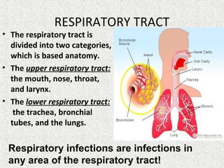
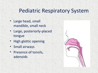
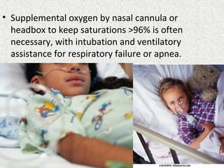
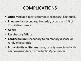
Тhe department of Pediatrics
of the V.N. KarazinV.N. Karazin
Kharkiv National UniversityKharkiv National University
Acute respiratory
diseases of upper respiratory
tract in children.
Acute bronchitis in children.
Lecturer: Ass.Prof. Rakovska L.
The plan of the lecture:
1. Definition of acute upper respiratory infections
2. Etiology, pathogenesis of common cold
3. Clinical manifestation, diagnosis, differential diagnosis,
treatment and prophylaxis of common cold
4. Acute pharyngitis, tonsillitis, laryngitis, epiglottitis in
children. Etiology, clinical signs, diagnostics.
5. Definition and classification of acute bronchitis in
children.
6. Etiology, pathogenesis, clinical signs, diagnosis,
differential diagnosis, treatment and prophylaxis of
acute bronchitis in children.
RESPIRATORY TRACT
• The respiratory tract is
divided into two categories,
which is based anatomy.
• The upper respiratory tract:
the mouth, nose, throat,
and larynx.
• The lower respiratory tract:
the trachea, bronchial
tubes, and the lungs.
Respiratory infections are infections in
any area of the respiratory tract!
Pediatric Respiratory System
• Large head, small
mandible, small neck
• Large, posteriorly-placed
tongue
• High glottic opening
• Small airways
• Presence of tonsils,
adenoids
Pediatric Respiratory System
• The muscle and elastic tissue is undeveloped
• Chest wall bones are soft.
• Horizontal ribs, primarily diaphragm
breathers.
• The position of diaphragm is high.
• The thorax is short and barrel-shaped (barrel
chest).
• Increased metabolic rate, increased O2
consumption
Pediatric Respiratory System
• Cillium movement is poor
• Large number of cells that produce mucus, the
mucosa is rich in vascularity.
• Immaturity of immune system: low levels of
secretory and serum immunoglobulin A,
interferon in the upper respiratory tract,
reduced also functional activity of T-cell
immunity.
• Upper respiratory
infections are
often referred to
as "colds.“
• Bronchitis and
pneumonia are
infections of the
lower respiratory
tract.
• Colds may also be termed coryza,
rhinitis, sniffles, nasopharyngitis, and
rhinopharyngitis, but usually also
involves the sinus mucosa and is more
correctly termed rhinosinusitis.
• Acute Viral Upper Respiratory Tract
Infections – is a large group of infectious
diseases which are caused by viruses,
transmitted by droplet way,
characterized by catarrhal syndrome
with predominant changes in upper
respiratory tract mucosa.
• The common cold is a self-limiting
illness caused by any one of more
than 250 viruses.
URTI. ETIOLOGY
• Viruses cause >90% of Upper Respiratory
Tract Infections (URTI):
• Rhinoviruses (>100 serotypes),
• Coronaviruses,
• Adenoviruses,
• Respiratory syncytial virus (RSV),
• Influenza viruses,
• Parainfluenza viruses (5 types)
• Rheoviruses (3 serotypes),
• Enteroviruses.
URTI. ETIOLOGY
• Rhinovirus is shed in large amounts, with
as many as 1 million infectious virions
present per mL of nasal washings.
Rhinovirus; Coryza Viruses; Common Cold Virus
http://www.lookfordiagnosis.com/mesh_info.php?term=Rhinovirus&lang=1
Around 30-50% of
colds are caused
by rhinoviruses.
URTI. ETIOLOGY
• Mycoplasma pneumoniae can occasionally
present with common cold symptoms before
developing into more extensive respiratory
disease.
• Other pathogens include Histoplasma
capsulatum, Chlamydia psittaci, and Coxiella
burnetii.
• Despite what is reported in folklore, no
good clinical evidence suggests that
colds are acquired by exposure to cold
weather, getting wet, or becoming
chilled.
URTI. EPIDEMIOLOGY
• Most frequent from autumn to spring.
• Receptivity in early age children, from 6 month to 5
years is high (40-80%).
• Most children have about 4 - 6 acute respiratory
infections each year, and 10% to 15% of children have
at least 12 colds each year.
• Very common are 2 peaks: starting nursery (2–3 years)
and starting primary school (4–5 years).
• The annual number of colds decreases with age, to 2 to
3 colds each year by adulthood.
• These infections tend to be even more frequent in
urban communities than in rural areas.
URTI. Mechanism of transmission
• A source of infection are
patients with URTI, and
virus-carriers.
• Droplet with inhalation of
small or large airborne
drops during coughing,
sneezing, speaking by
contact with contaminated
hands, toys ets. (hand-to-
face or -mouth contact).
• Also fecal-oral (Adenovirus,
Rheovirus infection).
URTI. Incubation period and
progression of disease
• The upper respiratory viral replication cycle
begins 8 to 12 hours after initial infection.
Symptoms usually begin 1 to 5 days after
initial infection. Symptoms peak 2–3 days
after symptom onset, whereas influenza
symptom onset is constant and immediate.
• The symptoms usually resolve spontaneously
in 7 to 10 days but some can last for up to
three weeks
URTI. PATHOGENESIS
viral inoculation
is the nasal mucosa,
although the conjunctiva
local reproduction
of the virus
start of
immune reactions
inflammatory process,
destructive changes
URTI. Pathophysyology
nasal discharge
local
inflammatory
response
sneezingthroat irritation
nasal
congestion
URTI.
Localizing Symptoms
• Nose dryness or irritation
• nasal congestion and nasal
obstruction
• rhinorhea (runny nose),
• sneezing
• sore throat or throat
irritation ("scratchy" throat)
• hoarse voice (hoarseness)
• cough (dry, non-productive
cough)
• 'pink eye'
• Nasal discharge may be clear initially but
often turns yellow-green within a few
days.
• Nasal obstruction can interfere with
sleep and/or feeding.
URTI. COMMON SYMPTOMS
• Fever (may be absent
or mild)
• muscle aches and
weakness
• fatigue, malaise,
• headaches,
• shiver,
• irritability, restlessness
• loss of appetite
• sleep disturbance
URTI. PHYSICAL EXAMINATIONS
• a change in the color or consistency of nasal
secretions,
• edema and erythema of nasal mucosa,
oropharyngeal erythema without any
exudate, or ulceration
• tonsils are mainly intact (exept adenoviral
infection)
• mildly enlarged nontender cervical lymph
nodes
URTI. Complications
Secondary bacterial infections:
• pneumonia,
• sinusitis,
• otitis media.
Wheezing .
• Colds may cause acute exacerbations of
asthma, emphysema or chronic bronchitis.
The most common risk factors for
complications:
• Minimal breastfeeding
• Prematurity, especially birth at less than 35 weeks’
gestation
• Age younger than 3 months at the time of infection
• Chronic lung disease
• Congenital heart disease
• Congenital immunodeficiency
• Severe neuromuscular disease
• Allergic rhinitis,
• Anatomic facial variations,
• Dental infections, and inhalation of irritants.
• Common cold is a clinical diagnosis.
An initial diagnosis often is made
from symptoms alone.
URTI. LABORATORY STUDIES
Laboratory studies often are not helpful.
• Identification of virus: Commercial rapid tests
are available for detection of respiratory
syncytial virus (RSV) and influenza; monospot
test (EBV).
• CBC – mainly leucopenia with a
lymphomonocytosis or normocytosis
• A nasal smear for eosinophils may be
useful in the evaluation for allergic rhinitis.
• Throat swab. Used in complicated
tonsillitis/pharyngitis.
URTI. DIFFERENTIAL DIAGNOSIS
• allergic rhinitis,
• foreign body (especially with unilateral nasal
discharge),
• structural abnormalities of the nose or
sinuses,.
• bacterial sinusitis,
• streptococcal pharyngitis
• tonsillitis .
URTI. DIFFERENTIAL DIAGNOSIS
Rare causes of rhinorrhea:
•choanal atresia or stenosis,
•congenital syphilis (with "snuffles"),
•nasopharyngeal malignancy,
•Wegener granulomatosis.
• Pharyngitis is an inflammation of the
pharynx.
• Etiology: adenoviruses, rhinovirus, EBV
(mononucleosis), enteroviruses (herpangina),
and primary HIV infection; group A b-
haemolytic streptococcus (Streptococcus
pyogenes), Chlamydophila pneumoniae, and M.
pneumoniae.
Pharyngitis
• Streptococcal
pharyngitis is relatively
uncommon before 2 to
3 years of age, but the
incidence increases in
young school-age
children, then declines
in late adolescence and
adulthood.
• Pharyngitis . Symptoms.
• sore throat, cough, dysphagia and fever. The
pharynx, soft palate, and tonsillar fauces are
inflamed and swollen.
• If involvement of the tonsils is prominent, the
term tonsillitis or tonsillopharyngitis is often
used.
• Tonsillitis: typically involves inflammation of
the tonsils.
• Etiology: adenovirus, EBV, group A b-haemolytic
streptococcus.
• Symptoms: fever, sore throat, tonsils are
enlarged, red, swollen and covered with a white,
yellow, blood-tinged exudate. The anterior
cervical lymph nodes are enlarged and tender to
touch.
• Throat culture is the diagnostic "gold standard"
for establishing the presence of streptococcal
tonsillopharyngitis.
Tonsillitis
URTI. TREATMENT
• If the child has a runny nose or cold
(without a cough, difficult breathing, an
ear problem or a sore throat), the child
needs home care only.
• Advise the mother how to give home care
and treat any fever, if present.
URTI. TREATMENT
There is no specific therapy
for the common cold.
Management consists of
symptomatic therapies.
• Bed rest up to
normalization of body
temperature.
• Vitaminized milk-vegetable
food.
• Drinking warm fluids to
maintain hydration (tea)
URTI. TREATMENT
• Clear the young infant's nose if it interferes with
feeding.
• Using nasal saline drops with bulb syringe nostril
aspiration (this treatment can help infants with
congestion and obstruction).
• Nasal decongestants (xylometazoline,
oxymetazoline, or phenylephrine), are available as
intranasal drops or nasal sprays, but are not
approved for use in children younger than 2 years
old.
URTI. TREATMENT
• Antihistamines.
First-generation antihistamines reduce
rhinorrhea by 25% to 30%, which seems to be
related to the anticholinergic effect rather
than the antihistaminic properties. Second-
generation or "nonsedating" antihistamines
have no effect on common cold symptoms.
• Cough suppressants in case of dry cough
(dextramethorphan, Glaucin, Libexin, Tusuprex,
etc).
Control of fever
• If axillaries temperature < 38,5 C in children
more then 3 month antipyretic treatment is
usually unnecessary. In children before 3 month
and in case of perinatal CNS damage, severe
heart diseases, febrile convulsions in the history
antipyretic treatment is necessary if axillaries
temperature < 38,0 C.
• Acetaminophen (paracetamol 10-15 mg/kg not
often than every 4 hours (not more than 5 times
per day) or ibuprophen 5-10 mg/kg per dose, not
often than 6 hours.
• Aspirin is contraindicated for
children before 12 years, because it
has been associated with Reye's
syndrome, a potentially fatal liver
disorder!!!
• Antibacterial therapy is not
beneficial for the common cold.
• Antibiotics kill bacteria, not viruses, and
are of no use in treating a cold.
• Antiviral treatment may be used in
severe causes
•‘Treat a cold it lasts a
week, don’t treat and it
lasts 7 days.’
Cold Prevention
• There are no proven methods for prevention of
colds other than good hand washing and
avoiding contact with infected persons. No
significant effect of vitamin C or echinacea for
prevention of the common cold has been
confirmed.
Cold Prevention
• There is no vaccine to prevent colds.
• There are two major reasons vaccines are
not being sought for colds.
– 1) almost every person who gets a cold recovers
without any complications,
– 2) with over 250 viral types, producing an
effective vaccine against most or all viral
types is nearly impossible with current
techniques.
Laryngitis: is an inflammation of
the larynx
• Etiology: viruses (adenovirus, influenza
virus, parainfluenza virus and rhinovirus)
and often a bacterial co-infection, including
either S. aureus, S. pneumoniae, or H.
influenzae type b.
Croup
Gradually onset:
symptoms of URTI followed after
several days by the
characteristic hoarseness and
aphonia, barking cough (Seal
bark) , inspiratory stridor, and
signs of respiratory distress
due to laryngeal obstruction.
• Worsening at night
(acute laryngotracheitis or
laryngotracheobronchitis) is a form of laryngitis
in infant and young children.
Croup. Etiology
• Common: Parainfluenza types 1, 2, and 3
• Less common: Influenza viruses A, B
• Rare: Respiratory syncytial virus (RSV),
adenovirus, measles
• In clinical practice, the term "croup"is
usually used to describe acute
laryngotracheitis (viral croup) and acute
spasmodic laryngitis (spasmodic croup).
• Spasmodic croup is believed to be caused by
viruses, although important allergic and
psychological factors probably contribute to
the illness in some patients.
Croup. Epidemiology
• Usually occurs in late fall and winter, but
sporadic cases throughout the year
• Peak incidence at 18 months (typical range, 1
to 6 years)
Croup. Pathogenesis
• Virus-induced inflammation of larynx, vocal
cords, and trachea
• Subglottic (narrowest part of a child's upper
airway) tracheal edema restricts airflow
Physical Examination.
Main symptoms of viral croup:
• Inspiratory stridor (from turbulent airflow);
• Barking cough, that worsens at night;
• Hoarse voice (from vocal cord edema)
• Hypoxia only with severe croup
• Non-toxic appearance
N.B. Sometimes you will hear a wet noise if the child's
nose is blocked. Clear the nose and listen again.
Often, a child who is not very ill will have stridor only
when he or she is crying or upset, so be sure to look
and listen for stridor when the child is calm.
Degree of stenosing laryngitis (croup)
• Stenosis of 1 degree – symptoms of croup only during
crying and physical activity. The respiratory failure is
absent.
• Stenosis of 2 degree – respiration noisy, the hoarse
voice, barking cough, chest retraction presents at rest
and increasing during crying and physical activity .The
respiratory failure is moderately expressed. Peripheral
cyanosis.
• Stenosis of 3 degree – significant symptoms at rest.
Signs of hypoxia. The respiratory failure
• Stenosis of 4 degree - a stage of an asphyxia.
Additional Studies for diagnosis
50% of cases reveal abnormal
neck radiographs:
• Posteroanterior view:
Tapered subglottic narrowing
("steeple sign’’
•Lateral view: Overdistention
of hypopharynx
Croup. Differential Diagnosis
• Upper Respiratory Tract Infections
• Acute epiglottitis;
• Retropharyngeall, peritonsillar abscess;
• Bacterial tracheitis;
• Infectious mononucleosis;
• Diphtheria
• Angioneurotic edema;
• Subglottic stenosis;
• Tracheomalacia;
•
Croup. Differential Diagnosis
• Epiglottitis: is bacterial inflammation of the
epiglottis.
• Acute epiglottitis is rapidly progressive infection
of the epiglottis and contiguous structures that
may cause life-threatening airway obstruction.
• Etiology: H. influenzae type b.
Epiglottitis: Signs/Symptoms
• Rapid onset, severe distress in hours
• High fever
• Intense sore throat, difficulty swallowing
• Drooling
• Stridor
• Sits up, leans forward, extends neck slightly
• One-third present unconscious, in shock
Epiglottitis:
OMEGA SIGN
(Dr.FaceB..k)
Croup. Management (1 degree)
• Cool mist treatment
• Mechanism of action: soothes inflamed
mucosa, decreases viscosity of tracheal
secretions; may activate larynx mechano-
receptors to produce reflex slowing of
respiratory flow rate
• No clinical trials have demonstrated efficacy is
effective
• Caution: May intensify bronchospasm
Croup. Management (2 degree)
• Corticosteroids: Dexamethasone 0.6 mg/kg
PO or IM (max 10 mg), clinical improvement in
4 to 6 hours
• For mild-moderate croup, dexamethasone
0,15-0.30 mg/kg
For any patient with increased work of
breathing
• Mechanism of action: Decreases laryngeal
mucosal edema via anti-inflammatory action
Croup. Management (3 degree)
• Nebulized epinephrine:
Dose: 0,25-0,75 mL of 2,25% racemic
epinephrine solution in 2.5 mL of normal
saline
• For severe or worsening respiratory distress
• Mechanism of action: a-agonist capillary
arteriole constriction decreases mucosal
edema
• "Rebound" phenomenon (2 to 4 hours after
treatment) rare if dexamethasone also given
Croup. Management (4 degree)
• Helium-oxygen therapy (usually 70% He:30%
O 2 )
• Helium (low-density and low-viscosity gas)
improves laminar gas flow and decreases
mechanical work of breathing
• Endotracheal intubation may be required for
severe croup
Bronchitis
Bronchitis is an inflammatory disease of
bronchi of various etiology: infectious
(mostly viral), allergic, physical and
chemical (so-called irritative bronchitis).
Classification of bronchitis
• For the nature of inflammation: catarrhal, purulent,
atrophic bronchitis.
• For the course: acute, recurring, chronic bronchitis.
• Acute bronchitis: acute simple bronchitis.
• acute obstructive bronchitis.
• acute bronchiolitis
• Recurring bronchitis: Phases: exacerbation, remission
• Chronic bronchitis:
• 1. Primary, Secondary chronic bronchitis.
• 2. Phases: exacerbation, remission
• Chronic obliterating bronchiolitis:
• Phases: exacerbation, remission
ICD-10
• J20. Acute bronchitis
• J20.0. Acute bronchitis, caused by Mycoplasma pneumonia
• J20.1. Acute bronchitis, caused by Haemophilus influenza
• J 20.2. Acute bronchitis, caused by Streptococcus
• J 20.3. Acute bronchitis, caused by Coxsacke virus
• J 20.4. Acute bronchitis, caused by parainfluenza virus
• J 20.5. Acute bronchitis, caused by RSV
• J 20.6. Acute bronchitis, caused by rhinovirus
• J 20.7. Acute bronchitis, caused by ECHO virus
• J 20.8. Acute bronchitis, caused by other specified agents
• J 20.9. Acute bronchitis, unspecified.
• J21. Acute bronchiolitis
• J21.0 Acute bronchiolitis due to respiratory syncytial virus
• J21.1 Acute bronchiolitis due to human metapneumovirus
• J21.8 Acute bronchiolitis due to other specified organisms
Criteria of acute bronchitis
I.Clinical:
• cough – dry and rough at the beginning of
disease, gradually becoming productive;
• symptoms of intoxication are not expressed
greatly and quickly disappear;
• No symptoms of respiratory insufficiency
Criteria of acute bronchitis
Physical examination
• Percussion:
slight tympanic
resonance,
• Auscultation
rhonchi, dry and
various bubbling
rales, heard on
both sides of lungs.
Obstructive bronchitis –
• is a variant of acute
bronchitis, which
proceeds with
respiratory tract
obstruction because of
bronchospasm,
mucous edema,
hypersecretion and
pressure from
without.
Obstructive bronchitis.
Clinical sings
• Paroxysmal, “spastic” cough,
• Expiratory dyspnea, tachypnea
• Chest retraction, increased use
of accessory muscles
• Wheezing,
• Oral crepitations,
• Tympanic percussion sounds,
• Prolonged expiratory phase
• Dry and various bubbling rales.
Criteria of acute bronchitis
X-ray:
• strengthened lung figure, at the same time
absence of focal shadow;
• Rarefied lung pattern in lateral divisions of the
lungs and its thickening in the medial
divisions (occult emphysema)
Criteria of acute bronchitis
III.Laboratory:
• CBC:
–normal leukocyte count or leukopoenia,
lymphocytosis, monocytosis.
–ESR is not increased.
Differential diagnosis of acute bronchitis:
•acute bronchiolitis,
•pneumonia;
•obstructive bronchitis – with bronchial
asthma paroxysm, GERD.
Acute bronchitis. Treatment.
• General:
– antipyretics, and good fluid intake.
• Specific mainly viral, no antibiotics.
• In case of cough with mucous – expectorant
and mucolytic drugs
(ambroxol, acetylcysteine,
carbocysteine, etc)
Indications for use of antibiotics in
acute bronchitis
• The age below 6 months
• Severe clinical course of bronchitis (neurotoxicosis, etc)
• The compromised premorbid background (premature
birth, chronic diseases of lung or heart,
immunodeficiency)
• Suspected concomitant bacterial infection:
– Fever with elevation of body temperature above 39 C
– Flaccidity, refusal of food
– Pronounced intoxication
– Dyspnea
– Asymmetry of rales
– Leukocytosis, increased ESR
• The following antibiotics are used:
• Protected penicillins (Amoxiillin-clavunate)
• Cephalosporins (Cephalexin, Cephadrixyl,
Cephasolin, Cephotaxim, Cephtriaxon)
• Macrolids (Azitromycin, claritromycin)
Bronchiolitis
• is an acute viral lower respiratory tract
infection that results in an inflammatory
obstruction of the peripheral airways. It is
characterized by obstructive respiratory
insufficiency and cyclic course. Bronchiolitis is
mainly a disease of the first months of life.
• It is potentially life-threatening.
Bronchiolitis. Etiology
• Common: Respiratory syncytial virus
(RSV; 70% of cases)
• Less common: Parainfluenza, influenza,
adenovirus , metapneumoviruses
• Rare: rhinovirus, Mycoplasma
pneumoniae, Chlamydia, and
ureaplasma
epithelial
damage
inflammation
of the bronchioles
secretion
of mucous
necrosis of
Сiliated
epithelium
oedema
of the
submucosa
airway
obstruction
Pathophysiology
Bronchiolitis
CLINICAL MANIFESTATIONS
• Progressive respiratory illness
• In early phase it is similar to the common cold with
cough, coryza, and rhinorrhea, and low-grade fever .
• It progresses over 3 to 7 days to noisy, raspy
breathing and audible wheezing. Irritability, which
may reflect the increased work of breathing and may
increase itself with increased respiratory effort.
• In contrast to the classic progression of disease,
young infants infected with RSV may not have a
prodrome and may have apnea as the first sign of
infection.
Bronchiolitis. Symptoms
• Cyanosis nasolabial triangle,
• Tachypnea with usually shallow respirations.
• Wheeze, increased expiratory phase, and rales
• In more severe cases infants may become too
breathless to feed
• Asphyxial paroxysms are possible for infants of the
first months of the life);
• Persistent spastic cough.
• Tachycardia: especially when hypoxemia present.
• Fever: usually mild.
• Vomiting (posttussive).
Physical signs of bronchiolar
obstruction
• Intercostal retractions of flexible parts of breast
(subcostal/ intercostal recession)
• Percussion of the chest: hyperresonance (high
tympanic resonance)
• Auscultation: prolonged breathing out, diffuse
wheezes and crackles throughout the breathing
cycle on the both sides of lungs and a lot of fine
bubbling rales, crepitation on inspiration.
• Hypoxia is common in severely affected patients
LABORATORY AND IMAGING STUDIES
Routine laboratory tests lack specificity for
diagnosing bronchiolitis and are not required
to confirm the diagnosis.
The chest radiograph:
• hyperinflation due to small airways obstruction,
• air trapping,
• flattened or depressed diaphragms.
• The lung fields may appear normal or collapse
and/or consolidation may be seen occasionally.
• An X- ray of a child
with RSV showing the
typical bilateral
perihilar fullness of
bronchiolitis.
Laboratory:
• CBC: no specific findings. May be normal or
decreased white blood cells count,
lymphocytosis or mild leukocytosis.
• Rapid viral identification: usually a
“respiratory panel” is available to include
common seasonal respiratory pathogens by
antigen detection.
• Viral culture: results delayed but may be
useful to identify causative organism.
• Pulse oximetry is
generally adequate
for monitoring
oxygen saturation.
• Arterial blood gas in
severe cases of
bronchiolitis.
• Visual assessment of
oxygenation
correlates poorly
with actual blood gas
values.
Bronchiolitis
DIFFERENTIAL DIAGNOSIS
• Pneumonia
• Acute bronchitis.
• Asthma
• Foreign body in the airway,
• Congenital airway obstructive lesion,
• Cystic fibrosis,
• Bronchopulmonary dysplasia,
• Cardiogenic asthma
Indications for hospitalization:
• young age (<6 months old),
• moderate to marked respiratory distress (sleeping
respiratory rate of >50 to 60 breaths/min),
• hypoxemia (Po2 <60 mm Hg or oxygen saturation <92%
on room air),
• apnea,
• feeding difficulty ,
• lack of appropriate care available at
home.
• Children with chronic disease
(bronchopulmonary dysplasia,
congenital heart disease,
neuromuscular weakness, or immunodeficiency)
Bronchiolitis
TREATMENT
• Supportive therapy
monitoring, control of fever,
feeding via NG tube,
adequate fluid intake with IV support if
necessary (avoid excessive hydration),
upper airway aspiration (suctioning),
oxygen administration
• Supplemental oxygen by nasal cannula or
headbox to keep saturations >96% is often
necessary, with intubation and ventilatory
assistance for respiratory failure or apnea.
•Most infants require only
supportive care for their
self-limited illness.
Pharmacologic treatment
• Bronchodilators: ß-2-agonist or alpha-
adrenergic agents (epinephrine) by inhalation.
• Corticosteroids: prednisone 1-2 mg/kg IV, IM
The benefit of bronchodilators and
corticosteroids is controversial.
• Antiviral treatment:
• Ribavirin, an antiviral agent that suppresses
viral RNA polymerase activity, may shorten
symptoms, and its use should be considered
for patients who are at risk for severe or fatal
infections (chronic lung disease or
immunosuppressive conditions).
• Antibiotics not indicated unless
secondary bacterial infection
detected.
Differentiating Viral from Bacterial
Infections
Variable Viral Bacterial
Leukocytosis Uncommon* Common
Shift to left
(↑ bands)
Uncommon Common
Neutropenia Possible Suggests
overwhelming
infection
↑ ESR Unusual Common
↑ CRP Unusual Common
Prognosis
• Acute severe obstructive symptoms usually
resolve in 3–5 days, but cough may last up to
10-14 days. Complete recovery expected for
most patients.
• Mortality is generally very low (<1 %), but is
higher in infants <6 months or with underlying
chronic conditions. Mortality rises to 33% if
ventilation is required.
COMPLICATIONS
• Otitis media: is most common (secondary; bacterial).
• Pneumonia: secondary, bacterial; occurs in < 1% of
hospitalized cases.
• Apnea
• Respiratory failure
• Cardiac failure: secondary to pulmonary disease or
rarely myocarditis
• Bronchiolitis obliterans: rare; usually associated with
adenovirus-induced bronchiolitis/pneumonia
PREVENTION
• Immunoprophylaxis:
• RSV–intravenous immunoglobulin (RSV-IVIg) with
high RSV antibody concentration and
palivizumab, an injectable RSV monoclonal
antibody, provide passive prophylaxis and are
recommended during the winter months for
prevention of RSV disease for patients at risk for
severe disease.
Recurrent bronchitis –
• is the disease with relapsing of acute
bronchitis 2 and more times a year during 1-2
years. The absence of clinical obstruction and
duration of clinical manifestation for 2 weeks
and longer every relapse are common.
• Phases of pathologic process: exacerbation,
remission.
Chronic bronchitis
• is a chronic spread inflammatory damage of
bronchi with rebuilding of mucous secretory
apparatus and sclerotic degeneration of deep
layers of bronchial wall.
• Phases of pathological process: exacerbation,
remission.
Chronic bronchitis
Clinical symptoms:
• - productive cough for several months during 2
years;
• - permanent various râles;
• - 2-3 relapses in a year during 2 years;
• - the signs of lungs ventilation disturbances in
remission phase.
Note: none of the above mentioned signs may be
regarded alone as reliable evidence of chronic
bronchitis. The sings must be considered in
complex in view of possible development of
chronic process.
Chronic bronchitis. Diagnostics.
X-ray, especially bronchography :
•increasing and deformity of lungs figure, the
disturbances of root of the lungs structure.
Bronchoscopy
•endobronchitis.
Primary chronic bronchitis is diagnosed after
exclusion of cystic fibrosis, bronchial asthma,
lungs and cardiovascular malformations, ciliary
dysgenesis.
Thank you for your
attention!

cold, bronchitis

  • 1.
    Тhe department ofPediatrics of the V.N. KarazinV.N. Karazin Kharkiv National UniversityKharkiv National University Acute respiratory diseases of upper respiratory tract in children. Acute bronchitis in children. Lecturer: Ass.Prof. Rakovska L.
  • 2.
    The plan ofthe lecture: 1. Definition of acute upper respiratory infections 2. Etiology, pathogenesis of common cold 3. Clinical manifestation, diagnosis, differential diagnosis, treatment and prophylaxis of common cold 4. Acute pharyngitis, tonsillitis, laryngitis, epiglottitis in children. Etiology, clinical signs, diagnostics. 5. Definition and classification of acute bronchitis in children. 6. Etiology, pathogenesis, clinical signs, diagnosis, differential diagnosis, treatment and prophylaxis of acute bronchitis in children.
  • 3.
    RESPIRATORY TRACT • Therespiratory tract is divided into two categories, which is based anatomy. • The upper respiratory tract: the mouth, nose, throat, and larynx. • The lower respiratory tract: the trachea, bronchial tubes, and the lungs. Respiratory infections are infections in any area of the respiratory tract!
  • 4.
    Pediatric Respiratory System •Large head, small mandible, small neck • Large, posteriorly-placed tongue • High glottic opening • Small airways • Presence of tonsils, adenoids
  • 5.
    Pediatric Respiratory System •The muscle and elastic tissue is undeveloped • Chest wall bones are soft. • Horizontal ribs, primarily diaphragm breathers. • The position of diaphragm is high. • The thorax is short and barrel-shaped (barrel chest). • Increased metabolic rate, increased O2 consumption
  • 6.
    Pediatric Respiratory System •Cillium movement is poor • Large number of cells that produce mucus, the mucosa is rich in vascularity. • Immaturity of immune system: low levels of secretory and serum immunoglobulin A, interferon in the upper respiratory tract, reduced also functional activity of T-cell immunity.
  • 7.
    • Upper respiratory infectionsare often referred to as "colds.“ • Bronchitis and pneumonia are infections of the lower respiratory tract.
  • 8.
    • Colds mayalso be termed coryza, rhinitis, sniffles, nasopharyngitis, and rhinopharyngitis, but usually also involves the sinus mucosa and is more correctly termed rhinosinusitis.
  • 9.
    • Acute ViralUpper Respiratory Tract Infections – is a large group of infectious diseases which are caused by viruses, transmitted by droplet way, characterized by catarrhal syndrome with predominant changes in upper respiratory tract mucosa.
  • 10.
    • The commoncold is a self-limiting illness caused by any one of more than 250 viruses.
  • 11.
    URTI. ETIOLOGY • Virusescause >90% of Upper Respiratory Tract Infections (URTI): • Rhinoviruses (>100 serotypes), • Coronaviruses, • Adenoviruses, • Respiratory syncytial virus (RSV), • Influenza viruses, • Parainfluenza viruses (5 types) • Rheoviruses (3 serotypes), • Enteroviruses.
  • 12.
    URTI. ETIOLOGY • Rhinovirusis shed in large amounts, with as many as 1 million infectious virions present per mL of nasal washings. Rhinovirus; Coryza Viruses; Common Cold Virus http://www.lookfordiagnosis.com/mesh_info.php?term=Rhinovirus&lang=1 Around 30-50% of colds are caused by rhinoviruses.
  • 13.
    URTI. ETIOLOGY • Mycoplasmapneumoniae can occasionally present with common cold symptoms before developing into more extensive respiratory disease. • Other pathogens include Histoplasma capsulatum, Chlamydia psittaci, and Coxiella burnetii.
  • 14.
    • Despite whatis reported in folklore, no good clinical evidence suggests that colds are acquired by exposure to cold weather, getting wet, or becoming chilled.
  • 15.
    URTI. EPIDEMIOLOGY • Mostfrequent from autumn to spring. • Receptivity in early age children, from 6 month to 5 years is high (40-80%). • Most children have about 4 - 6 acute respiratory infections each year, and 10% to 15% of children have at least 12 colds each year. • Very common are 2 peaks: starting nursery (2–3 years) and starting primary school (4–5 years). • The annual number of colds decreases with age, to 2 to 3 colds each year by adulthood. • These infections tend to be even more frequent in urban communities than in rural areas.
  • 16.
    URTI. Mechanism oftransmission • A source of infection are patients with URTI, and virus-carriers. • Droplet with inhalation of small or large airborne drops during coughing, sneezing, speaking by contact with contaminated hands, toys ets. (hand-to- face or -mouth contact). • Also fecal-oral (Adenovirus, Rheovirus infection).
  • 17.
    URTI. Incubation periodand progression of disease • The upper respiratory viral replication cycle begins 8 to 12 hours after initial infection. Symptoms usually begin 1 to 5 days after initial infection. Symptoms peak 2–3 days after symptom onset, whereas influenza symptom onset is constant and immediate. • The symptoms usually resolve spontaneously in 7 to 10 days but some can last for up to three weeks
  • 18.
    URTI. PATHOGENESIS viral inoculation isthe nasal mucosa, although the conjunctiva local reproduction of the virus start of immune reactions inflammatory process, destructive changes
  • 19.
  • 20.
    URTI. Localizing Symptoms • Nosedryness or irritation • nasal congestion and nasal obstruction • rhinorhea (runny nose), • sneezing • sore throat or throat irritation ("scratchy" throat) • hoarse voice (hoarseness) • cough (dry, non-productive cough) • 'pink eye'
  • 21.
    • Nasal dischargemay be clear initially but often turns yellow-green within a few days. • Nasal obstruction can interfere with sleep and/or feeding.
  • 22.
    URTI. COMMON SYMPTOMS •Fever (may be absent or mild) • muscle aches and weakness • fatigue, malaise, • headaches, • shiver, • irritability, restlessness • loss of appetite • sleep disturbance
  • 23.
    URTI. PHYSICAL EXAMINATIONS •a change in the color or consistency of nasal secretions, • edema and erythema of nasal mucosa, oropharyngeal erythema without any exudate, or ulceration • tonsils are mainly intact (exept adenoviral infection) • mildly enlarged nontender cervical lymph nodes
  • 24.
    URTI. Complications Secondary bacterialinfections: • pneumonia, • sinusitis, • otitis media. Wheezing . • Colds may cause acute exacerbations of asthma, emphysema or chronic bronchitis.
  • 25.
    The most commonrisk factors for complications: • Minimal breastfeeding • Prematurity, especially birth at less than 35 weeks’ gestation • Age younger than 3 months at the time of infection • Chronic lung disease • Congenital heart disease • Congenital immunodeficiency • Severe neuromuscular disease • Allergic rhinitis, • Anatomic facial variations, • Dental infections, and inhalation of irritants.
  • 26.
    • Common coldis a clinical diagnosis. An initial diagnosis often is made from symptoms alone.
  • 27.
    URTI. LABORATORY STUDIES Laboratorystudies often are not helpful. • Identification of virus: Commercial rapid tests are available for detection of respiratory syncytial virus (RSV) and influenza; monospot test (EBV). • CBC – mainly leucopenia with a lymphomonocytosis or normocytosis • A nasal smear for eosinophils may be useful in the evaluation for allergic rhinitis. • Throat swab. Used in complicated tonsillitis/pharyngitis.
  • 28.
    URTI. DIFFERENTIAL DIAGNOSIS •allergic rhinitis, • foreign body (especially with unilateral nasal discharge), • structural abnormalities of the nose or sinuses,. • bacterial sinusitis, • streptococcal pharyngitis • tonsillitis .
  • 29.
    URTI. DIFFERENTIAL DIAGNOSIS Rarecauses of rhinorrhea: •choanal atresia or stenosis, •congenital syphilis (with "snuffles"), •nasopharyngeal malignancy, •Wegener granulomatosis.
  • 30.
    • Pharyngitis isan inflammation of the pharynx. • Etiology: adenoviruses, rhinovirus, EBV (mononucleosis), enteroviruses (herpangina), and primary HIV infection; group A b- haemolytic streptococcus (Streptococcus pyogenes), Chlamydophila pneumoniae, and M. pneumoniae.
  • 31.
    Pharyngitis • Streptococcal pharyngitis isrelatively uncommon before 2 to 3 years of age, but the incidence increases in young school-age children, then declines in late adolescence and adulthood.
  • 32.
    • Pharyngitis .Symptoms. • sore throat, cough, dysphagia and fever. The pharynx, soft palate, and tonsillar fauces are inflamed and swollen. • If involvement of the tonsils is prominent, the term tonsillitis or tonsillopharyngitis is often used.
  • 33.
    • Tonsillitis: typicallyinvolves inflammation of the tonsils. • Etiology: adenovirus, EBV, group A b-haemolytic streptococcus. • Symptoms: fever, sore throat, tonsils are enlarged, red, swollen and covered with a white, yellow, blood-tinged exudate. The anterior cervical lymph nodes are enlarged and tender to touch. • Throat culture is the diagnostic "gold standard" for establishing the presence of streptococcal tonsillopharyngitis.
  • 34.
  • 35.
    URTI. TREATMENT • Ifthe child has a runny nose or cold (without a cough, difficult breathing, an ear problem or a sore throat), the child needs home care only. • Advise the mother how to give home care and treat any fever, if present.
  • 36.
    URTI. TREATMENT There isno specific therapy for the common cold. Management consists of symptomatic therapies. • Bed rest up to normalization of body temperature. • Vitaminized milk-vegetable food. • Drinking warm fluids to maintain hydration (tea)
  • 37.
    URTI. TREATMENT • Clearthe young infant's nose if it interferes with feeding. • Using nasal saline drops with bulb syringe nostril aspiration (this treatment can help infants with congestion and obstruction). • Nasal decongestants (xylometazoline, oxymetazoline, or phenylephrine), are available as intranasal drops or nasal sprays, but are not approved for use in children younger than 2 years old.
  • 38.
    URTI. TREATMENT • Antihistamines. First-generationantihistamines reduce rhinorrhea by 25% to 30%, which seems to be related to the anticholinergic effect rather than the antihistaminic properties. Second- generation or "nonsedating" antihistamines have no effect on common cold symptoms. • Cough suppressants in case of dry cough (dextramethorphan, Glaucin, Libexin, Tusuprex, etc).
  • 39.
    Control of fever •If axillaries temperature < 38,5 C in children more then 3 month antipyretic treatment is usually unnecessary. In children before 3 month and in case of perinatal CNS damage, severe heart diseases, febrile convulsions in the history antipyretic treatment is necessary if axillaries temperature < 38,0 C. • Acetaminophen (paracetamol 10-15 mg/kg not often than every 4 hours (not more than 5 times per day) or ibuprophen 5-10 mg/kg per dose, not often than 6 hours.
  • 40.
    • Aspirin iscontraindicated for children before 12 years, because it has been associated with Reye's syndrome, a potentially fatal liver disorder!!!
  • 41.
    • Antibacterial therapyis not beneficial for the common cold. • Antibiotics kill bacteria, not viruses, and are of no use in treating a cold. • Antiviral treatment may be used in severe causes
  • 42.
    •‘Treat a coldit lasts a week, don’t treat and it lasts 7 days.’
  • 43.
    Cold Prevention • Thereare no proven methods for prevention of colds other than good hand washing and avoiding contact with infected persons. No significant effect of vitamin C or echinacea for prevention of the common cold has been confirmed.
  • 44.
    Cold Prevention • Thereis no vaccine to prevent colds. • There are two major reasons vaccines are not being sought for colds. – 1) almost every person who gets a cold recovers without any complications, – 2) with over 250 viral types, producing an effective vaccine against most or all viral types is nearly impossible with current techniques.
  • 45.
    Laryngitis: is aninflammation of the larynx • Etiology: viruses (adenovirus, influenza virus, parainfluenza virus and rhinovirus) and often a bacterial co-infection, including either S. aureus, S. pneumoniae, or H. influenzae type b.
  • 46.
    Croup Gradually onset: symptoms ofURTI followed after several days by the characteristic hoarseness and aphonia, barking cough (Seal bark) , inspiratory stridor, and signs of respiratory distress due to laryngeal obstruction. • Worsening at night (acute laryngotracheitis or laryngotracheobronchitis) is a form of laryngitis in infant and young children.
  • 47.
    Croup. Etiology • Common:Parainfluenza types 1, 2, and 3 • Less common: Influenza viruses A, B • Rare: Respiratory syncytial virus (RSV), adenovirus, measles
  • 48.
    • In clinicalpractice, the term "croup"is usually used to describe acute laryngotracheitis (viral croup) and acute spasmodic laryngitis (spasmodic croup). • Spasmodic croup is believed to be caused by viruses, although important allergic and psychological factors probably contribute to the illness in some patients.
  • 49.
    Croup. Epidemiology • Usuallyoccurs in late fall and winter, but sporadic cases throughout the year • Peak incidence at 18 months (typical range, 1 to 6 years)
  • 50.
    Croup. Pathogenesis • Virus-inducedinflammation of larynx, vocal cords, and trachea • Subglottic (narrowest part of a child's upper airway) tracheal edema restricts airflow
  • 51.
    Physical Examination. Main symptomsof viral croup: • Inspiratory stridor (from turbulent airflow); • Barking cough, that worsens at night; • Hoarse voice (from vocal cord edema) • Hypoxia only with severe croup • Non-toxic appearance N.B. Sometimes you will hear a wet noise if the child's nose is blocked. Clear the nose and listen again. Often, a child who is not very ill will have stridor only when he or she is crying or upset, so be sure to look and listen for stridor when the child is calm.
  • 52.
    Degree of stenosinglaryngitis (croup) • Stenosis of 1 degree – symptoms of croup only during crying and physical activity. The respiratory failure is absent. • Stenosis of 2 degree – respiration noisy, the hoarse voice, barking cough, chest retraction presents at rest and increasing during crying and physical activity .The respiratory failure is moderately expressed. Peripheral cyanosis. • Stenosis of 3 degree – significant symptoms at rest. Signs of hypoxia. The respiratory failure • Stenosis of 4 degree - a stage of an asphyxia.
  • 53.
    Additional Studies fordiagnosis 50% of cases reveal abnormal neck radiographs: • Posteroanterior view: Tapered subglottic narrowing ("steeple sign’’ •Lateral view: Overdistention of hypopharynx
  • 55.
    Croup. Differential Diagnosis •Upper Respiratory Tract Infections • Acute epiglottitis; • Retropharyngeall, peritonsillar abscess; • Bacterial tracheitis; • Infectious mononucleosis; • Diphtheria • Angioneurotic edema; • Subglottic stenosis; • Tracheomalacia; •
  • 56.
    Croup. Differential Diagnosis •Epiglottitis: is bacterial inflammation of the epiglottis. • Acute epiglottitis is rapidly progressive infection of the epiglottis and contiguous structures that may cause life-threatening airway obstruction. • Etiology: H. influenzae type b.
  • 57.
    Epiglottitis: Signs/Symptoms • Rapidonset, severe distress in hours • High fever • Intense sore throat, difficulty swallowing • Drooling • Stridor • Sits up, leans forward, extends neck slightly • One-third present unconscious, in shock
  • 58.
  • 59.
  • 60.
    Croup. Management (1degree) • Cool mist treatment • Mechanism of action: soothes inflamed mucosa, decreases viscosity of tracheal secretions; may activate larynx mechano- receptors to produce reflex slowing of respiratory flow rate • No clinical trials have demonstrated efficacy is effective • Caution: May intensify bronchospasm
  • 61.
    Croup. Management (2degree) • Corticosteroids: Dexamethasone 0.6 mg/kg PO or IM (max 10 mg), clinical improvement in 4 to 6 hours • For mild-moderate croup, dexamethasone 0,15-0.30 mg/kg For any patient with increased work of breathing • Mechanism of action: Decreases laryngeal mucosal edema via anti-inflammatory action
  • 62.
    Croup. Management (3degree) • Nebulized epinephrine: Dose: 0,25-0,75 mL of 2,25% racemic epinephrine solution in 2.5 mL of normal saline • For severe or worsening respiratory distress • Mechanism of action: a-agonist capillary arteriole constriction decreases mucosal edema • "Rebound" phenomenon (2 to 4 hours after treatment) rare if dexamethasone also given
  • 63.
    Croup. Management (4degree) • Helium-oxygen therapy (usually 70% He:30% O 2 ) • Helium (low-density and low-viscosity gas) improves laminar gas flow and decreases mechanical work of breathing • Endotracheal intubation may be required for severe croup
  • 64.
    Bronchitis Bronchitis is aninflammatory disease of bronchi of various etiology: infectious (mostly viral), allergic, physical and chemical (so-called irritative bronchitis).
  • 65.
    Classification of bronchitis •For the nature of inflammation: catarrhal, purulent, atrophic bronchitis. • For the course: acute, recurring, chronic bronchitis. • Acute bronchitis: acute simple bronchitis. • acute obstructive bronchitis. • acute bronchiolitis • Recurring bronchitis: Phases: exacerbation, remission • Chronic bronchitis: • 1. Primary, Secondary chronic bronchitis. • 2. Phases: exacerbation, remission • Chronic obliterating bronchiolitis: • Phases: exacerbation, remission
  • 66.
    ICD-10 • J20. Acutebronchitis • J20.0. Acute bronchitis, caused by Mycoplasma pneumonia • J20.1. Acute bronchitis, caused by Haemophilus influenza • J 20.2. Acute bronchitis, caused by Streptococcus • J 20.3. Acute bronchitis, caused by Coxsacke virus • J 20.4. Acute bronchitis, caused by parainfluenza virus • J 20.5. Acute bronchitis, caused by RSV • J 20.6. Acute bronchitis, caused by rhinovirus • J 20.7. Acute bronchitis, caused by ECHO virus • J 20.8. Acute bronchitis, caused by other specified agents • J 20.9. Acute bronchitis, unspecified. • J21. Acute bronchiolitis • J21.0 Acute bronchiolitis due to respiratory syncytial virus • J21.1 Acute bronchiolitis due to human metapneumovirus • J21.8 Acute bronchiolitis due to other specified organisms
  • 67.
    Criteria of acutebronchitis I.Clinical: • cough – dry and rough at the beginning of disease, gradually becoming productive; • symptoms of intoxication are not expressed greatly and quickly disappear; • No symptoms of respiratory insufficiency
  • 68.
    Criteria of acutebronchitis Physical examination • Percussion: slight tympanic resonance, • Auscultation rhonchi, dry and various bubbling rales, heard on both sides of lungs.
  • 69.
    Obstructive bronchitis – •is a variant of acute bronchitis, which proceeds with respiratory tract obstruction because of bronchospasm, mucous edema, hypersecretion and pressure from without.
  • 70.
    Obstructive bronchitis. Clinical sings •Paroxysmal, “spastic” cough, • Expiratory dyspnea, tachypnea • Chest retraction, increased use of accessory muscles • Wheezing, • Oral crepitations, • Tympanic percussion sounds, • Prolonged expiratory phase • Dry and various bubbling rales.
  • 71.
    Criteria of acutebronchitis X-ray: • strengthened lung figure, at the same time absence of focal shadow; • Rarefied lung pattern in lateral divisions of the lungs and its thickening in the medial divisions (occult emphysema)
  • 72.
    Criteria of acutebronchitis III.Laboratory: • CBC: –normal leukocyte count or leukopoenia, lymphocytosis, monocytosis. –ESR is not increased.
  • 73.
    Differential diagnosis ofacute bronchitis: •acute bronchiolitis, •pneumonia; •obstructive bronchitis – with bronchial asthma paroxysm, GERD.
  • 74.
    Acute bronchitis. Treatment. •General: – antipyretics, and good fluid intake. • Specific mainly viral, no antibiotics. • In case of cough with mucous – expectorant and mucolytic drugs (ambroxol, acetylcysteine, carbocysteine, etc)
  • 75.
    Indications for useof antibiotics in acute bronchitis • The age below 6 months • Severe clinical course of bronchitis (neurotoxicosis, etc) • The compromised premorbid background (premature birth, chronic diseases of lung or heart, immunodeficiency) • Suspected concomitant bacterial infection: – Fever with elevation of body temperature above 39 C – Flaccidity, refusal of food – Pronounced intoxication – Dyspnea – Asymmetry of rales – Leukocytosis, increased ESR
  • 76.
    • The followingantibiotics are used: • Protected penicillins (Amoxiillin-clavunate) • Cephalosporins (Cephalexin, Cephadrixyl, Cephasolin, Cephotaxim, Cephtriaxon) • Macrolids (Azitromycin, claritromycin)
  • 77.
    Bronchiolitis • is anacute viral lower respiratory tract infection that results in an inflammatory obstruction of the peripheral airways. It is characterized by obstructive respiratory insufficiency and cyclic course. Bronchiolitis is mainly a disease of the first months of life. • It is potentially life-threatening.
  • 78.
    Bronchiolitis. Etiology • Common:Respiratory syncytial virus (RSV; 70% of cases) • Less common: Parainfluenza, influenza, adenovirus , metapneumoviruses • Rare: rhinovirus, Mycoplasma pneumoniae, Chlamydia, and ureaplasma
  • 79.
    epithelial damage inflammation of the bronchioles secretion ofmucous necrosis of Сiliated epithelium oedema of the submucosa airway obstruction Pathophysiology
  • 80.
    Bronchiolitis CLINICAL MANIFESTATIONS • Progressiverespiratory illness • In early phase it is similar to the common cold with cough, coryza, and rhinorrhea, and low-grade fever . • It progresses over 3 to 7 days to noisy, raspy breathing and audible wheezing. Irritability, which may reflect the increased work of breathing and may increase itself with increased respiratory effort. • In contrast to the classic progression of disease, young infants infected with RSV may not have a prodrome and may have apnea as the first sign of infection.
  • 81.
    Bronchiolitis. Symptoms • Cyanosisnasolabial triangle, • Tachypnea with usually shallow respirations. • Wheeze, increased expiratory phase, and rales • In more severe cases infants may become too breathless to feed • Asphyxial paroxysms are possible for infants of the first months of the life); • Persistent spastic cough. • Tachycardia: especially when hypoxemia present. • Fever: usually mild. • Vomiting (posttussive).
  • 82.
    Physical signs ofbronchiolar obstruction • Intercostal retractions of flexible parts of breast (subcostal/ intercostal recession) • Percussion of the chest: hyperresonance (high tympanic resonance) • Auscultation: prolonged breathing out, diffuse wheezes and crackles throughout the breathing cycle on the both sides of lungs and a lot of fine bubbling rales, crepitation on inspiration. • Hypoxia is common in severely affected patients
  • 83.
    LABORATORY AND IMAGINGSTUDIES Routine laboratory tests lack specificity for diagnosing bronchiolitis and are not required to confirm the diagnosis. The chest radiograph: • hyperinflation due to small airways obstruction, • air trapping, • flattened or depressed diaphragms. • The lung fields may appear normal or collapse and/or consolidation may be seen occasionally.
  • 84.
    • An X-ray of a child with RSV showing the typical bilateral perihilar fullness of bronchiolitis.
  • 85.
    Laboratory: • CBC: nospecific findings. May be normal or decreased white blood cells count, lymphocytosis or mild leukocytosis. • Rapid viral identification: usually a “respiratory panel” is available to include common seasonal respiratory pathogens by antigen detection. • Viral culture: results delayed but may be useful to identify causative organism.
  • 86.
    • Pulse oximetryis generally adequate for monitoring oxygen saturation. • Arterial blood gas in severe cases of bronchiolitis. • Visual assessment of oxygenation correlates poorly with actual blood gas values.
  • 87.
    Bronchiolitis DIFFERENTIAL DIAGNOSIS • Pneumonia •Acute bronchitis. • Asthma • Foreign body in the airway, • Congenital airway obstructive lesion, • Cystic fibrosis, • Bronchopulmonary dysplasia, • Cardiogenic asthma
  • 88.
    Indications for hospitalization: •young age (<6 months old), • moderate to marked respiratory distress (sleeping respiratory rate of >50 to 60 breaths/min), • hypoxemia (Po2 <60 mm Hg or oxygen saturation <92% on room air), • apnea, • feeding difficulty , • lack of appropriate care available at home. • Children with chronic disease (bronchopulmonary dysplasia, congenital heart disease, neuromuscular weakness, or immunodeficiency)
  • 89.
    Bronchiolitis TREATMENT • Supportive therapy monitoring,control of fever, feeding via NG tube, adequate fluid intake with IV support if necessary (avoid excessive hydration), upper airway aspiration (suctioning), oxygen administration
  • 90.
    • Supplemental oxygenby nasal cannula or headbox to keep saturations >96% is often necessary, with intubation and ventilatory assistance for respiratory failure or apnea.
  • 91.
    •Most infants requireonly supportive care for their self-limited illness.
  • 92.
    Pharmacologic treatment • Bronchodilators:ß-2-agonist or alpha- adrenergic agents (epinephrine) by inhalation. • Corticosteroids: prednisone 1-2 mg/kg IV, IM The benefit of bronchodilators and corticosteroids is controversial.
  • 93.
    • Antiviral treatment: •Ribavirin, an antiviral agent that suppresses viral RNA polymerase activity, may shorten symptoms, and its use should be considered for patients who are at risk for severe or fatal infections (chronic lung disease or immunosuppressive conditions).
  • 94.
    • Antibiotics notindicated unless secondary bacterial infection detected.
  • 95.
    Differentiating Viral fromBacterial Infections Variable Viral Bacterial Leukocytosis Uncommon* Common Shift to left (↑ bands) Uncommon Common Neutropenia Possible Suggests overwhelming infection ↑ ESR Unusual Common ↑ CRP Unusual Common
  • 96.
    Prognosis • Acute severeobstructive symptoms usually resolve in 3–5 days, but cough may last up to 10-14 days. Complete recovery expected for most patients. • Mortality is generally very low (<1 %), but is higher in infants <6 months or with underlying chronic conditions. Mortality rises to 33% if ventilation is required.
  • 97.
    COMPLICATIONS • Otitis media:is most common (secondary; bacterial). • Pneumonia: secondary, bacterial; occurs in < 1% of hospitalized cases. • Apnea • Respiratory failure • Cardiac failure: secondary to pulmonary disease or rarely myocarditis • Bronchiolitis obliterans: rare; usually associated with adenovirus-induced bronchiolitis/pneumonia
  • 98.
    PREVENTION • Immunoprophylaxis: • RSV–intravenousimmunoglobulin (RSV-IVIg) with high RSV antibody concentration and palivizumab, an injectable RSV monoclonal antibody, provide passive prophylaxis and are recommended during the winter months for prevention of RSV disease for patients at risk for severe disease.
  • 99.
    Recurrent bronchitis – •is the disease with relapsing of acute bronchitis 2 and more times a year during 1-2 years. The absence of clinical obstruction and duration of clinical manifestation for 2 weeks and longer every relapse are common. • Phases of pathologic process: exacerbation, remission.
  • 100.
    Chronic bronchitis • isa chronic spread inflammatory damage of bronchi with rebuilding of mucous secretory apparatus and sclerotic degeneration of deep layers of bronchial wall. • Phases of pathological process: exacerbation, remission.
  • 101.
    Chronic bronchitis Clinical symptoms: •- productive cough for several months during 2 years; • - permanent various râles; • - 2-3 relapses in a year during 2 years; • - the signs of lungs ventilation disturbances in remission phase. Note: none of the above mentioned signs may be regarded alone as reliable evidence of chronic bronchitis. The sings must be considered in complex in view of possible development of chronic process.
  • 102.
    Chronic bronchitis. Diagnostics. X-ray,especially bronchography : •increasing and deformity of lungs figure, the disturbances of root of the lungs structure. Bronchoscopy •endobronchitis. Primary chronic bronchitis is diagnosed after exclusion of cystic fibrosis, bronchial asthma, lungs and cardiovascular malformations, ciliary dysgenesis.
  • 103.
    Thank you foryour attention!

Editor's Notes

  • #15 but these factors can reduce the protective mechanisms
  • #16 Colds occur throughout the year
  • #19 Both secretory immunoglobulin A and serum antibodies are involved in resolving the illness and protecting from reinfection.
  • #20 Damage to the nasal epithelium does not occur, and inflammation is mediated by the production of cytokines and other mediators. Nasal mucociliary transport is markedly reduced during the illness and may be impaired for weeks.
  • #21 Nose dryness or irritation is often the first symptom, and is followed within hours by profuse watery rhinorrhea, nasal congestion, and sneezing.
  • #23 The most common complaints and symptoms, associated with a cold usually are mild.
  • #29 Allergic rhinitis is characterized by absence of fever, eosinophils in the nasal discharge, and other allergic manifestations, such as allergic shiners, nasal polyps, a transverse crease on the nasal bridge, and pale, edematous, nasal mucosa.