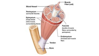
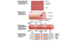
Unit 6
The
Muscular System
DeFord
Section 3
Skeletal Muscle Structure
Although muscle fiber makes up most of the muscle
tissue, a large amount of connective tissue, blood vessels,
and nerves are also present.
• Connective tissue covers and supports each muscle fiber and
reinforces the muscle as a whole.
• The heath of muscle depends on a sufficient nerve and blood supply.
Each skeletal muscle has a nerve ending that controls its activity.
• Active muscles use a lot of energy and require a continuous supply
of oxygen and nutrients, which are supplied by arteries. Muscles
produce large amounts of metabolic waste that must be removed by
veins.
Gross Anatomy of Skeletal Muscles
Cells are surrounded and bundled by connective tissue!
Connective Tissue Organization:
• Endomysium – encloses a single muscle fiber
• Perimysium – wraps around a fascicle (bundle) of
muscle fibers
• Epimysium – covers the entire skeletal muscle
• Fascia – outer layer of the epimysium
Blood Vessel
Perimysium
(surrounds fascicle)
Epimysium
(wraps around
entire muscle,
surrounded by fascia)
Muscle
Fiber (cell)
Fascicle
(bundle of muscle
fibers, surrounded by
perimysium)
Endomysium
(encloses each muscle
fiber)
Tendon
Bone
Arrangement of Fascicles
The fascicles of muscles can be arranged in a variety of
ways – the most common are:
• Parallel – fascicles are parallel to the long axis of the muscle. Some
of these are also classified as fusiform muscles.
• Pennate – the fascicles are short and attach to a central tendon.
Can be unipennate, bipennate or multipennate.
• Convergent – the muscle has a broad origin but the fascicles
converge to one tendon.
• Circular – muscle has the fascicles arranged in concentric circles.
Skeletal Muscle Attachments
Skeletal muscles attach to bones, cartilage, and
connective tissue coverings.
•Epimysium blends into a connective tissue attachment
•Tendons – cord-like structures
•Mostly collagen fibers
•Often cross a joint due to toughness and small size
•Aponeuroses – sheet-like structures
•Attach muscles indirectly to bones, cartilages, or
connective tissue coverings
Microanatomy of Skeletal Muscles
• Sarcolemma – specialized cell membrane
• Sarcoplasm – muscle cell cytoplasm
• Sarcoplasmic Reticulum – specialized smooth
endoplasmic reticulum
• used to store and maintain calcium ion levels
• surrounds the myofibril
Microanatomy of Skeletal Muscles
• T Tubules – tubules running through the muscle fibers
through which calcium ions flow
• Sarcomeres – repeating contractile unit of the myofibril
• Myofibril – long contraction organelle aligned to
give distinct bands
• I band = light band that contains
only thin filaments called actin.
• A band = dark band that contains
thick filaments called myosin.
• Z-lines at each end provide anchors for thin
filaments
APU6.3 Skeletal Muscle Structure

APU6.3 Skeletal Muscle Structure

  • 1.
    Unit 6 The Muscular System DeFord Section3 Skeletal Muscle Structure
  • 2.
    Skeletal Muscle Structure Althoughmuscle fiber makes up most of the muscle tissue, a large amount of connective tissue, blood vessels, and nerves are also present. • Connective tissue covers and supports each muscle fiber and reinforces the muscle as a whole. • The heath of muscle depends on a sufficient nerve and blood supply. Each skeletal muscle has a nerve ending that controls its activity. • Active muscles use a lot of energy and require a continuous supply of oxygen and nutrients, which are supplied by arteries. Muscles produce large amounts of metabolic waste that must be removed by veins.
  • 3.
    Gross Anatomy ofSkeletal Muscles Cells are surrounded and bundled by connective tissue! Connective Tissue Organization: • Endomysium – encloses a single muscle fiber • Perimysium – wraps around a fascicle (bundle) of muscle fibers • Epimysium – covers the entire skeletal muscle • Fascia – outer layer of the epimysium
  • 4.
    Blood Vessel Perimysium (surrounds fascicle) Epimysium (wrapsaround entire muscle, surrounded by fascia) Muscle Fiber (cell) Fascicle (bundle of muscle fibers, surrounded by perimysium) Endomysium (encloses each muscle fiber) Tendon Bone
  • 5.
    Arrangement of Fascicles Thefascicles of muscles can be arranged in a variety of ways – the most common are: • Parallel – fascicles are parallel to the long axis of the muscle. Some of these are also classified as fusiform muscles. • Pennate – the fascicles are short and attach to a central tendon. Can be unipennate, bipennate or multipennate. • Convergent – the muscle has a broad origin but the fascicles converge to one tendon. • Circular – muscle has the fascicles arranged in concentric circles.
  • 7.
    Skeletal Muscle Attachments Skeletalmuscles attach to bones, cartilage, and connective tissue coverings. •Epimysium blends into a connective tissue attachment •Tendons – cord-like structures •Mostly collagen fibers •Often cross a joint due to toughness and small size •Aponeuroses – sheet-like structures •Attach muscles indirectly to bones, cartilages, or connective tissue coverings
  • 8.
    Microanatomy of SkeletalMuscles • Sarcolemma – specialized cell membrane • Sarcoplasm – muscle cell cytoplasm • Sarcoplasmic Reticulum – specialized smooth endoplasmic reticulum • used to store and maintain calcium ion levels • surrounds the myofibril
  • 9.
    Microanatomy of SkeletalMuscles • T Tubules – tubules running through the muscle fibers through which calcium ions flow • Sarcomeres – repeating contractile unit of the myofibril • Myofibril – long contraction organelle aligned to give distinct bands • I band = light band that contains only thin filaments called actin. • A band = dark band that contains thick filaments called myosin. • Z-lines at each end provide anchors for thin filaments

Editor's Notes

  • #4  Endo MY sium. Peri MY sium. Epi MY sium Fascia is the wrapping around the entire muscle. Fascia is to muscle like the clear casing is on a sausage; It is a connective tissue that is keeping it all wrapped together as one unit.
  • #8 Every muscle has the same basic setup…attaches in 2, 3, or 4 places! Every muscle is attached to bones in AT LEAST two locations (via tendons) Where does the bicep attach? Humerus and scapula Think about a bungee cord as the muscle – attaches in 2 places (bridge, person)
  • #10 Muscle fibers consist of bundles of threadlike structures called myofibrils. Each myofibril is made up of two types of protein filaments – thick ones and thin ones. The thick filaments are made up of a protein called myosin. The thin filaments are made up of a protein called actin. Myosin and actin filaments are arranged to form overlapping patterns, which are responsible for the light and dark bands that can be seen in skeletal muscles (the striated appearance of the muscle). The thin actin filaments are anchored at their midpoints to a structure called the Z line. The region from one Z-line to the next is called a sarcomere, which is the functional unit of muscle contractions.