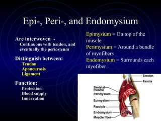
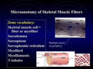
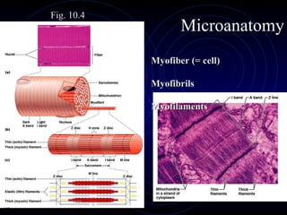
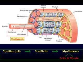
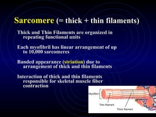
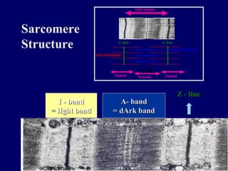
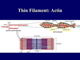
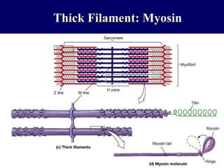
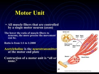
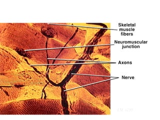
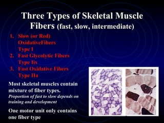
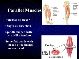
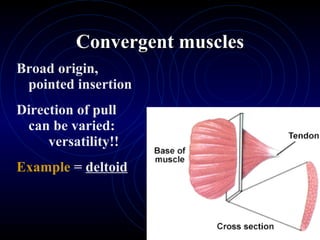
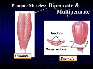
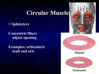
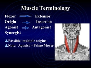
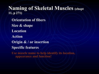
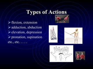
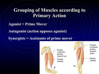
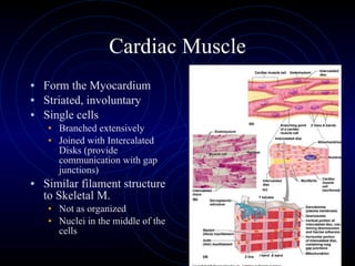
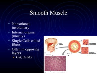
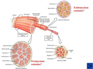
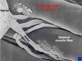
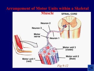
repetitio est mater studiorum repetition is the mother of learning
Describe the distinguishing characteristics of the different muscle tissues Discuss the organization of skeletal muscle Explain the micro-anatomy of a skeletal muscle fiber Describe the fascicle arrangement in different types of muscle Review general muscle terminology Ch 10:  Skeletal  Muscle Tissue  (Myology) main objectives:
Types of Muscle Tissue One of the 4 primary tissue types Skeletal = Striated =  Voluntary 40 % of weight Cardiac (involuntary) Smooth (involuntary, nonstriated Made up of many tissue types. > 700 skeletal muscles Word roots: sarco myo Musculus = little mouse
Function of Skeletal Muscles Skeletal movement  Posture and body position Support of soft tissues Joint Stabilization Guarding of entrances & exits Maintenance of body temperature Protection Facial expression
Gross to Microscopic Anatomy Each skeletal muscle cell (fiber) is wrapped by 3 layers of connective tissue.
Epi-, Peri-, and Endomysium Are interwoven  -  Continuous with tendon, and eventually the periosteum Distinguish between: Tendon Aponeurosis Ligament Function:  Protection Blood supply Innervation Epimysium  = On top of the muscle Perimysium  = Around a bundle of myofibers Endomysium  = Surrounds each myofiber
Nerve   and Blood Supply Skeletal muscles are rich in nerves and  blood vessels. Chemical communication at neuromuscular junction Synaptic terminal of axon meets motor end plate of muscle cell Coiled capillaries  are able to adapt to changes in length of muscle fiber
Coiled Capillaries
Origin and Insertion Origin:  The part that stays still Usually proximal Insertion:  The part that moves Usually distal
Microanatomy of Skeletal Muscle Fibers Some vocabulary: Skeletal muscle cell = fiber or myofiber  Sarcolemma  Sarcoplasm  Sarcoplasmic reticulum Myofibril Myofilaments T-tubules Multiple nuclei on periphery
Microanatomy Myofiber (= cell)  Myofibrils  Myofilaments Fig. 10.4
Myofiber (cell)    Myofibrils   Myofilaments Actin & Myosin Myofilaments
Sarcomere   (= thick + thin filaments) Thick and Thin Filaments are organized in repeating functional units Each myofibril has linear arrangement of up to 10,000 sarcomeres Banded appearance ( striation ) due to arrangement of thick and thin filaments Interaction of thick and thin filaments responsible for skeletal muscle fiber contraction
Sarcomere Structure  Z - line A- band = dArk band I - band = lIght band
Thin Filament: Actin
Thick Filament: Myosin
Motor Unit = All muscle fibers that are controlled by a single motor neuron (axon) The lower the ratio of muscle fibers to neurons, the more precise the movement can be. Ratio is from 1:1 to 1:2000 Acetylcholine  is the  neurotransmitter  at the motor end plate Contraction of a motor unit is “all or none.”
Neuromuscular Junction LM  x230
Muscle Control Muscle tone = Resting tension of skeletal muscles  (continuous contraction of some motor units to maintain some muscle tension) Recruitment or  Multiple motor unit summation for greater force Some directional control depending on which motor units are stimulated
Muscle Hypertrophy vs. Atrophy Hypertrophy   due to anaerobic exercise Leads to increased muscle size -  how? Atrophy   if supply of myofilaments exceeds demand. Muscle fibers become smaller and weaker.  Eventual death of muscle fibers is irreversible! Importance of Physical Therapy
Three Types of Skeletal Muscle Fibers  (fast, slow, intermediate) Slow (or Red) OxidativeFibers Type I Fast Glycolytic Fibers Type Iix Fast Oxidative Fibers Type IIa Most skeletal muscles contain mixture of fiber types.  Proportion of fast to slow depends on training and development One motor unit only contains one fiber type
1.  Slow (or Red) Oxidative Fibers Type I Slow but continuous contraction for extended periods Smaller diameter (~ half) contain myoglobin  more capillaries  more mitochondria Do not fatigue as fast due to aerobic production of ATP
Fast contraction after nervous stimulation Large diameter large glycogen reserve fewer mitochondria densely packed  myofibrils Fatigue fast due to mainly anaerobic respiration 2.  Fast Glycolytic Fibers Type IIx
3.  Fast Oxidative Fibers Type IIa Have attributes in between fast and slow types
Organization (shape) of Skeletal Muscle Fibers Effect of individual muscle contraction   determined by: arrangement of muscle fibers method of attachment to skeleton   Bundles of muscle fibers =fascicle Muscle fibers within 1 fascicle are parallel
Parallel Muscles Extensor vs. flexor Origin vs. Insertion Spindle shaped with cord-like tendons Some flat bands with broad attachments on each end
Convergent muscles Broad origin, pointed insertion Direction of pull can be varied: versatility!! Example  =  deltoid
Pennate Muscles: Unipennate One or more tendons  run though muscle body Fascicles in oblique angle to tendon Can generate more tension Example  = biceps brachii
Pennate Muscles:  Bipennate &    Multipennate Example Example
Circular Muscles  = Sphincters Concentric fibers adjust opening Examples: orbicularis oculi and oris
Muscle Terminology Flexor   Extensor Origin   Insertion Agonist Antagonist Synergist Possible: multiple origins   Note:  Agonist = Prime Mover
Naming of Skeletal Muscles  (chapt 11, p 271) Orientation of fibers Size & shape  Location Action Origin & / or insertion Specific features Use muscle name to help identify its location, appearance and function!
Types of Actions flexion, extension adduction, abduction elevation, depression pronation, supination etc., etc. . . . .
Grouping of Muscles according to Primary Action Agonist = Prime Mover Antagonist (action opposes agonist) Synergists = Assistants of prime mover
Cardiac Muscle Form the Myocardium Striated, involuntary Single cells Branched extensively Joined with Intercalated Disks (provide communication with gap junctions) Similar filament structure to Skeletal M. Not as organized Nuclei in the middle of the cells
Smooth Muscle Nonstriated, involuntary Internal organs (mostly) Single Cells called fibers Often in opposing layers Gut, bladder
 
Organization of Skeletal Muscle Fig 9-1 Perimysium contains? Endomysium contains?
Neuromuscular Junction
Arrangement of Motor Units within a Skeletal Muscle Fig 9-12
Slow vs. Fast Fibers
Rectus femoris, Deltoid

Chapter10 muscletissuemarieb

  • 1.
    repetitio est materstudiorum repetition is the mother of learning
  • 2.
    Describe the distinguishingcharacteristics of the different muscle tissues Discuss the organization of skeletal muscle Explain the micro-anatomy of a skeletal muscle fiber Describe the fascicle arrangement in different types of muscle Review general muscle terminology Ch 10: Skeletal Muscle Tissue (Myology) main objectives:
  • 3.
    Types of MuscleTissue One of the 4 primary tissue types Skeletal = Striated = Voluntary 40 % of weight Cardiac (involuntary) Smooth (involuntary, nonstriated Made up of many tissue types. > 700 skeletal muscles Word roots: sarco myo Musculus = little mouse
  • 4.
    Function of SkeletalMuscles Skeletal movement Posture and body position Support of soft tissues Joint Stabilization Guarding of entrances & exits Maintenance of body temperature Protection Facial expression
  • 5.
    Gross to MicroscopicAnatomy Each skeletal muscle cell (fiber) is wrapped by 3 layers of connective tissue.
  • 6.
    Epi-, Peri-, andEndomysium Are interwoven - Continuous with tendon, and eventually the periosteum Distinguish between: Tendon Aponeurosis Ligament Function: Protection Blood supply Innervation Epimysium = On top of the muscle Perimysium = Around a bundle of myofibers Endomysium = Surrounds each myofiber
  • 7.
    Nerve and Blood Supply Skeletal muscles are rich in nerves and blood vessels. Chemical communication at neuromuscular junction Synaptic terminal of axon meets motor end plate of muscle cell Coiled capillaries are able to adapt to changes in length of muscle fiber
  • 8.
  • 9.
    Origin and InsertionOrigin: The part that stays still Usually proximal Insertion: The part that moves Usually distal
  • 10.
    Microanatomy of SkeletalMuscle Fibers Some vocabulary: Skeletal muscle cell = fiber or myofiber Sarcolemma Sarcoplasm Sarcoplasmic reticulum Myofibril Myofilaments T-tubules Multiple nuclei on periphery
  • 11.
    Microanatomy Myofiber (=cell) Myofibrils Myofilaments Fig. 10.4
  • 12.
    Myofiber (cell) Myofibrils Myofilaments Actin & Myosin Myofilaments
  • 13.
    Sarcomere (= thick + thin filaments) Thick and Thin Filaments are organized in repeating functional units Each myofibril has linear arrangement of up to 10,000 sarcomeres Banded appearance ( striation ) due to arrangement of thick and thin filaments Interaction of thick and thin filaments responsible for skeletal muscle fiber contraction
  • 14.
    Sarcomere Structure Z - line A- band = dArk band I - band = lIght band
  • 15.
  • 16.
  • 17.
    Motor Unit =All muscle fibers that are controlled by a single motor neuron (axon) The lower the ratio of muscle fibers to neurons, the more precise the movement can be. Ratio is from 1:1 to 1:2000 Acetylcholine is the neurotransmitter at the motor end plate Contraction of a motor unit is “all or none.”
  • 18.
  • 19.
    Muscle Control Muscletone = Resting tension of skeletal muscles (continuous contraction of some motor units to maintain some muscle tension) Recruitment or Multiple motor unit summation for greater force Some directional control depending on which motor units are stimulated
  • 20.
    Muscle Hypertrophy vs.Atrophy Hypertrophy due to anaerobic exercise Leads to increased muscle size - how? Atrophy if supply of myofilaments exceeds demand. Muscle fibers become smaller and weaker. Eventual death of muscle fibers is irreversible! Importance of Physical Therapy
  • 21.
    Three Types ofSkeletal Muscle Fibers (fast, slow, intermediate) Slow (or Red) OxidativeFibers Type I Fast Glycolytic Fibers Type Iix Fast Oxidative Fibers Type IIa Most skeletal muscles contain mixture of fiber types. Proportion of fast to slow depends on training and development One motor unit only contains one fiber type
  • 22.
    1. Slow(or Red) Oxidative Fibers Type I Slow but continuous contraction for extended periods Smaller diameter (~ half) contain myoglobin more capillaries more mitochondria Do not fatigue as fast due to aerobic production of ATP
  • 23.
    Fast contraction afternervous stimulation Large diameter large glycogen reserve fewer mitochondria densely packed myofibrils Fatigue fast due to mainly anaerobic respiration 2. Fast Glycolytic Fibers Type IIx
  • 24.
    3. FastOxidative Fibers Type IIa Have attributes in between fast and slow types
  • 25.
    Organization (shape) ofSkeletal Muscle Fibers Effect of individual muscle contraction determined by: arrangement of muscle fibers method of attachment to skeleton Bundles of muscle fibers =fascicle Muscle fibers within 1 fascicle are parallel
  • 26.
    Parallel Muscles Extensorvs. flexor Origin vs. Insertion Spindle shaped with cord-like tendons Some flat bands with broad attachments on each end
  • 27.
    Convergent muscles Broadorigin, pointed insertion Direction of pull can be varied: versatility!! Example = deltoid
  • 28.
    Pennate Muscles: UnipennateOne or more tendons run though muscle body Fascicles in oblique angle to tendon Can generate more tension Example = biceps brachii
  • 29.
    Pennate Muscles: Bipennate & Multipennate Example Example
  • 30.
    Circular Muscles = Sphincters Concentric fibers adjust opening Examples: orbicularis oculi and oris
  • 31.
    Muscle Terminology Flexor Extensor Origin Insertion Agonist Antagonist Synergist Possible: multiple origins Note: Agonist = Prime Mover
  • 32.
    Naming of SkeletalMuscles (chapt 11, p 271) Orientation of fibers Size & shape Location Action Origin & / or insertion Specific features Use muscle name to help identify its location, appearance and function!
  • 33.
    Types of Actionsflexion, extension adduction, abduction elevation, depression pronation, supination etc., etc. . . . .
  • 34.
    Grouping of Musclesaccording to Primary Action Agonist = Prime Mover Antagonist (action opposes agonist) Synergists = Assistants of prime mover
  • 35.
    Cardiac Muscle Formthe Myocardium Striated, involuntary Single cells Branched extensively Joined with Intercalated Disks (provide communication with gap junctions) Similar filament structure to Skeletal M. Not as organized Nuclei in the middle of the cells
  • 36.
    Smooth Muscle Nonstriated,involuntary Internal organs (mostly) Single Cells called fibers Often in opposing layers Gut, bladder
  • 37.
  • 38.
    Organization of SkeletalMuscle Fig 9-1 Perimysium contains? Endomysium contains?
  • 39.
  • 40.
    Arrangement of MotorUnits within a Skeletal Muscle Fig 9-12
  • 41.
  • 42.