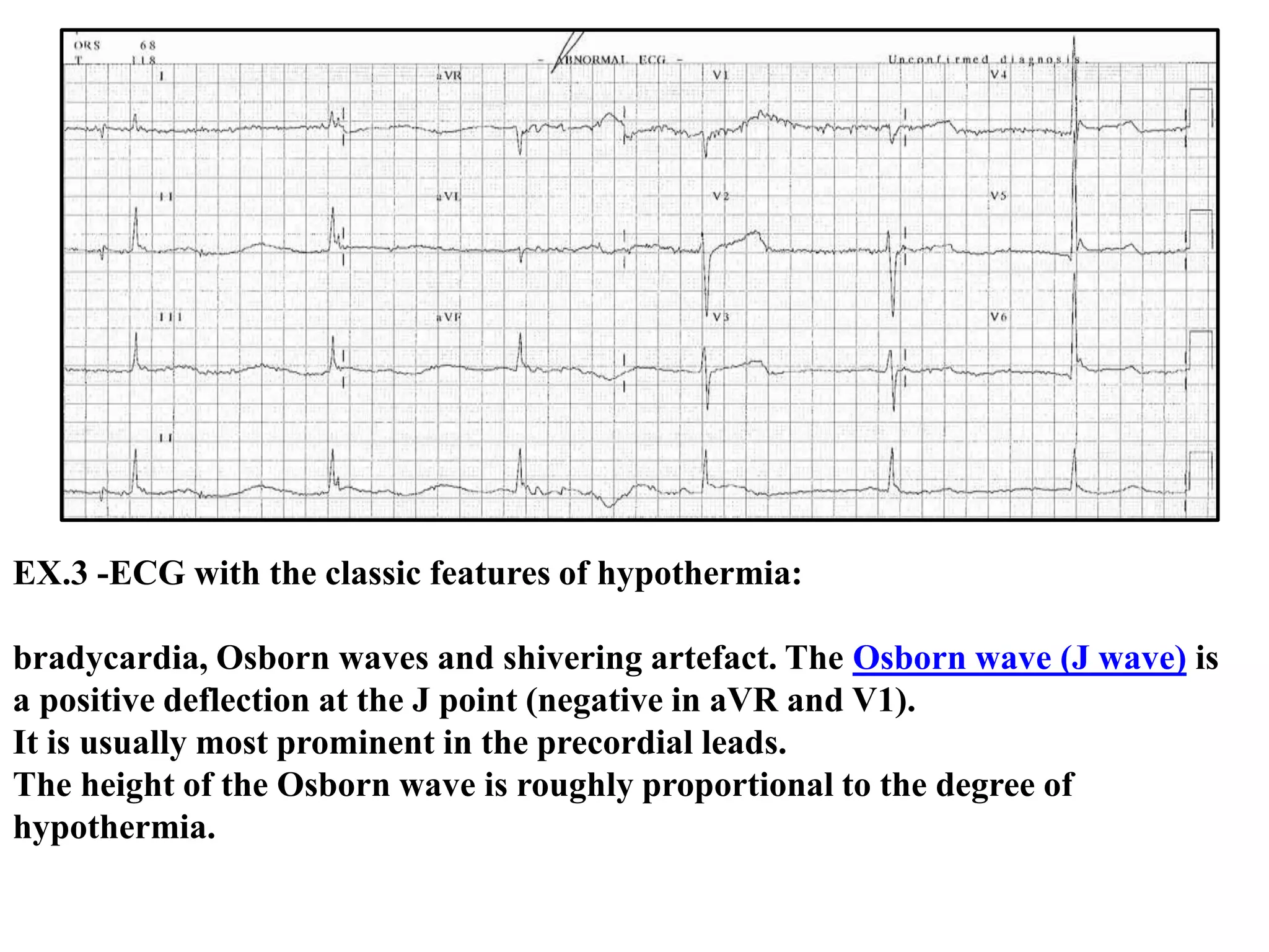
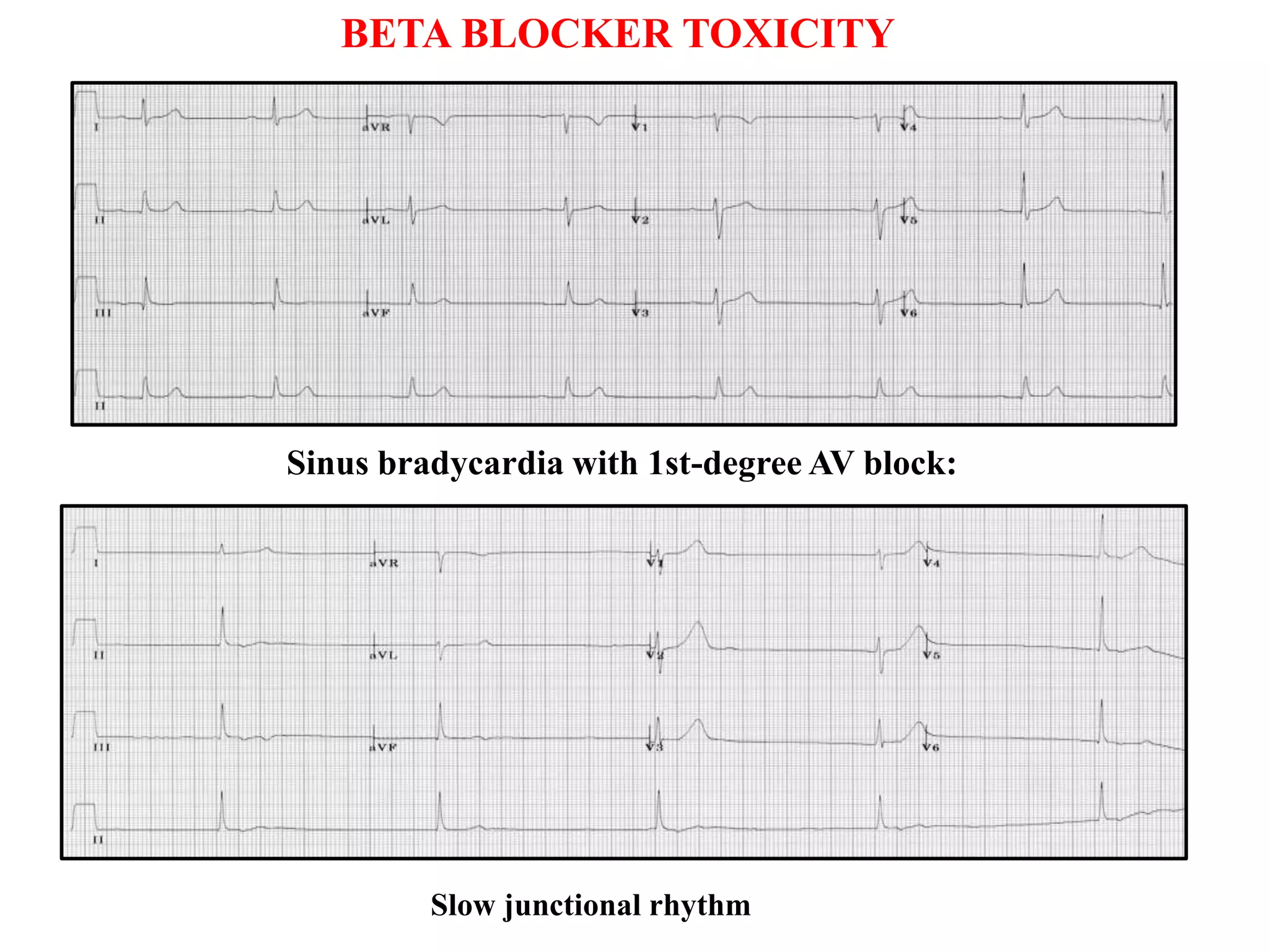
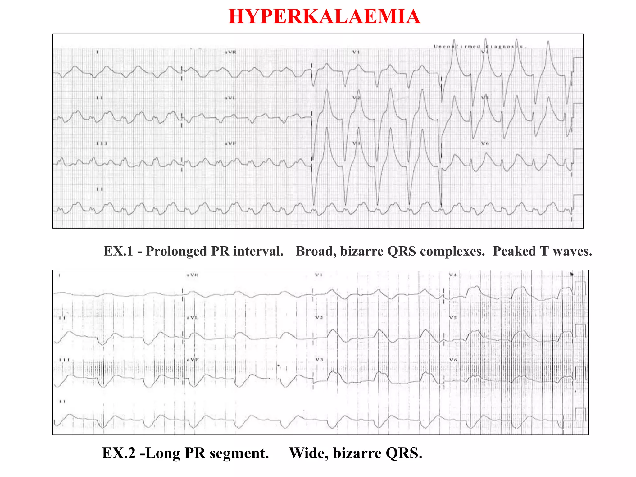
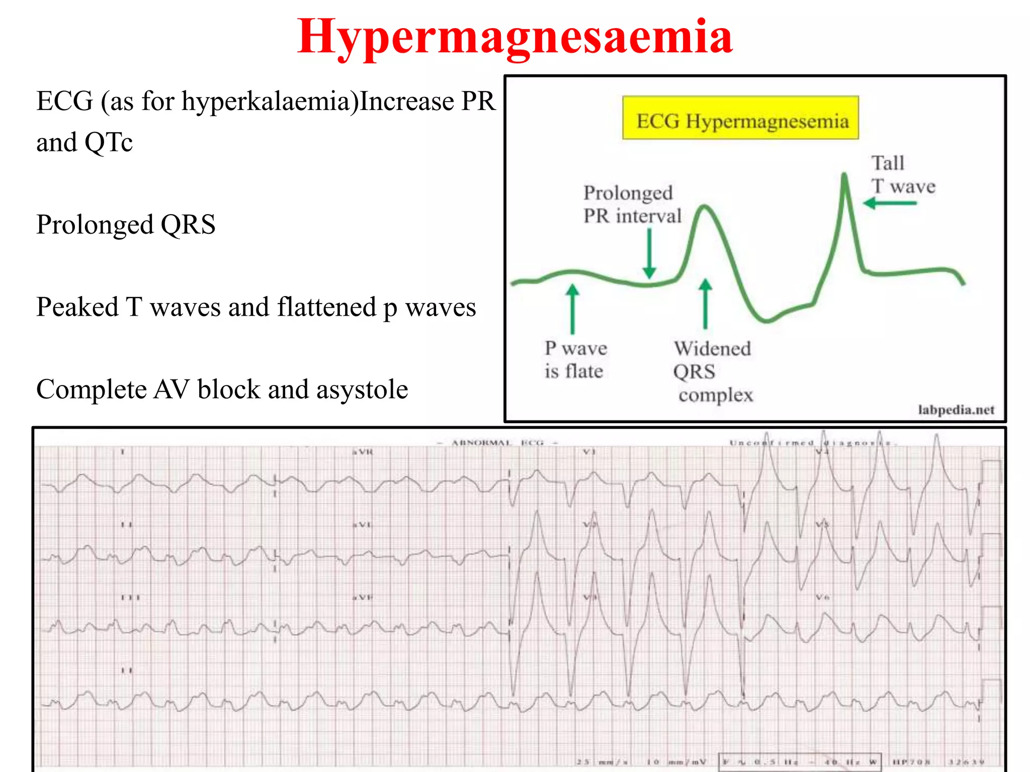
Hypothermia is defined as a core body temperature of < 35 degrees centigrade
Mild hypothermia is 32-35 degrees
Moderate hypothermia is 29-32 degrees
Severe hypothermia is < 29 degrees
ECG Changes in Hypothermia
Bradyarrhythmias (see below)
Osborne Waves (= J waves)
Prolonged PR, QRS and QT intervals
Shivering artefact
Ventricular ectopics
Cardiac arrest due to VT, VF or asystole
Bradyarrhythmias
Sinus bradycardia (may be marked)
Atrial fibrillation with slow ventricular response
Slow junctional rhythms
Varying degrees of AV block (1st-3rd)
EX.1-Marked sinus bradycardia (30bpm) secondary to
hypothermia
EX.2- Atrial fibrillation with slow ventricular
response due to hypothermia
EX.3 -ECG with the classic features of hypothermia:
bradycardia, Osborn waves and shivering artefact. The Osborn wave (J wave) is
a positive deflection at the J point (negative in aVR and V1).
It is usually most prominent in the precordial leads.
The height of the Osborn wave is roughly proportional to the degree of
hypothermia.
EX.4- Prolonged QTc (620ms) due to severe hypothermia
EX.5- Shivering Artefact
Shivering artefact is seen as a “fuzziness” of the ECG baseline.
It is not specific to hypothermia and may be seen with other
conditions associated with tremor (e.g. Parkinson’s disease).
Shivering artefact in a patient with hypothermia (note also the
Osborn waves, bradycardia, prolonged QT)
The ECG findings in hyperthermia include:
Sinus tachycardia Diffuse non specific ST segment changes
Non specific ischemic changes Prolonged QT interval
HYPERTHERMIA
Hyperthermia is when core temperature exceeds that normally maintained by
homeostatic mechanisms
Fever or pyrexia is an elevation of body temperature above the normal range of 36.5–
37.5 °C (97.7–99.5 °F) due to an increase in the temperature regulatory set point
Uncontrolled hyperthermia differs from fever in that the body temperature is elevated
above the theromoregulatory set point due to excessive heat production and/or
insufficient heat dissipation
ECG Abnormalities with Raised Intracranial Pressure
Raised ICP is associated with certain characteristic ECG changes:
Widespread giant T-wave inversions (“cerebral T waves”).
QT prolongation.
Bradycardia (the Cushing reflex – indicates imminent brainstem herniation).
Other possible ECG changes that may be seen:
ST segment elevation / depression — this may mimic myocardial ischaemia or pericarditis.
Increased U wave amplitude.
Other rhythm disturbances: sinus tachycardia, junctional rhythms, premature ventricular
contractions, atrial fibrillation.
In some cases, these ECG abnormalities may be associated with echocardiographic evidence of
regional ventricular wall motion abnormality (so-called “neurogenic stunned myocardium”)
Causes
ECG changes due to raised ICP are most commonly seen with massive intracranial haemorrhage:
Subarachnoid haemorrhage
Intraparenchymal haemorrhage (haemorrhagic stroke)
They may also be seen with:
Massive ischaemic stroke causing cerebral oedema (e.g. MCA occlusion)
Traumatic brain injury
Cerebral metastases (rarely)
Widespread, giant T-wave inversions (“cerebral T waves”) secondary
to subarachnoid haemorrhage.
The QT interval is also grossly prolonged (600 ms).
Subarachnoid Haemorrhage
Widespread T-wave inversions with slight ST depression secondary to subarachnoid
haemorrhage.
The QT interval is prolonged (greater than half the R-R interval).
This ECG pattern could easily be mistaken for myocardial ischaemia as the T-wave
morphology is very similar, although obviously the clinical picture would be very different
(coma versus chest pain).
Traumatic Brain Injury
This ECG was taken from a previously healthy 18-year old girl with severe traumatic brain
injury and massively raised intracranial pressure (30-40 mmHg).
There is widespread ST elevation with a pericarditis-like morphology and no reciprocal change
(except in aVR and V1).
She had no cardiac injury / abnormality to explain the ST elevation.
The ST segments normalised as the intracranial pressure came under control (following
treatment with thiopentone and hypertonic saline).
The most common ECG changes seen with thyrotoxicosis are:
Sinus tachycardia
Atrial fibrillation with rapid ventricular response
High left-ventricular voltage
Supraventricular arrhythmias (premature atrial beats, paroxysmal supraventricular
tachycardia, multifocal atrial tachycardia, atrial flutter)
Non-specific ST and T wave changes
Ventricular extrasystoles
Myxoedema coma
This is the admission ECG of a 79-year old man who was referred to
ICU with coma, hypothermia, severe bradycardia and hypotension
refractory to inotropes.
TSH was markedly elevated with an undetectable T4.
The ECG shows marked bradycardia (30 bpm) with low QRS
voltages (esp. in the limb leads) and widespread T-wave inversions,
typical of severe myxoedema.
Sodium Channel Blocking Medications
Tricyclic antidepressants (= most common)
Type Ia antiarrhythmics (quinidine, procainamide)
Type Ic antiarrhythmics (flecainide, encainide)
Local anaesthetics (bupivacaine, ropivacaine)
Antimalarials (chloroquine, hydroxychloroquine)
Dextropropoxyphene
Propranolol
Carbamazepine
Quinine
ECG Features of Sodium-Channel Blockade
Features consistent with sodium-channel blockade:
Interventricular conduction delay — QRS > 100 ms in lead II
Right axis deviation of the terminal QRS:
Terminal R wave > 3 mm in aVR
R/S ratio > 0.7 in aVR
Patients with tricyclic overdose will also usually demonstrate sinus
tachycardiasecondary to muscarinic (M1) receptor blockade.
Typical ECG of TCA
toxicity demonstrating:
Sinus tachycardia with first-degree AV block (P waves hidden in the T waves, best seen in
V1-2).
Broad QRS complexes.
Positive R’ wave in aVR.
There is subtle ECG evidence of fast sodium channel blockade: Note
the QRS widening (135 ms), 1st degree heart block (PR 240ms) and
small secondary R wave in aVR.
Carbamazepine Cardiotoxicity
Quetiapine Toxicity
Brisk sinus tachycardia (HR = 120 bpm)
Prolonged QTc interval (QTc = 560ms; the absolute QT interval is
more than half the R-R interval)
The classic digoxin toxic dysrhythmia combines:
supraventricular tachycardia (due to increased automaticity)
slow ventricular response (due to decreased AV conduction)
Other common dysrhythmias associated with digoxin toxicity include:
Frequent PVCs (the most common abnormality), including ventricular
bigeminy and trigeminy
Sinus bradycardia
Slow Atrial Fibrillation
Any type of AV block (1st degree, 2nd degree & 3rd degree)
Regularised AF = AF with complete heart block and a junctional or
ventricular escape rhythm
Ventricular tachycardia, including polymorphic and bidirectional VT
Bigeminy
Atrial tachycardia with high-grade AV block and PVCs
Coarse atrial fibrillation with 3rd degree AV block and a junctional escape rhythm
DIGOXIN TOXICITY
“Paroxysmal” atrial tachycardia with block and frequent PVCs
Atrial flutter with a slow ventricular rate due to digoxin toxicity.
DIGOXIN TOXICITY
Sinus bradycardia with 1st-degree AV block:
Slow junctional rhythm
BETA BLOCKER TOXICITY
Complete heart block
sotalol overdose: Sinus bradycardia. Very long QT interval (~600ms).
BETA BLOCKER TOXICITY
Hypercalcaemia
EX.1 - Osborn waves in severe hypercalcaemia (4.1 mmol/L)
EX.2-Hypercalcaemia causing marked shortening of the QT interval (260ms).
HYPOCALCAEMIA
Hypocalcaemia causes QTc prolongation primarily by prolonging the ST segment.
The T wave is typically left unchanged.
Dysrhythmias are uncommon, although atrial fibrillation has been reported.
Torsades de pointes may occur
Serum potassium > 5.5 mEq/L - repolarization abnormalities:
Peaked T waves (usually the earliest sign of hyperkalaemia)
Serum potassium > 6.5 mEq/L - progressive paralysis of the atria:
P wave widens and flattens
PR segment lengthens
P waves eventually disappear
Serum potassium > 7.0 mEq/L - conduction abnormalities andbradycardia:
Prolonged QRS interval with bizarre QRS morphology
High-grade AV block with slow junctional and ventricular escape rhythms
Any kind of conduction block (bundle branch blocks, fascicular blocks)
Sinus bradycardia or slow AF
Development of a sine wave appearance (a pre-terminal rhythm)
Serum potassium level of > 9.0 mEq/L causes cardiac arrest due to:
Asystole
Ventricular fibrillation
PEA with bizarre, wide complex rhythm
EX.1 - Prolonged PR interval. Broad, bizarre QRS complexes. Peaked T waves.
EX.2 -Long PR segment. Wide, bizarre QRS.
HYPERKALAEMIA
HYPERKALAEMIA
EX.3 -Slow junctional rhythm. Intraventricular conduction delay. Peaked T waves.
EX4-Broad complex rhythm with atypical LBBB morphology. Left axis deviation. Absent
P waves.
ECG changes when K+ < 2.7 mmol/l
Increased amplitude and width of the P wave
Prolongation of the PR interval
T wave flattening and inversion
ST depression
Prominent U waves (best seen in the precordial leads)
Apparent long QT interval due to fusion of the T and U waves (=
long QU interval)
With worsening hypokalaemia…
Frequent supraventricular and ventricular ectopics
Supraventricular tachyarrhythmias: AF, atrial flutter, atrial
tachycardia
Potential to develop life-threatening ventricular arrhythmias, e.g.
VT, VF and Torsades de Pointes
EX.2 -Hypokalaemia causing Torsades de Pointes
Note the atrial ectopic causing ‘R on T’ (or is it ‘R on U’?) that initiates the paroxysm of
TdP
EX.1 -ST depression. T wave inversion. Prominent U waves. Long QU interval.
HYPOKALAEMIA
Hypomagnesaemia Overview
Normal serum magnesium = 0.8 – 1.0 mmol/L.
Hypomagnesaemia = <0.8 mmol/L
ECG changes in Hypomagnesaemia
prolonged QTc.
Atrial and ventricular ectopy, atrial tachyarrhythmias
and torsades de pointes
Hypomagnesaemia
EX.1- Hypomagnesaemia causing long QTc (510ms)
Hypermagnesaemia
ECG (as for hyperkalaemia)Increase PR
and QTc
Prolonged QRS
Peaked T waves and flattened p waves
Complete AV block and asystole
ECG abnormalities in drugs, temperature, electrolyte , metabolic and temperature changes.

ECG abnormalities in drugs, temperature, electrolyte , metabolic and temperature changes.

  • 3.
    Hypothermia is definedas a core body temperature of < 35 degrees centigrade Mild hypothermia is 32-35 degrees Moderate hypothermia is 29-32 degrees Severe hypothermia is < 29 degrees ECG Changes in Hypothermia Bradyarrhythmias (see below) Osborne Waves (= J waves) Prolonged PR, QRS and QT intervals Shivering artefact Ventricular ectopics Cardiac arrest due to VT, VF or asystole Bradyarrhythmias Sinus bradycardia (may be marked) Atrial fibrillation with slow ventricular response Slow junctional rhythms Varying degrees of AV block (1st-3rd)
  • 4.
    EX.1-Marked sinus bradycardia(30bpm) secondary to hypothermia
  • 5.
    EX.2- Atrial fibrillationwith slow ventricular response due to hypothermia
  • 6.
    EX.3 -ECG withthe classic features of hypothermia: bradycardia, Osborn waves and shivering artefact. The Osborn wave (J wave) is a positive deflection at the J point (negative in aVR and V1). It is usually most prominent in the precordial leads. The height of the Osborn wave is roughly proportional to the degree of hypothermia.
  • 7.
    EX.4- Prolonged QTc(620ms) due to severe hypothermia
  • 8.
    EX.5- Shivering Artefact Shiveringartefact is seen as a “fuzziness” of the ECG baseline. It is not specific to hypothermia and may be seen with other conditions associated with tremor (e.g. Parkinson’s disease). Shivering artefact in a patient with hypothermia (note also the Osborn waves, bradycardia, prolonged QT)
  • 9.
    The ECG findingsin hyperthermia include: Sinus tachycardia Diffuse non specific ST segment changes Non specific ischemic changes Prolonged QT interval HYPERTHERMIA Hyperthermia is when core temperature exceeds that normally maintained by homeostatic mechanisms Fever or pyrexia is an elevation of body temperature above the normal range of 36.5– 37.5 °C (97.7–99.5 °F) due to an increase in the temperature regulatory set point Uncontrolled hyperthermia differs from fever in that the body temperature is elevated above the theromoregulatory set point due to excessive heat production and/or insufficient heat dissipation
  • 11.
    ECG Abnormalities withRaised Intracranial Pressure Raised ICP is associated with certain characteristic ECG changes: Widespread giant T-wave inversions (“cerebral T waves”). QT prolongation. Bradycardia (the Cushing reflex – indicates imminent brainstem herniation). Other possible ECG changes that may be seen: ST segment elevation / depression — this may mimic myocardial ischaemia or pericarditis. Increased U wave amplitude. Other rhythm disturbances: sinus tachycardia, junctional rhythms, premature ventricular contractions, atrial fibrillation. In some cases, these ECG abnormalities may be associated with echocardiographic evidence of regional ventricular wall motion abnormality (so-called “neurogenic stunned myocardium”) Causes ECG changes due to raised ICP are most commonly seen with massive intracranial haemorrhage: Subarachnoid haemorrhage Intraparenchymal haemorrhage (haemorrhagic stroke) They may also be seen with: Massive ischaemic stroke causing cerebral oedema (e.g. MCA occlusion) Traumatic brain injury Cerebral metastases (rarely)
  • 12.
    Widespread, giant T-waveinversions (“cerebral T waves”) secondary to subarachnoid haemorrhage. The QT interval is also grossly prolonged (600 ms). Subarachnoid Haemorrhage
  • 13.
    Subarachnoid Haemorrhage Widespread T-waveinversions with slight ST depression secondary to subarachnoid haemorrhage. The QT interval is prolonged (greater than half the R-R interval). This ECG pattern could easily be mistaken for myocardial ischaemia as the T-wave morphology is very similar, although obviously the clinical picture would be very different (coma versus chest pain).
  • 14.
    Traumatic Brain Injury ThisECG was taken from a previously healthy 18-year old girl with severe traumatic brain injury and massively raised intracranial pressure (30-40 mmHg). There is widespread ST elevation with a pericarditis-like morphology and no reciprocal change (except in aVR and V1). She had no cardiac injury / abnormality to explain the ST elevation. The ST segments normalised as the intracranial pressure came under control (following treatment with thiopentone and hypertonic saline).
  • 16.
    The most commonECG changes seen with thyrotoxicosis are: Sinus tachycardia Atrial fibrillation with rapid ventricular response High left-ventricular voltage Supraventricular arrhythmias (premature atrial beats, paroxysmal supraventricular tachycardia, multifocal atrial tachycardia, atrial flutter) Non-specific ST and T wave changes Ventricular extrasystoles
  • 17.
    Myxoedema coma This isthe admission ECG of a 79-year old man who was referred to ICU with coma, hypothermia, severe bradycardia and hypotension refractory to inotropes. TSH was markedly elevated with an undetectable T4. The ECG shows marked bradycardia (30 bpm) with low QRS voltages (esp. in the limb leads) and widespread T-wave inversions, typical of severe myxoedema.
  • 19.
    Sodium Channel BlockingMedications Tricyclic antidepressants (= most common) Type Ia antiarrhythmics (quinidine, procainamide) Type Ic antiarrhythmics (flecainide, encainide) Local anaesthetics (bupivacaine, ropivacaine) Antimalarials (chloroquine, hydroxychloroquine) Dextropropoxyphene Propranolol Carbamazepine Quinine ECG Features of Sodium-Channel Blockade Features consistent with sodium-channel blockade: Interventricular conduction delay — QRS > 100 ms in lead II Right axis deviation of the terminal QRS: Terminal R wave > 3 mm in aVR R/S ratio > 0.7 in aVR Patients with tricyclic overdose will also usually demonstrate sinus tachycardiasecondary to muscarinic (M1) receptor blockade.
  • 20.
    Typical ECG ofTCA toxicity demonstrating: Sinus tachycardia with first-degree AV block (P waves hidden in the T waves, best seen in V1-2). Broad QRS complexes. Positive R’ wave in aVR.
  • 21.
    There is subtleECG evidence of fast sodium channel blockade: Note the QRS widening (135 ms), 1st degree heart block (PR 240ms) and small secondary R wave in aVR. Carbamazepine Cardiotoxicity
  • 22.
    Quetiapine Toxicity Brisk sinustachycardia (HR = 120 bpm) Prolonged QTc interval (QTc = 560ms; the absolute QT interval is more than half the R-R interval)
  • 23.
    The classic digoxintoxic dysrhythmia combines: supraventricular tachycardia (due to increased automaticity) slow ventricular response (due to decreased AV conduction) Other common dysrhythmias associated with digoxin toxicity include: Frequent PVCs (the most common abnormality), including ventricular bigeminy and trigeminy Sinus bradycardia Slow Atrial Fibrillation Any type of AV block (1st degree, 2nd degree & 3rd degree) Regularised AF = AF with complete heart block and a junctional or ventricular escape rhythm Ventricular tachycardia, including polymorphic and bidirectional VT
  • 24.
    Bigeminy Atrial tachycardia withhigh-grade AV block and PVCs Coarse atrial fibrillation with 3rd degree AV block and a junctional escape rhythm DIGOXIN TOXICITY
  • 25.
    “Paroxysmal” atrial tachycardiawith block and frequent PVCs Atrial flutter with a slow ventricular rate due to digoxin toxicity. DIGOXIN TOXICITY
  • 26.
    Sinus bradycardia with1st-degree AV block: Slow junctional rhythm BETA BLOCKER TOXICITY
  • 27.
    Complete heart block sotaloloverdose: Sinus bradycardia. Very long QT interval (~600ms). BETA BLOCKER TOXICITY
  • 29.
    Hypercalcaemia EX.1 - Osbornwaves in severe hypercalcaemia (4.1 mmol/L) EX.2-Hypercalcaemia causing marked shortening of the QT interval (260ms).
  • 30.
    HYPOCALCAEMIA Hypocalcaemia causes QTcprolongation primarily by prolonging the ST segment. The T wave is typically left unchanged. Dysrhythmias are uncommon, although atrial fibrillation has been reported. Torsades de pointes may occur
  • 31.
    Serum potassium >5.5 mEq/L - repolarization abnormalities: Peaked T waves (usually the earliest sign of hyperkalaemia) Serum potassium > 6.5 mEq/L - progressive paralysis of the atria: P wave widens and flattens PR segment lengthens P waves eventually disappear Serum potassium > 7.0 mEq/L - conduction abnormalities andbradycardia: Prolonged QRS interval with bizarre QRS morphology High-grade AV block with slow junctional and ventricular escape rhythms Any kind of conduction block (bundle branch blocks, fascicular blocks) Sinus bradycardia or slow AF Development of a sine wave appearance (a pre-terminal rhythm) Serum potassium level of > 9.0 mEq/L causes cardiac arrest due to: Asystole Ventricular fibrillation PEA with bizarre, wide complex rhythm
  • 32.
    EX.1 - ProlongedPR interval. Broad, bizarre QRS complexes. Peaked T waves. EX.2 -Long PR segment. Wide, bizarre QRS. HYPERKALAEMIA
  • 33.
    HYPERKALAEMIA EX.3 -Slow junctionalrhythm. Intraventricular conduction delay. Peaked T waves. EX4-Broad complex rhythm with atypical LBBB morphology. Left axis deviation. Absent P waves.
  • 34.
    ECG changes whenK+ < 2.7 mmol/l Increased amplitude and width of the P wave Prolongation of the PR interval T wave flattening and inversion ST depression Prominent U waves (best seen in the precordial leads) Apparent long QT interval due to fusion of the T and U waves (= long QU interval) With worsening hypokalaemia… Frequent supraventricular and ventricular ectopics Supraventricular tachyarrhythmias: AF, atrial flutter, atrial tachycardia Potential to develop life-threatening ventricular arrhythmias, e.g. VT, VF and Torsades de Pointes
  • 35.
    EX.2 -Hypokalaemia causingTorsades de Pointes Note the atrial ectopic causing ‘R on T’ (or is it ‘R on U’?) that initiates the paroxysm of TdP EX.1 -ST depression. T wave inversion. Prominent U waves. Long QU interval. HYPOKALAEMIA
  • 36.
    Hypomagnesaemia Overview Normal serummagnesium = 0.8 – 1.0 mmol/L. Hypomagnesaemia = <0.8 mmol/L ECG changes in Hypomagnesaemia prolonged QTc. Atrial and ventricular ectopy, atrial tachyarrhythmias and torsades de pointes
  • 37.
  • 38.
    Hypermagnesaemia ECG (as forhyperkalaemia)Increase PR and QTc Prolonged QRS Peaked T waves and flattened p waves Complete AV block and asystole