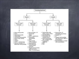
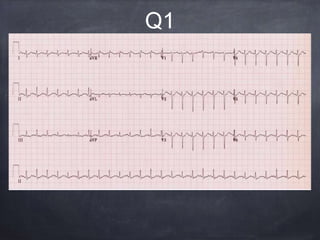
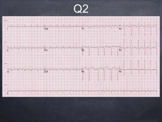
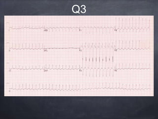
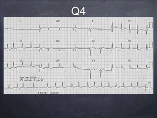
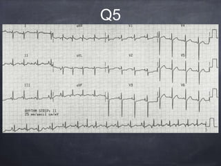
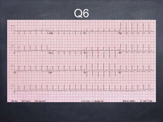
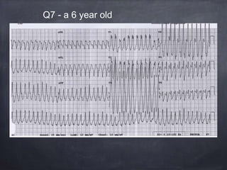
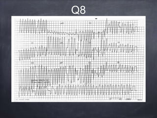
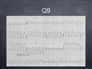
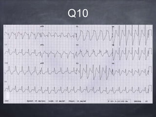
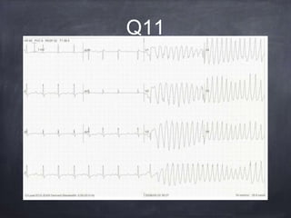
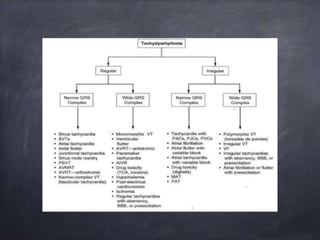
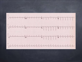
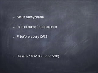
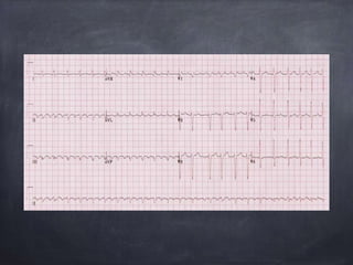
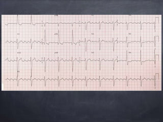
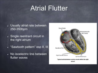
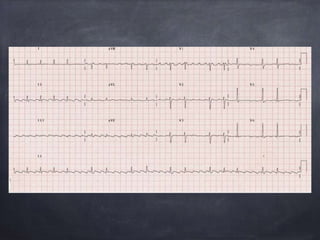
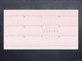
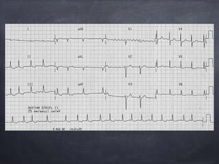
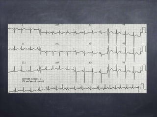
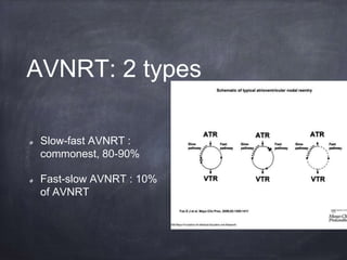
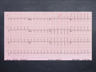
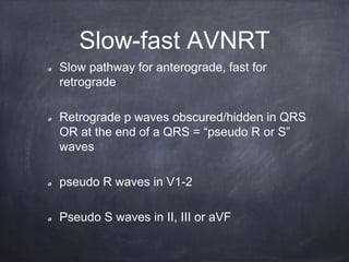
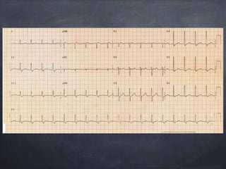
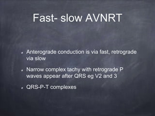
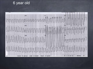
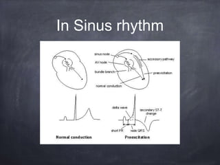
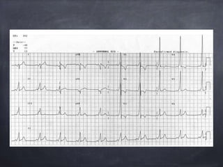
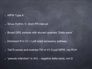
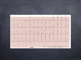
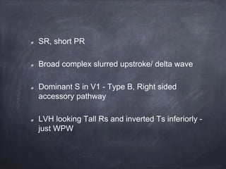
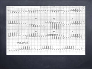
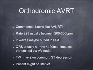
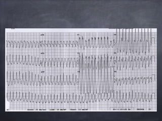
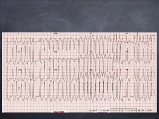
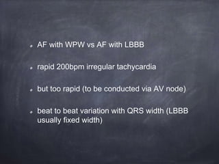
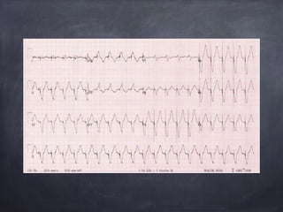
Tachyarrythmias
Ali Mackay
QUIZ!!
Q1
Q2
Q3
Q4
Q5
Q6
Q7 - a 6 year old
Q8
Q9
Q10
Q11
Tachyarrythmias
Ali Mackay
Sinus tachycardia
“camel hump” appearance
P before every QRS
Usually 100-160 (up to 220)
Shock
Hypoxia
Anaemia
Drug intox/wd esp
Tetanus
Pheochromocytoma
Cardiac Failure
Fever/infection (8
beats per 1°↑)
Pain, anxiety
Thyrotoxicosis
Pregnancy
Delerium
ST - Causes
Atrial flutter with 4:1 block
Vent rate 65
Usually atrial rate between
250-350bpm
Single reentrant circuit in
the right atrium
“Sawtooth pattern” esp II, III
No isoelectric line between
flutter waves
Atrial Flutter
Atrial flutter: 2 types
Anticlockwise reentry (70%)
Flutter waves inverted in II, III and aVF (Type
1)
Clockwise reentry (30%) upright
in same leads (Type 2)
rarely occurs in absence of underlying heart
disease
2:1 = vent rate 150 (commonest)
3:1, 4:1
Causes- atrial flutter
IHD
Acute MI
CCF
PE
Myocarditis
Chest trauma
Digoxin toxicity
Atrial flutter with variable block
May mimic Atrial fibrilllation
alternating 3:1 and 4:1
Or rapid SVT with rate related ST depression
Very rapid, regular narrow complex
tachycardia
Undulating baseline
Usually occurs due to sympathetic stimulation
with an accessory pathway. Eg AV nodal
drugs given to WPW
Can progress to VF
Atrial flutter 1:1
DC cardiovert!
Treatment?
Atrial flutter with high grade block
Marked AV block 5:1 up to 8:1
V. low vent rate ?AV nodal blocking drug
-? digoxin
Atrial fibrillation
irregularly irregular
course fibrillatory waves in V1
ST “sagging” in V6, II, III and aVF
? digoxin effect
Common in COPD
rapid, irregular with multiple P wave
morphologies
Bonus: Right axis deviation, RVH (dominant R
wave in V1, deep S wave in V6) - cor
pulmonale
Multifocal Atrial
Tachycardia (MAT)
Narrow complex tachy - about 150bpm
no P waves
Slow -fast AVNRT (Atrioventricular Nodal
Reentrant Tachycardia)
Type of paroxysmal supraventricular
tachycardia (PSVT)
Commonest type of PSVT 80-90%
Caused by reentry circuit in or around the AV
node: slow and fast pathway.
AVNRT
AVNRT: 2 types
Slow-fast AVNRT :
commonest, 80-90%
Fast-slow AVNRT : 10%
of AVNRT
Slow-fast AVNRT
Slow pathway for anterograde, fast for
retrograde
Retrograde p waves obscured/hidden in QRS
OR at the end of a QRS = “pseudo R or S”
waves
pseudo R waves in V1-2
Pseudo S waves in II, III or aVF
Anterograde conduction is via fast, retrograde
via slow
Narrow complex tachy with retrograde P
waves appear after QRS eg V2 and 3
QRS-P-T complexes
Fast- slow AVNRT
6 year old
280bpm, broad complexes
regular broad complex tachycardia
could be VT but.... only 6 years old!
AVRT
Also a PSVT
But an AVNT (Atrioventricular Reentry
Tachycardia)
Wolff Parkinson-White
Accessory internodal tracts - bundle of kent
Reentry circuit formed by normal conduction via
AV node and accessory pathway
Wolff- Parkinson White
In Sinus rhythm
WPW Type A
Sinus rhythm, V. short PR interval
Broad QRS compex with slurred upstroke “Delta wave”
Dominant R in V1 = Left sided accessory pathway
Tall R waves and inverted TW in V1-3 just WPW, not RVH
“pseudo infarction” in AVL - negative delta wave, not Q
SR, short PR
Broad complex slurred upstroke/ delta wave
Dominant S in V1 - Type B, Right sided
accessory pathway
LVH looking Tall Rs and inverted Ts inferiorly -
just WPW
AVRT
Orthodromic AVRT
Commonest. Looks like AVNRT!
Rate 225 usually between 200-300bpm
P waves maybe buried in QRS
QRS usually narrow <120ms - impulses
transmitted via AV node
TW inversion common, ST depression
Patient might be stable!
Antidromic AVRT
In a 6 year old
Therefore unlikely to be VT but very difficult to
distinguish
Reverted with vagal manouvres
AF with WPW vs AF with LBBB
rapid 200bpm irregular tachycardia
but too rapid (to be conducted via AV node)
beat to beat variation with QRS width (LBBB
usually fixed width)
Can occur in 20% of WPW
Accessory pathway means AF can be
transmitted to ventricles 1:1 fashion
Rate >200bpm
irregular
Wide QRSm change in shape and morphology
AF and WPW
NO:
Adenosine
Beta blockers
CCBs
Digoxin
Lignocaine
YES:
DC cardioversion
?Amiodarone
Treatment of AF and
WPW
Also AF with WPW vs polymorphic VT?
300bpm in some places
AV nodal conduction in V1 and 2 with
Narrow/atrial complexes
Absence of normal BBB Morphology
Extreme access deviation
Very broad QRS complex >160ms
Capture beats
Fusion beats
Brugada’s sign
Josephson’s sign V6
RSR complexes with a taller L rabbit ear (specific) V1
Features of VT
Capture beat
And -ve in
III and aVF
Extreme axis
deviation.
QRS +ve in aVR
Fusion beat
Extreme axis deviation
Fusion beats
Capture beats
Brugada’s sign - in V6 - time from onset of
QRS to nadir of S wave is >100ms
Monomorphic VT
Josephson
sign
RSR pattern
Monomorphic VT
Uniform QRS - very broad >200ms
Josephson’s sign in lead III
Age >35
Structural heart disease
Ischaemic heart disease
Prev MI
CCF
Cardiomyopathy
FHx Sudden cardiac death
Clinical features
suggestive of VT
Sinus tachy, grossly prolonged PR interval - P
waves hidden in Prev T or QRS complex
QRS v broad about 200ms - but broad in the
terminal portion of the QRS
No positive brugada criteria
+ve R in AVR
TCA toxicity
Sinus rhythm, inverted T waves, u waves
Long Q-U interval = hyopkalaemia
Premature atrial beat
R-on-T phenomenom
Torsades du pointes
For TdP to be diagnosed, ECG confirmation of
Long QT must be sought (before or after
rhythm is reverted)
Use the QTc
Estimates the QT interval at a HR of 60
Bazett’s formula easiest: QTc/√RR
(Where RR interval (secs) = 60/HR)
Prolonged: Men >440ms
Women >460ms
>500ms: at inc risk of TdP
Long QT
↓ K
↓Mg
↓Ca
↓Temp
↑ICP
Post cardiac arrest
Drugs: quinidine
phenothiazines
antihistamines TCAs
Chloral hydrate
erythromycin
azithromycin
fluconazole
Congenital Long QT
Myocardial ischaemia
Causes of Long QT
Paced ventricular rhythm with pacing spikes
Tachydysrhythmias
Tachydysrhythmias

Tachydysrhythmias

  • 1.
  • 3.
  • 4.
  • 5.
  • 6.
  • 7.
  • 8.
  • 9.
  • 10.
    Q7 - a6 year old
  • 11.
  • 12.
  • 13.
  • 14.
  • 15.
  • 18.
    Sinus tachycardia “camel hump”appearance P before every QRS Usually 100-160 (up to 220)
  • 19.
    Shock Hypoxia Anaemia Drug intox/wd esp Tetanus Pheochromocytoma CardiacFailure Fever/infection (8 beats per 1°↑) Pain, anxiety Thyrotoxicosis Pregnancy Delerium ST - Causes
  • 22.
    Atrial flutter with4:1 block Vent rate 65
  • 23.
    Usually atrial ratebetween 250-350bpm Single reentrant circuit in the right atrium “Sawtooth pattern” esp II, III No isoelectric line between flutter waves Atrial Flutter
  • 24.
    Atrial flutter: 2types Anticlockwise reentry (70%) Flutter waves inverted in II, III and aVF (Type 1) Clockwise reentry (30%) upright in same leads (Type 2) rarely occurs in absence of underlying heart disease 2:1 = vent rate 150 (commonest) 3:1, 4:1
  • 25.
    Causes- atrial flutter IHD AcuteMI CCF PE Myocarditis Chest trauma Digoxin toxicity
  • 27.
    Atrial flutter withvariable block May mimic Atrial fibrilllation alternating 3:1 and 4:1
  • 29.
    Or rapid SVTwith rate related ST depression Very rapid, regular narrow complex tachycardia Undulating baseline Usually occurs due to sympathetic stimulation with an accessory pathway. Eg AV nodal drugs given to WPW Can progress to VF Atrial flutter 1:1
  • 30.
  • 32.
    Atrial flutter withhigh grade block Marked AV block 5:1 up to 8:1 V. low vent rate ?AV nodal blocking drug -? digoxin
  • 34.
    Atrial fibrillation irregularly irregular coursefibrillatory waves in V1 ST “sagging” in V6, II, III and aVF ? digoxin effect
  • 36.
    Common in COPD rapid,irregular with multiple P wave morphologies Bonus: Right axis deviation, RVH (dominant R wave in V1, deep S wave in V6) - cor pulmonale Multifocal Atrial Tachycardia (MAT)
  • 38.
    Narrow complex tachy- about 150bpm no P waves Slow -fast AVNRT (Atrioventricular Nodal Reentrant Tachycardia)
  • 39.
    Type of paroxysmalsupraventricular tachycardia (PSVT) Commonest type of PSVT 80-90% Caused by reentry circuit in or around the AV node: slow and fast pathway. AVNRT
  • 40.
    AVNRT: 2 types Slow-fastAVNRT : commonest, 80-90% Fast-slow AVNRT : 10% of AVNRT
  • 42.
    Slow-fast AVNRT Slow pathwayfor anterograde, fast for retrograde Retrograde p waves obscured/hidden in QRS OR at the end of a QRS = “pseudo R or S” waves pseudo R waves in V1-2 Pseudo S waves in II, III or aVF
  • 44.
    Anterograde conduction isvia fast, retrograde via slow Narrow complex tachy with retrograde P waves appear after QRS eg V2 and 3 QRS-P-T complexes Fast- slow AVNRT
  • 45.
  • 46.
    280bpm, broad complexes regularbroad complex tachycardia could be VT but.... only 6 years old! AVRT
  • 47.
    Also a PSVT Butan AVNT (Atrioventricular Reentry Tachycardia) Wolff Parkinson-White
  • 48.
    Accessory internodal tracts- bundle of kent Reentry circuit formed by normal conduction via AV node and accessory pathway Wolff- Parkinson White
  • 49.
  • 51.
    WPW Type A Sinusrhythm, V. short PR interval Broad QRS compex with slurred upstroke “Delta wave” Dominant R in V1 = Left sided accessory pathway Tall R waves and inverted TW in V1-3 just WPW, not RVH “pseudo infarction” in AVL - negative delta wave, not Q
  • 53.
    SR, short PR Broadcomplex slurred upstroke/ delta wave Dominant S in V1 - Type B, Right sided accessory pathway LVH looking Tall Rs and inverted Ts inferiorly - just WPW
  • 54.
  • 56.
    Orthodromic AVRT Commonest. Lookslike AVNRT! Rate 225 usually between 200-300bpm P waves maybe buried in QRS QRS usually narrow <120ms - impulses transmitted via AV node TW inversion common, ST depression Patient might be stable!
  • 58.
    Antidromic AVRT In a6 year old Therefore unlikely to be VT but very difficult to distinguish Reverted with vagal manouvres
  • 60.
    AF with WPWvs AF with LBBB rapid 200bpm irregular tachycardia but too rapid (to be conducted via AV node) beat to beat variation with QRS width (LBBB usually fixed width)
  • 61.
    Can occur in20% of WPW Accessory pathway means AF can be transmitted to ventricles 1:1 fashion Rate >200bpm irregular Wide QRSm change in shape and morphology AF and WPW
  • 62.
  • 64.
    Also AF withWPW vs polymorphic VT? 300bpm in some places AV nodal conduction in V1 and 2 with Narrow/atrial complexes
  • 65.
    Absence of normalBBB Morphology Extreme access deviation Very broad QRS complex >160ms Capture beats Fusion beats Brugada’s sign Josephson’s sign V6 RSR complexes with a taller L rabbit ear (specific) V1 Features of VT
  • 67.
    Capture beat And -vein III and aVF Extreme axis deviation. QRS +ve in aVR Fusion beat
  • 68.
    Extreme axis deviation Fusionbeats Capture beats Brugada’s sign - in V6 - time from onset of QRS to nadir of S wave is >100ms Monomorphic VT
  • 70.
  • 71.
    Monomorphic VT Uniform QRS- very broad >200ms Josephson’s sign in lead III
  • 72.
    Age >35 Structural heartdisease Ischaemic heart disease Prev MI CCF Cardiomyopathy FHx Sudden cardiac death Clinical features suggestive of VT
  • 74.
    Sinus tachy, grosslyprolonged PR interval - P waves hidden in Prev T or QRS complex QRS v broad about 200ms - but broad in the terminal portion of the QRS No positive brugada criteria +ve R in AVR TCA toxicity
  • 76.
    Sinus rhythm, invertedT waves, u waves Long Q-U interval = hyopkalaemia Premature atrial beat R-on-T phenomenom Torsades du pointes
  • 77.
    For TdP tobe diagnosed, ECG confirmation of Long QT must be sought (before or after rhythm is reverted)
  • 78.
    Use the QTc Estimatesthe QT interval at a HR of 60 Bazett’s formula easiest: QTc/√RR (Where RR interval (secs) = 60/HR) Prolonged: Men >440ms Women >460ms >500ms: at inc risk of TdP Long QT
  • 79.
    ↓ K ↓Mg ↓Ca ↓Temp ↑ICP Post cardiacarrest Drugs: quinidine phenothiazines antihistamines TCAs Chloral hydrate erythromycin azithromycin fluconazole Congenital Long QT Myocardial ischaemia Causes of Long QT
  • 81.
    Paced ventricular rhythmwith pacing spikes