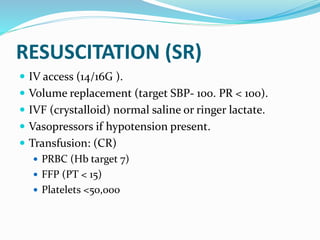
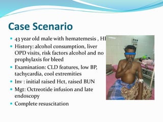
Presented By
Dr.(Maj) Ajay Kandpal MD DNB
An Axe and 2 Men
Basis Of Presentation
 Sleisenger and Fordtran’s GI & Liver Diseases 10th
Edition
 Yamada’s Textbook of Gastroenterology 6th Edition
 Harrison textbook of Medicine 19th Edition
 Strength of recommendation from ACG guidelines
2012
 Some other Articles
Outline Of Presentation
 Definitions
 Introduction
 Approach considerations
 Risk stratification and Management of UGI bleed
Definitions
 Upper GI Bleeding : always proximal to ligament of
Treitz
 Hematemesis : Vomiting of red blood or coffee ground
vomitus, indicative of bleeding from the esophagus,
stomach or duodenum.
 Malena: Black tarry stools resulting from degradation of
blood to hematin or other hemochromes by intestinal
bacteria.
 Hematochezia : Bright red blood per rectum and
suggests active UGI or small bowel bleeding or distal
colonic or anorectal bleeding
Severe GI Bleeding?
 Hematemesis ,melena, hematochezia or positive
nasogastric lavage
 Accompanied by shock
 Orthostatic hypotension
 Decrease in the haematocrit value by atleast 6%
 Decrease in Hb of atleast 2 grams/dl or transfusion of
two units of packed red blood cells.
Some Facts
 Approximately 50% of admissions for GI bleeding are
for upper GI (UGI) bleeding (from the esophagus,
stomach, and duodenum).
 Self limiting in 80%.
 Remaining 20% have a mortality of 30-40%.
 5-10% mortality despite advances
Causes
COMMON CAUSES
 Duodenal ulceration
 Gastric ulceration
 Esophagitis, gastritis,
duodenitis
 Varices
 Mallory Weiss tear
LESS COMMON CAUSES
 Gastro oesophageal
cancer
 Leiomyoma
 PHG
 Dieulafoy lesion
Causes
Approach Considerations
 Clinical suspicion
 Arriving at a confirmed diagnosis
 Management of UGI bleed
Clinical Suspicion
 Overt bleed
 History points
 Examination findings
 PR findings
History
Suspected source /cause History
Esophagus erosion GERD/heartburn/alcohol/odynophagia
/ pill ingestion/NG tube placement
Peptic ulcer Epigastric discomfort/asprin/NSAID
use/ history of PUD
Mallory weiss tear Alcohol binge
Retching/vomiting
Cameron lesion Large hiatal hernia
Gastric angiodysplasia CKD
Variceal bleed CLD/cirrhosis/heavy alcohol use
Malignancies Dysphagia/wt loss/early satitation
Nasopharynx Radiation/recurrent epistaxis/
malignancy
Lungs Hemoptysis
Physical Examination
SEVERE BLEED (SR)
 Tachycardia
 Systolic blood pressure of less than 90 mm Hg
 Cool extremities
 Syncope
 Ongoing brisk hematemesis or the occurrence of
maroon or bright-red stools, which requires rapid
blood transfusion.
Review Article
Upper Gastrointestinal Bleeding – A Review of the Literature
(Part 1)
Author(s): Bashir S, Roy P
Vol. 5, No. 2 (2008-05 - 2008-06)
HEPATOLOGICAL SIGNS
SKIN AND MUCOSAL SIGNS
 Skin,lips,buccal mucosa
suggestive of HHT or osler
weber rendu disease
 Pigmented skin lesions
HSP
 Pigmented lip lesions
Peutz Jeghers syndrome
 New onset acanthosis
nigricans
RHEUMATOLOGICAL SIGNS
Secondary to NSAIDS
HEMATOLOGICAL SIGNS
• Petechial hemorrhages
• Bruising
• Suggests bleeding diathesis
• Drug induced aplastic anemia
Variceal Non variceal
History
CLD, Jaundice,decompesation
Alcohol consumption
Drugs,pain abdomen
,heartburn,retrosternal discomfort
NCPF recurrent bacterial
infection,umbilical sepsis
Examination
Jaundice,abd.distention.signs of liver
cell failue
No such signs .look for
rheumatological,petechial signs
Epigastric tenderness
Painless bleeding Painful bleeding
Large amount ,FRESH BLEED Mild to moderate, COFFEE
GROUND
Decompensates rapidly
Clues From Initial Investigation
Parameter Implications
Hb /HCT 24-72hrs
Need of transfusuion
MCV Less than 80
Less than 80 with negative SOB
Platelets CLD
Hematological disorder
PT/INR Coagulopathy
Disproportionate rise of
BUN
Points To Decide Line Of
Management
 Severe or non severe
 Overt/ hidden
 Etiology
 Co-morbid conditions
 Availability of resources
Where To Manage
 Severe UGI bleed in ICU. (SR)
 Acute GI bleed with hemodynamic stability in ward.
 Mild Acute GI bleed with hemodynamic stability and
routine investigations normal, on OPD basis.(CR)
 SBP>110, pulse<100
 Hb13/12. BUN <18.2
 No malena, syncope, cardiac failure , liver disease
 Chronic bleed OPD basis.
How to Manage
 Resuscitation
 Initial medical therapy
 Endoscopy
 Endoscopic hemosatasis
 Post endoscopic considerations
 Imaging and surgery
RESUSCITATION (SR)
 IV access (14/16G ).
 Volume replacement (target SBP- 100. PR < 100).
 IVF (crystalloid) normal saline or ringer lactate.
 Vasopressors if hypotension present.
 Transfusion: (CR)
 PRBC (Hb target 7)
 FFP (PT < 15)
 Platelets <50,000
 Monitoring
 Vitals
 Hct levels 4-6 hrly till stablise
 Urinary catheter
 ET intubation
 MI evaluation
Initial Medical Therapy
 Depends on cause
 PUD – high dose PPI(CR)
 Reduce endoscopic stigmata of bleed
 Reduced need of endoscopy
 No improvement in transfusion reqmt/ rebleed
rate/surgery/survival
 Portal htn/variceal bleed
 Octreotide infusion
Endoscopy
 Requirement
 Stable hemodynamic condition
 Intubation if required
 Correction of coagulopathy and thromboctopenia
 Gastric lavage (CR)
 IV erthromycin/metoclopramide 30-90 min before (CR)
 Timing
 Cirrohosis/aorto enteric fistula /rebleed in hosp---6hrs
(CR)
 Stable with no ongong bleed – 12 hrs (CR)
Pre-Endoscopic Risk Stratification
 Clinical condition
 Co morbidities
 Scoring systems for non- varicel bleed
Pre- Endoscopy Scoring System
 Blatchford Score : BP, heart rate, syncope,melena,
liver disease,heart failure, Hb%, BUN.
Assess need of intervention to control bleed (score>6)
 Clinical Rockall score : SBP, heart rate, age,
comorbidities.
 AIMS 65 score : Albumin <3gm/dl, INR >1.5,
Impaired mental status, SBP<90mmHg, age>65 yrs.
Score less than 2 is low mortality, short stay.less
hopitalization
Endoscopic Risk Stratification
 Visual scales
 Scoring systems
Post Endoscopy Scoring System
(SR)
 Complete Rockall score : Clinical Rockall score +
endoscopic stigmata of recent bleeding and
endoscopic diagnosis.
• Score 0-2: very less risk of rebleeding
• Score 9 and above: high risk of rebleeding and mortality.
• Correlates well with mortality but not as well with
rebleeding.
FORREST Classification for Peptic
Ulcers
CLASSIFICATION ENDOSCOPIC PIC RISK OF REBLEEDING
FORREST IA ACTIVE SPURTING
AND BLEEDING
85 TO 100%
FORREST IB OOZING BLEEDING 10- 25%
FORREST 2A PIGMENTED
PROTRUBERANCE OR
NBVV
50%
FORRSET 2B ADHERENT CLOT 30 -35%
FORRSET 2C FLAT PIGMENTED
SPOT
<8%
FORRSET 3 CLEAN BASED ULCER <3%
Risk For Bleed
 The most important predictor of hemorrhage is the
size of varices, with the highest risk of first
hemorrhage (15% per year) occurring in patients with
large varices.
 Other predictors of hemorrhage are decompensated
cirrhosis (Child B ,C).
 The endoscopic presence of red wale marks.
Red Wale Signs Of A Varix In
Endoscopy
 Suggests recent haemorrhage
 Longitudinal streak located on
esophageal varix
 Cherry red spot (circular and
red)
Endoscopic Hemostasis
 Thermal probe
 multipolar electrocoagulation probe(MPEC)
 Non contact argon plasma coagulation
 Injection therapy
 Epinephrine 1:10000, submucosal
 N-butyl-2-cyanoacrylate, which is recommended as a first-
line endoscopic therapy
 Sclerosant like Ethanolamine or alcohol
 Hemoclips
 Band ligation
 Hemostatic spray
Endoscopic Therapy Rec.
 Oozing /spurting or NBVV (SR)
 Adherent clot with comorbidities increasing risk of
bleed (CR)
 Not to clean base ulcer or flat pigmented spot (SR)
 Epinephrine should not be used alone, if used then in
combination of other therapy(SR)
 Use of IV PPI after hemostasis in ulcer with Oozing
/spurting or NBVV (SR)
 Oral PPI clean base ulcer or flat pigmented spot (SR)
 Relook endoscopy for rebleed or high stigmata of
rebleed in initial endoscopy (SR)
Post Endoscopy
Post Endoscopy Considerations
 PUD
 PPI
 H Pylori eradication (SR)
 Iron replacement
 Aspirin (CR)
 Primary prevention : avoid
 Secondary prevention : start in 1-3 days
 NSAID (SR)
 Not if possible
 Cox2 inhibitors plus PPI
 VARICEAL BLEED
 Octreotide /Terlipressin
 NSBB
 TIPSS
Second Look Endoscopy
 Routine second look endoscopy is not
recommended for most patients with peptic ulcer
bleeding, except in those in whom the initial
endoscopic examination was suboptimal.
Rebleeding After Endoscopic
Treatment
 An urgent repeat endoscopy should be performed
within 6 hrs of rebleeding.
 75% of patients achieve endoscopic hemostasis &
donot require surgery.
 High dose PPI for more than 72 hrs should be
considered.
 Factors predicting failure of endoscopic retreatment
include ulcer >2cm & hypotension on initial
presentation.
ACTIVELY BLEEDING VARICES
TIPS OR SURGERY(PC SHUNT OR
ESO.TRANSECTION)
REPEAT BAND LIGATION OR
SCLEROSIS
BLEEDING CONTINUES
PERIPHERAL VENOUS VASOPRESSIN
ENDOSCOPIC BAND LIGATION OR
SCLEROSIS
BALLOON TAMPONADE
TIPS
BLEEDING CONTINUES
BLEEDING
CONTROLLED
CHRONIC
MANAGEMENT
OF VARICES
THAT HAVE BLED
CONSIDER:COURSE
OF ENDOSCOPIC
BAND LIGATION OR
INJECTION
SCLEROSIS OR
PORTOCAVAL
SHUNT SURGERY
(or)
and /or
or
Imaging
 Role only if both endoscopy and colonoscopy fails
 Mesentric arteriography 0.5ml/min
 Radionuclide scaning 0.04ml/min
Surgery
 Indicated only for massive continuous hemorrhage
with failed endoscopy and colonoscopy
Prevention
 Risk factors
 NSAID
 Binge drinking
 CLD
 Primary prophylaxis
 Secondary prophylaxis
Case Scenario
 43 year old male with hematemesis , HE
 History: alcohol consumption, liver
OPD visits, risk factors alcohol and no
prophylaxis for bleed
 Examination: CLD features, low BP,
tachycardia, cool extremities
 Inv : initial raised Hct, raised BUN
 Mgt: Octreotide infusion and late
endoscopy
 Complete resuscitation

Approach to UGI bleed Dr Kandy

  • 1.
  • 2.
  • 3.
    Basis Of Presentation Sleisenger and Fordtran’s GI & Liver Diseases 10th Edition  Yamada’s Textbook of Gastroenterology 6th Edition  Harrison textbook of Medicine 19th Edition  Strength of recommendation from ACG guidelines 2012  Some other Articles
  • 4.
    Outline Of Presentation Definitions  Introduction  Approach considerations  Risk stratification and Management of UGI bleed
  • 5.
    Definitions  Upper GIBleeding : always proximal to ligament of Treitz  Hematemesis : Vomiting of red blood or coffee ground vomitus, indicative of bleeding from the esophagus, stomach or duodenum.  Malena: Black tarry stools resulting from degradation of blood to hematin or other hemochromes by intestinal bacteria.  Hematochezia : Bright red blood per rectum and suggests active UGI or small bowel bleeding or distal colonic or anorectal bleeding
  • 6.
    Severe GI Bleeding? Hematemesis ,melena, hematochezia or positive nasogastric lavage  Accompanied by shock  Orthostatic hypotension  Decrease in the haematocrit value by atleast 6%  Decrease in Hb of atleast 2 grams/dl or transfusion of two units of packed red blood cells.
  • 7.
    Some Facts  Approximately50% of admissions for GI bleeding are for upper GI (UGI) bleeding (from the esophagus, stomach, and duodenum).  Self limiting in 80%.  Remaining 20% have a mortality of 30-40%.  5-10% mortality despite advances
  • 8.
    Causes COMMON CAUSES  Duodenalulceration  Gastric ulceration  Esophagitis, gastritis, duodenitis  Varices  Mallory Weiss tear LESS COMMON CAUSES  Gastro oesophageal cancer  Leiomyoma  PHG  Dieulafoy lesion
  • 9.
  • 10.
    Approach Considerations  Clinicalsuspicion  Arriving at a confirmed diagnosis  Management of UGI bleed
  • 11.
    Clinical Suspicion  Overtbleed  History points  Examination findings  PR findings
  • 12.
    History Suspected source /causeHistory Esophagus erosion GERD/heartburn/alcohol/odynophagia / pill ingestion/NG tube placement Peptic ulcer Epigastric discomfort/asprin/NSAID use/ history of PUD Mallory weiss tear Alcohol binge Retching/vomiting Cameron lesion Large hiatal hernia Gastric angiodysplasia CKD Variceal bleed CLD/cirrhosis/heavy alcohol use Malignancies Dysphagia/wt loss/early satitation Nasopharynx Radiation/recurrent epistaxis/ malignancy Lungs Hemoptysis
  • 13.
    Physical Examination SEVERE BLEED(SR)  Tachycardia  Systolic blood pressure of less than 90 mm Hg  Cool extremities  Syncope  Ongoing brisk hematemesis or the occurrence of maroon or bright-red stools, which requires rapid blood transfusion.
  • 14.
    Review Article Upper GastrointestinalBleeding – A Review of the Literature (Part 1) Author(s): Bashir S, Roy P Vol. 5, No. 2 (2008-05 - 2008-06)
  • 15.
  • 16.
    SKIN AND MUCOSALSIGNS  Skin,lips,buccal mucosa suggestive of HHT or osler weber rendu disease  Pigmented skin lesions HSP  Pigmented lip lesions Peutz Jeghers syndrome  New onset acanthosis nigricans
  • 17.
    RHEUMATOLOGICAL SIGNS Secondary toNSAIDS HEMATOLOGICAL SIGNS • Petechial hemorrhages • Bruising • Suggests bleeding diathesis • Drug induced aplastic anemia
  • 18.
    Variceal Non variceal History CLD,Jaundice,decompesation Alcohol consumption Drugs,pain abdomen ,heartburn,retrosternal discomfort NCPF recurrent bacterial infection,umbilical sepsis Examination Jaundice,abd.distention.signs of liver cell failue No such signs .look for rheumatological,petechial signs Epigastric tenderness Painless bleeding Painful bleeding Large amount ,FRESH BLEED Mild to moderate, COFFEE GROUND Decompensates rapidly
  • 19.
    Clues From InitialInvestigation Parameter Implications Hb /HCT 24-72hrs Need of transfusuion MCV Less than 80 Less than 80 with negative SOB Platelets CLD Hematological disorder PT/INR Coagulopathy Disproportionate rise of BUN
  • 20.
    Points To DecideLine Of Management  Severe or non severe  Overt/ hidden  Etiology  Co-morbid conditions  Availability of resources
  • 21.
    Where To Manage Severe UGI bleed in ICU. (SR)  Acute GI bleed with hemodynamic stability in ward.  Mild Acute GI bleed with hemodynamic stability and routine investigations normal, on OPD basis.(CR)  SBP>110, pulse<100  Hb13/12. BUN <18.2  No malena, syncope, cardiac failure , liver disease  Chronic bleed OPD basis.
  • 22.
    How to Manage Resuscitation  Initial medical therapy  Endoscopy  Endoscopic hemosatasis  Post endoscopic considerations  Imaging and surgery
  • 23.
    RESUSCITATION (SR)  IVaccess (14/16G ).  Volume replacement (target SBP- 100. PR < 100).  IVF (crystalloid) normal saline or ringer lactate.  Vasopressors if hypotension present.  Transfusion: (CR)  PRBC (Hb target 7)  FFP (PT < 15)  Platelets <50,000
  • 24.
     Monitoring  Vitals Hct levels 4-6 hrly till stablise  Urinary catheter  ET intubation  MI evaluation
  • 25.
    Initial Medical Therapy Depends on cause  PUD – high dose PPI(CR)  Reduce endoscopic stigmata of bleed  Reduced need of endoscopy  No improvement in transfusion reqmt/ rebleed rate/surgery/survival  Portal htn/variceal bleed  Octreotide infusion
  • 26.
    Endoscopy  Requirement  Stablehemodynamic condition  Intubation if required  Correction of coagulopathy and thromboctopenia  Gastric lavage (CR)  IV erthromycin/metoclopramide 30-90 min before (CR)  Timing  Cirrohosis/aorto enteric fistula /rebleed in hosp---6hrs (CR)  Stable with no ongong bleed – 12 hrs (CR)
  • 27.
    Pre-Endoscopic Risk Stratification Clinical condition  Co morbidities  Scoring systems for non- varicel bleed
  • 28.
    Pre- Endoscopy ScoringSystem  Blatchford Score : BP, heart rate, syncope,melena, liver disease,heart failure, Hb%, BUN. Assess need of intervention to control bleed (score>6)  Clinical Rockall score : SBP, heart rate, age, comorbidities.  AIMS 65 score : Albumin <3gm/dl, INR >1.5, Impaired mental status, SBP<90mmHg, age>65 yrs. Score less than 2 is low mortality, short stay.less hopitalization
  • 30.
    Endoscopic Risk Stratification Visual scales  Scoring systems
  • 31.
    Post Endoscopy ScoringSystem (SR)  Complete Rockall score : Clinical Rockall score + endoscopic stigmata of recent bleeding and endoscopic diagnosis. • Score 0-2: very less risk of rebleeding • Score 9 and above: high risk of rebleeding and mortality. • Correlates well with mortality but not as well with rebleeding.
  • 33.
    FORREST Classification forPeptic Ulcers CLASSIFICATION ENDOSCOPIC PIC RISK OF REBLEEDING FORREST IA ACTIVE SPURTING AND BLEEDING 85 TO 100% FORREST IB OOZING BLEEDING 10- 25% FORREST 2A PIGMENTED PROTRUBERANCE OR NBVV 50% FORRSET 2B ADHERENT CLOT 30 -35% FORRSET 2C FLAT PIGMENTED SPOT <8% FORRSET 3 CLEAN BASED ULCER <3%
  • 34.
    Risk For Bleed The most important predictor of hemorrhage is the size of varices, with the highest risk of first hemorrhage (15% per year) occurring in patients with large varices.  Other predictors of hemorrhage are decompensated cirrhosis (Child B ,C).  The endoscopic presence of red wale marks.
  • 35.
    Red Wale SignsOf A Varix In Endoscopy  Suggests recent haemorrhage  Longitudinal streak located on esophageal varix  Cherry red spot (circular and red)
  • 36.
    Endoscopic Hemostasis  Thermalprobe  multipolar electrocoagulation probe(MPEC)  Non contact argon plasma coagulation  Injection therapy  Epinephrine 1:10000, submucosal  N-butyl-2-cyanoacrylate, which is recommended as a first- line endoscopic therapy  Sclerosant like Ethanolamine or alcohol  Hemoclips  Band ligation  Hemostatic spray
  • 37.
    Endoscopic Therapy Rec. Oozing /spurting or NBVV (SR)  Adherent clot with comorbidities increasing risk of bleed (CR)  Not to clean base ulcer or flat pigmented spot (SR)  Epinephrine should not be used alone, if used then in combination of other therapy(SR)  Use of IV PPI after hemostasis in ulcer with Oozing /spurting or NBVV (SR)  Oral PPI clean base ulcer or flat pigmented spot (SR)
  • 38.
     Relook endoscopyfor rebleed or high stigmata of rebleed in initial endoscopy (SR)
  • 39.
  • 40.
    Post Endoscopy Considerations PUD  PPI  H Pylori eradication (SR)  Iron replacement  Aspirin (CR)  Primary prevention : avoid  Secondary prevention : start in 1-3 days  NSAID (SR)  Not if possible  Cox2 inhibitors plus PPI
  • 41.
     VARICEAL BLEED Octreotide /Terlipressin  NSBB  TIPSS
  • 42.
    Second Look Endoscopy Routine second look endoscopy is not recommended for most patients with peptic ulcer bleeding, except in those in whom the initial endoscopic examination was suboptimal.
  • 43.
    Rebleeding After Endoscopic Treatment An urgent repeat endoscopy should be performed within 6 hrs of rebleeding.  75% of patients achieve endoscopic hemostasis & donot require surgery.  High dose PPI for more than 72 hrs should be considered.  Factors predicting failure of endoscopic retreatment include ulcer >2cm & hypotension on initial presentation.
  • 44.
    ACTIVELY BLEEDING VARICES TIPSOR SURGERY(PC SHUNT OR ESO.TRANSECTION) REPEAT BAND LIGATION OR SCLEROSIS BLEEDING CONTINUES PERIPHERAL VENOUS VASOPRESSIN ENDOSCOPIC BAND LIGATION OR SCLEROSIS BALLOON TAMPONADE TIPS BLEEDING CONTINUES BLEEDING CONTROLLED CHRONIC MANAGEMENT OF VARICES THAT HAVE BLED CONSIDER:COURSE OF ENDOSCOPIC BAND LIGATION OR INJECTION SCLEROSIS OR PORTOCAVAL SHUNT SURGERY (or) and /or or
  • 45.
    Imaging  Role onlyif both endoscopy and colonoscopy fails  Mesentric arteriography 0.5ml/min  Radionuclide scaning 0.04ml/min
  • 46.
    Surgery  Indicated onlyfor massive continuous hemorrhage with failed endoscopy and colonoscopy
  • 47.
    Prevention  Risk factors NSAID  Binge drinking  CLD  Primary prophylaxis  Secondary prophylaxis
  • 48.
    Case Scenario  43year old male with hematemesis , HE  History: alcohol consumption, liver OPD visits, risk factors alcohol and no prophylaxis for bleed  Examination: CLD features, low BP, tachycardia, cool extremities  Inv : initial raised Hct, raised BUN  Mgt: Octreotide infusion and late endoscopy  Complete resuscitation

Editor's Notes

  • #4 SR CR
  • #6 ONE DEF THEN OTHER DEF Red indicate fresh bleed, cofee old bleed Melena can signify bleeding that originates from a UGI, small bowel, or proximal colonic source and generally occurs when 50 to 100 mL or more of blood is delivered into the GI tract (usually the upper tract), with passage of characteristic stool occurring several hours after the bleeding event
  • #7 HEADING FIRST.. LATER SLIDE
  • #10 BEAUTIFY California university study
  • #13 BEAUTIFY early satitation in gastric ca
  • #14 BEAUTIFY
  • #15 BEAUTIFY
  • #16 BEAUTIFY
  • #17 BEAUTIFY
  • #19 NON VARICEAL CAUSE Peptic ulcer Dieulafoy’s Mallory weiss tear Cameron’s lesions Upper gi malignancy GAVE
  • #21 ONE BY ONE
  • #22 ICU IN CIRCLE AFTER ARROW WARD IN CIRCLE AFTER ARROW OPD IN CIRCLE AFTER ARROW
  • #24 Barcelona trial… rstrictive transfusion associated with better survival and less re bleed in CHILD A B but opposite for C NICE guidelines for correction of coagulopathy during AUGIB6 Coagulopathy Threshold Management Platelets <50 x109/L Platelet transfusion INR >1.5 FFP PCC (if on warfarin) aPTTr >1.5 FFP Fibrinogen <1.5g/L FFP Cryoprecipitate (if low despite FFP
  • #25 ET.. Altered sensorium with risk of aspiration MI evluation .. Chest pain, old 60 yrs , history of cad, ecg and serial troponin
  • #26 Dose of ppi 80 MG STAT THEN 8 MG PER HR INFUSION Octreotide 50 mcg bolus then infusion of 50 mcg/hr for 2-5 days
  • #27 ERTHROMYCIN 250 MG Lavage not required as per guideline
  • #28 Complete rockall is clinical plus endoscopic finding .. Corelate well with mortality but not with risk of rebleed
  • #37 Inj therapy adv wider avl , less damage than probes, less perf, safe in coagulopathy