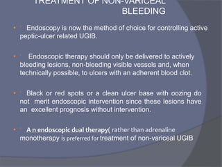
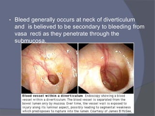
This document provides a comprehensive overview of gastrointestinal bleeding, detailing both upper and lower GI bleeding, their causes, presentation, diagnostic approaches, management strategies, and complications. It emphasizes the importance of identifying the source of bleeding and highlights the use of endoscopy, angiography, and surgical interventions based on the severity and type of bleeding. Preventive measures and specific conditions related to lower GI bleeding, such as diverticular disease and angiodysplasia, are also discussed.


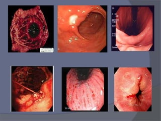
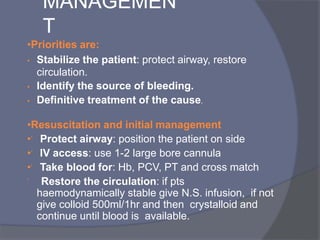


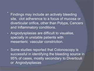
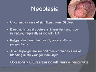
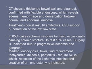
![Colonoscopy
• Most appropriate in the
setting of mild to
moderate bleeding, with
patient in stable condition
• Preparation with
Polyethylene Glycol
(PEG) orally or via NG
tube[4-8L] for 4-6 hrs,
with metaclopramide IV
improves visualisation](https://image.slidesharecdn.com/l2-gibleeding-240726163657-3f5d1723/85/Internal-Medicine-GI-bleeding-simple-pptx-56-320.jpg)
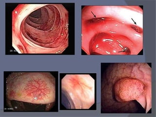
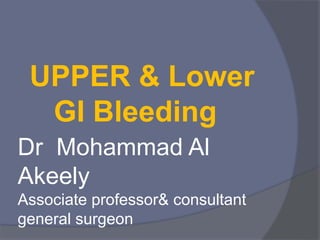
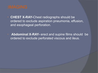
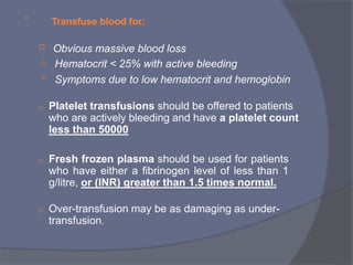
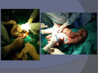
![Radionuclide Scanning
• Technician-99m [ 99mTc-labelled RBC ]
Most sensitive, but least accurate for localising
bleed
• Patients own blood is labelled and reinjected, which
is extravasated into GI tract lumen, creating a focus
that can be detected scintigraphically, Initially
images obtained serially, then at 4 hour intervals,
upto 24hrs.
• can detect bleed as slow as
0.1mL/min
• Unfortunately, spatial resolution is low with
reported accuracy of 40 - 60 %](https://image.slidesharecdn.com/l2-gibleeding-240726163657-3f5d1723/85/Internal-Medicine-GI-bleeding-simple-pptx-61-320.jpg)
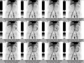
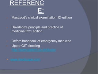
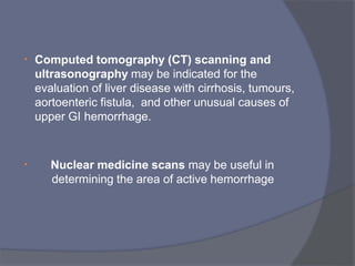
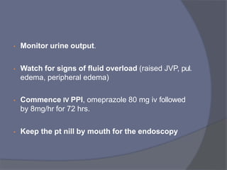
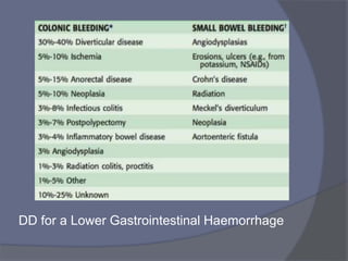
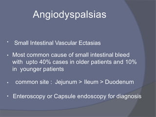
![Angiodysplasia
• Some reports state vascular lesions account upto 40% of LGI-B,
However, recent reports state much low incidence
• also called Arteriovenous Malformations [AVM’(]
• They are acquired degenerative lesions secondary to
progressive dilatation of normal blood vessels within the
submucosa of the intestine
• Age of incidence > 50 yrs with M=F, usually associated with
aortic stenosis and renal failure, esp in older patients
• Haemorrhage tends to arise from right side of colon, with
CECUM being most common location](https://image.slidesharecdn.com/l2-gibleeding-240726163657-3f5d1723/85/Internal-Medicine-GI-bleeding-simple-pptx-70-320.jpg)


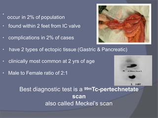
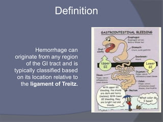
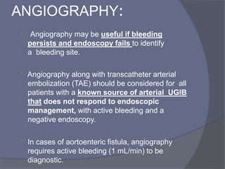
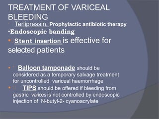
![Meckel’s Diverticulum
Bleeding is usually from an ulcerative lesion
on the ideal wall opposite the diverticulum,
resulting from the acid production by
ectopic gastric mucosa
• True diverticulum, which is a congenital remnant
of the omphalomesentric duct [2% of ppl]
•
• Surgical management includes Segmental
resection to incorporate the opposing ill
mucosa, which is typically the site for bleeding.](https://image.slidesharecdn.com/l2-gibleeding-240726163657-3f5d1723/85/Internal-Medicine-GI-bleeding-simple-pptx-87-320.jpg)