SlideShare a Scribd company logo
1 of 86
Dysmyelinating
disorders
White matter
disorders
Demyelination Dysmyelination Hypo-
myelination
Destruction of
normal myelin
Formation of
abnormal
myelin
Reduction of
the amount of
myelin
Basic principles of myelination on MRI
1. Un-myelinated White matter
• Hypointense on T1 W
• Hyperintense on T2 W
2. Myelinated White matter
 Hyperintense on T1 W
 Hypointense on T2W
Normal brain Myelination is a dynamic process that
begins during 5th fetal month & continuous through
out life
.
PROGRESS FAST UPTO 2 YRS. Some association fibres
remains unmyelinated until the age of 20-30 years
Dys-myelination refers to the abnormal
development or destruction of the myelin
sheath, a fatty layer that surrounds and
insulates nerve fibers in the brain and spinal
cord.
DEFINATION
Cognitivedefecits an early presentation-hereditary
diffuse leukoencephalopathy with axonal spheroids
(HDLS) .
Prominent features
behavioural changes, mood changes and loss of
realistic assessments of daily life experiences
Bulbar symptoms at onset –Alexander disease
Extrapyramidal signs and symptoms - dystonia
and/or dyskinesias , are less frequent
but might be a predominant manifestation in certain
disorders, including progressive leukoencephalopathy
with ovarian failure, various HLDs and AxD.
Seizures – MLD , krabbes ,Alexander disease
Adult Leukodystrophies:
A Step by- Step Diagnostic Approach Based on
imaging
Step 1 Identify Symmetric White
Matter Involvement
 Symmetric white matter involvement at MRI is a typical finding in patients with
leukodystrophies.
(a) Axial T2-weighted
MR image in a 23-year-old woman
shows symmetric white matter
involvement.
(b) Axial FLAIR MR image shows an
asymmetric pattern in a 46-year-old
woman shows asymmetric white
matter involvement
Step 2: Look for a White Matter
Involvement Pattern
X- linked Adrenoleukodystrophy
 Inherited disorder of peroxisomal metabolism.
 Altered peroxisome metabolism in ALD results from absent or deficient acyl CoA synthetase
leading to impaired β-oxidation of very-long-chain fatty acids (VLCFAs)
 ABCD1 gene mutation
Imaging
 MRI : White matter abnormalities are usually first seen in occipital regions, with
early involvement of the splenium of the corpus callosum and posterior limbs
of the internal capsule.
 The changes then progress to involve more anterior regions.
 In cerebral ALD, contrast enhancement at the periphery of the signal
abnormalities is said to be characteristic
Axial T2-weighted MR image shows parieto-occipital symmetric white
matter hyperintensity.
(b) Contrast-enhanced axial T1-weighted MR image in the shows
enhancement foci in the white
(c) Sagittal T2-weighted MR image shows thoracic cord atrophy.
The Loes Scoring System.
 The Loes scoring system is universally applied to quantify the extent
of cerebral involvement.
 Regions of the brain (such as parietooccipital white matter, anterior
temporal white matter, visual pathway, corpus callosum, auditory
pathway, basal ganglia, projection fibers, and cerebellum) are
subdivided and scored based on the extent of disease .
 With a score of 0 if normal, 0.5 if unilateral involvement is present,
and 1 if the lesion or atrophy is bilateral.
 Global atrophy is also assessed.
 A normal MRI scan has a score of 0, and the maximum severity score
is 34.
Abbreviation: MRI, magnetic resonance imaging.
Each region is given a score of 0 for normal, 0.5 for unilateral involvement, and 1
for bilateral involvement or atrophy. The maximum score is 34.
Parieto-occipital white matter
(maximum 4)
Basal ganglia (maximum 1)
Anterior temporal white matter
(maximum 4)
•Frontal white matter (maximum 4)
•Periventricular
•Central
•Subcortical
•Local atrophy
•Visual pathway (maximum 4)Optic
radiation
•Meyer’s loop
•Lateral geniculate body
•Optic tract
•Corpus callosum (maximum 5)
•Splenium
•Genu
•Body
•Splenium atrophy
•Genu atrophy
•Auditory pathway (maximum 4)Medial
geniculate body
•Brachium of inferior colliculus
•Lateral lemniscus
•Pons
•Global atrophy (maximum 4)Mild
•Moderate
•Severe
•Brainstem
•Cerebellum (maximum 2)White matter
•Atrophy
•Projection fibers (maximum 2)Internal
capsule
•Brain stem
MRI severity scale scoring (Loes et al 5 )
Metachromatic Leukodystrophy
 AR lysosomal storage disorder
 An enzyme deficiency of arylsulfatase A, which is encoded on the ARSA gene on
chromosome 22 q/or pathogenic mutations in PSAP (which encodes
prosaposin)
 Sulfatides accumulate in the central and peripheral nervous systems, kidneys,
testes, and visceral organs (gall bladder).
 MLD is an adulthood leukodystrophy that is frequently misdiagnosed as early-
onset dementia and/or a schizophrenic disorder, as the neurological symptoms
can occur late in the disease course.
A high index of suspicion for metachromatic leukodystrophy must exist for patients
presenting with a peripheral neuropathy or gall bladder abnormality in the absence of
other neurologic features.
The MRI may not reveal pathology early in the disease course; therefore, a normal MRI is
not sufficient to exclude metachromatic leukodystrophy.
TIGROID PATTERN
 A low arylsulfatase A level (less than 10% of normal values) is detected in
white blood cells or cultured fibroblasts, confirmed by the detection of
elevated sulfatides in the urine and ARSA sequencing for mutations.
 Phase 1/II trial of intrathecal aryl sulfatase A .
 Juvenile MLD – autologous HSCT .
 Genetherapy under trial .
Leukoencephalopathy with Axonal Spheroids and Pigmented Glia.—
• Autosomal dominant
• Colony-stimulating factor 1 receptor (CSF1R) gene mutation.
• The gene defect affects the tyrosine kinase domain of macrophage
colony-stimulating factor 1 receptor.
• Unlike other leukodystrophies, this disease manifests exclusively in
adults(20-50).
• P/W Behavioral changes, dementia, motor impairment.
(a) Luxol fast blue stain for myelin shows myelin loss and tissue vacuolation
with axonal spheroids [arrows in (a) and (b)], that are immunopositive for
amyloid precursor protein (b). Affected white matter has many lipid-laden
macrophages immunohistochemistry-positive for HLA-DR (c)
(a) Case 1 (MRI performed 1.2 years after start
of symptoms); localized white matter lesions
(arrow) in both frontal and parietal
hemispheres involving the corpus callosum
(arrow dashed). (b) Case 2 (MRI performed 1.9
years after start of symptoms); confluent white
matter lesions in both frontal and parietal
hemispheres with cortical atrophy in the
affected areas. (c) Case 3 (MRI performed 3.5
years after start of symptoms); localized
periventricular lesions (arrow) with
corresponding frontoparietal atrophy and
involvement of the corpus callosum (arrow
dashed). (d) Case 4 (MRI performed 2.5 years
after start of symptoms); bilateral frontoparietal
white matter changes (arrow) extending into
the corpus callosum (arrow dashed).
Axial T2-weighted MRI with increased signal intensity in the frontoparietal white matter sparing arcuate U fibres (A)
and in the corticospinal tracts (B, arrows). Sagittal proton density imaging with atrophy and increased signal
intensity in the posterior body of the corpus callosum (C, arrow). Restricted diffusion on diffusion weighted
imaging (D, arrows) that is dark on apparent diffusion coefficient mapping in the areas of T2 signal abnormality (E,
arrows). Haematoxylin–eosin staining (F) demonstrates unpigmented macrophages and large neuroaxonal
spheroids (arrows).
leukoencephalopathy with axonal spheroids and pigmented glia in a 43-year-old
woman. (a) Axial FLAIR MR image shows symmetric white matter involvement with
diffuse hyperintensity and cysts (arrow). (b) Axial diffusion-weighted MR image shows
areas of restricted diffusion, which was confirmed on the apparent diffusion coefficient
map (not shown). (c) Nonenhanced CT image shows bilateral deep and periventricular
cerebral calcification
CT images in 40 year old male (A-C) show multiple symmetric small-sized
discrete calcifications in the bilateral frontal and parietal subcortical and
periventricular white matter.
Small bilateral calcifications in the frontal
white matter adjacent to the anterior
horns of the lateral ventricles on an axial
CT image (arrows). B, Sagittal view
represents the symmetric and
characteristic stepping stone appearance
(arrows). C, Case 4. Small bilateral
calcifications in the parietal subcortical
white matter on an axial CT image
(arrows), but there are no calcifications in
the frontal area. D, Sagittal CT image
displays subtle calcifications in the
anterior pericallosal region bilaterally
(arrows)
Globoid Cell Leukodystrophy (Krabbe Disease)
 Globoid cell leukodystrophy, or Krabbe disease, is an autosomal recessive lysosomal
storage disorder caused by mutations in GALC on chromosome 14q31.
 GALC encodes the enzyme galactosylceramidase, which is essential in the degradation of
lipids (galactosylceramide and psychosine) during myelin turnover.
 Adult form (10% of cases)
 Presents with pyramidal tract dysfunction and spastic paraparesis.
 Can also develop cognitive decline, seizures & cortical blindness.
 20% of patients abnormal NCS (slowing of conduction velocity)
Krabbe disease in a 48-year-oldman. (a) Axial FLAIR image shows
bilateralparieto-occipital white matter involvement,extending to the splenium of
the corpus callosum.(b) Coronal T2-weighted MR image also shows a posterior
white matter involvement pattern extending to the corticospinal tracts
bilaterally(arrows).
Sjögren-Larsson Syndrome
Sjögren-Larsson syndrome is a rare autosomal recessive
disorder
Sjögren-Larsson syndrome is caused by inactivating
mutations in the aldehyde dehydrogenase 3 family
member A2 gene (ALDH3A2), which encodes for fatty
aldehyde dehydrogenase (FALDH) and results in abnormal
metabolism of long-chain aliphatic aldehydes and
alcohols.
Magnetic resonance imaging of the brain in a 4year old child with Sjogren Larsson
syndrome demonstrating hyperintense signal changes in the periventricular and deep white
matter on FLAIR axial view (A and B) and hypointense signal changes on T1weighted axial
image
Late-phase SjögrenLarsson syndrome in a 27-yearold man. (a) Axial
FLAIR MR image shows a periventricular white matter involvement
pattern. (b) Proton spectroscopic image of the frontal white matter,
obtained with a short echo time, shows abnormal peaks at 1.3 ppm
(arrow), indicating lipid deposition. Cho = choline, Cr = creatine, mI
= myoinositol, NAA = N-acetyl aspartate
L-2-hydroxyglutaric aciduria
• autosomal recessive inheritance [L2HGDH] gene
• Cerebellar ataxia and intellectual decline.
• Hearing loss is another possible symptom of the disease.
• Increased L2-hydroxyglutaric acid in urine is diagnostic.
MRI
• shows predominant subcortical white matter involvement, initially focal and evolving to become
confluent.
• Periventricular white matter is spared.
• Increased signal intensity on T2-weighted or FLAIR MR images may be observed in the globus pallidus,
and less importantly, in the caudate nucleus and putamen, with symmetric distribution. These same
signal intensity abnormalities also may be observed in the dentate nucleus .
L-2-hydroxyglutaric aciduria in a 29-year-old woman. (a) Axial FLAIR image shows a subcortical
pattern of white matter involvement. (b) Coronal T2-weighted MR image shows the same pattern and
bilateral hyperintensities in the dentate nuclei (arrow), which are commonly observed in patients with
this disease.
Axial T2-weighted MRIs show (A) bilateral symmetrical
white matter (WM) hyperintensity (red arrows) in the
centripetal pattern involving subcortical and deep WM,
with sparing of periventricular WM (white arrows). (B)
Hyperintense basal ganglia (white arrows) with more
hyperintensity along the outer rim of the putamen (outer
rim sign, red arrow). (C) Hyperintense dentate nucleus
(white arrows). (D) The fluid-attenuated inversion recovery
image shows rarefaction (white arrows).
CORONAL FLAIR
SHOWS SUBCORTICAL T2/ FLAIR
HYPERINTENSITY
MRI shows hyperintensity of the basal ganglia with a higher
signal intensity of the outer rim of the caudate nucleus (white
arrow) and putamen (black arrow); the entire globus
pallidus(open arrow) shows high signal intensity. (b) Axial T2-
weighted shows a homogeneously affected caudate nucleus
(black arrow), putamen (white arrow), and globus pallidus (open
arrow) in an advanced stage of disease.
Alexander disease
Etiology & inheritance:
 AD is sporadic leukoencephalopathy of unknown etiology.
 GFAP gene
 Mutations in the GFAP gene lead to the production of a structurally altered glial
fibrillary acidic protein. The altered protein is thought to impair the formation of
normal intermediate filaments.
 No definitive diagnostic biochemical test for AD & the diagnosis is made by
biopsy
(a) Sagittal T1-weighted magnetic resonance image
showing “tadpole” atrophy of brainstem; i.e., atrophy of
medulla oblongata and cervical cord (arrowhead) with
preserved basis pontis (arrow). (b) Axial T2 sequence
showing marked atrophy of medulla oblongata (arrows)
.
T2-weighted MR image shows symmetric demyelination in the
frontal lobe white matter.The internal and external capsules and
parietal white matter are also involved.
Alexander disease in a 5-year-old boy with macrocephaly
Adult onset autosomal dominant
leukodystrophy (ADLD)
 ADLD is caused by duplications of the LMNB1 gene on chromosome 5q23, which
result in overexpression of lamin B1 protein.
 Overexpression of this protein leads to disruption of myelin homeostasis and slowly
progressive, non-inflammatory demyelination, predominantly in deep white matter
structures and cerebral peduncle, mistaken for chronic progressive MS.
 Typically, patients present in 4th or 5th decade with autonomic abnormalities,
followed by pyramidal symptoms, ataxia and cognitive deterioration
MRI :
 Diffuse white matter T2 hyperintensities involving the frontal lobe, parietal
lobe and middle cerebellar peduncle.
 Atrophy of the brainstem and corpus callosum
FLAIR show symmetric hyperintense signals involving the deep white matter of
both hemispheres with relative sparing of the periventricular regions (A, arrows)
and hyperintensity of cerebellar peduncles and pontine nuclei (B, arrows). T1-
weighted images show diffuse spinal cord atrophy (C).
A, Frontoparietal changes are less severe in the periventricular region..C, Changes
in the middle cerebellar peduncles and in the pontine nuclei.
FIGURE 4. T1-weighted spin-echo image (T1W) (A), T2W (B), and cross-
sectional (C) MR image T1W on sagittal surface and cross-sectional MRI
showed diffuse shriking, with accompanying abnormal signal in white matter
of thoracic one to four level spinal cord on T2WI.
Cerebrotendinous Xanthomatosis
 Autosomal recessive disorder caused by mutations in the CYP27A1 gene, which
encodes sterol 27-hydroxylase.
 The deficiency of this enzyme results in the accumulation of cholesterol and
cholestanol leading to premature arteriosclerosis,neurotoxicity and the formation of
xanthomas in tendons, CNS, skin, and other organs.
 Adolescence with cataracts
 In adulthood - spastic paraparesis, pyramidal tract signs, cerebellar ataxia, bulbar symptoms
and peripheral neuropathy
 Childhood history of diarrhoea or failure to thrive.
 MRI : Non- specific supratentorial atrophy and deep periventricular white matter
changes
 Sparing of U fibres and corpus callosum are spared.
 classic picture - high signal intensity within the cerebellar white matter and low signal
intensity in the dentate nucleus on T2
MRI brain of 20-year-old female, (a) T2 sagittal
section showing mild cerebellar atrophy. (b and
c) T2 and FLAIR axial section showing
bilaterally symmetrical hyperintensities
involving the dentate nuclei and the deep
cerebellar white matter (arrow), which appears
iso to hypointense on T1 (arrow in d).
a) Achilles tendon xanthoma. Magnetic resonance
imaging brain showing (b) symmetrical cerebellar
atrophy and T2 hyperintensity of dendate nuclei, (c)
fluid-attenuated inversion recovery hyperintensities in
globus pallidus and white matter, (d) fluid-attenuated
inversion recovery hyperintensity in substantia nigra
Canavan disease
 Etiology & inheritance:
Synonym :Spongyform leukodystrophy,
van Bogaert-Bertrand disease.
 Caused by deficiency of N acetylaspartylase that results in the
accumulation of N-acetyl aspartic acid in urine, plasma & brain.
 Inheritance is autosomal recessive.
Figure 1: (a) MRI of brain, T2
axial image, showing marked
symmetrical hyperintensity of
cerebral white matter with
involvement of the subcortical
arcuate fibres; (b) T2-weighted
axial section at the level of
cerebellum showing
hyperintensities in dentate
nuclei; (c) Axial section showing
extensive symmetrical diffusion
restriction in the subcortical
white matter; (d) Single voxel
MRS from left parietal white
matter showing NAA peak with
normal creatine and choline
peaks
CANAVAN
Ashwin 1y/M
Atrophic brainstem with diffuse white matter hyperintensities
Adult polyglucosan body disease
 APBD typically presents in the fifth to the sixth decade of life with a
combination of upper and lower motor neuron impairment
resembling amyotrophic lateral sclerosis,along with cerebellar ataxia
or Parkinson disease-like symptoms with extrapyramidal movement
disorders.
 In some patients, polyneuropathic symptoms with hyporeflexia,
distal symmetric sensory loss, muscle atrophy and fasciculations can
be prominent, with slowed nerve conduction velocity and
denervation potentials on electrophysiological testing.
 Cognitive deficits, reflecting white matter involvement, tend to be
very mild.
The affected gene in APBD, GBE1, encodes a glycogen- branching enzyme (GBE1),
dysfunction of which leads to accumulation of polyglucosan bodies in the central
and peripheral nerves.
Triheptanoin diet( 7 carbon triglyceride ) therapy under trial
MRI
 Diffuse periventricular white matter changes predominantly the occipital and
temporal lobes and the mesencephalon and cerebellum.
 Most prominent in the periventricular region, posterior limb of the internal capsule
and external capsule .
 Later stages - thinning of the corpus callosum diffuse cerebral, cerebellar and spinal
cord atrophy
Brain MRI (FLAIR) shows Symmetric hyperintense white
matter lesions in the (A) periventricular area, (B) optic
radiations, (C) mesencephalic white matter, (D) superior
cerebellar peduncles, and (E) medulla. No contrast
enhancement was noted (not shown).
Cervical spine MRI(A) T2 fluid-attenuated inversion
recovery and (B) T2-weighted images show marked
atrophy of the medulla and cervical spinal cord. (C) T2-
weighted images show hyperintensities in the
corticospinal tracts and (D) increased signal in the dorsal
columns (arrowhead).
Differential diagnoses for adulthood
leukodystrophies
 Inherited vasculopathies with white matter involvement –CADASIL ,CARASIL
 Inherited CNS diseases with grey and white matter involvement- fragile x ataxia
syndrome ,DRPLA
 Inborn errors of metabolism
 Other disorders with white matter involvement like Wilson,s Disease
 Acquired inflammatory, toxic, and traumatic white-matter predominant central
nervous system disorders
Assessment and Diagnosis
 Initial assessment should focus on the exclusion of common acquired causes and
severe small vessel disease(round 1).
 If these initial tests are negative and the patient is suspected to have a genetic
disorder, then the first line of testing should include white cell enzyme activities, a
VLCFA profile(in men), plasma cholestanol and bile alcohols and plasma amino acids,
to exclude the classical leukodystrophies which can present in adulthood
1)Hydrocortisone supplementation for Addison syndrome in
adrenoleukodystrophy
2) cholecystectomy for gall bladder dysfunction, polyps and to
prevent cancer in metachromatic leukodystrophy
3) avoid head trauma and ensure prompt treatment of fever
and infection to avoid triggering deterioration
Mangement
Evolving Therapeutic Approach
Multimodal therapy approaches have the
highest potential not only of halting but
also repairing the complex and
multifactorial pathology of
leukodystrophies.
The CRISPR (clustered regularly
interspaced palindromic
repeats)-Cas (CRISPR-associated protein)
approach
is a promising method for precise gene
editing,
LEUKODYSTROPHY FINAL.pptx sms medical jaipur

More Related Content

What's hot

Approach to dementia
Approach to dementiaApproach to dementia
Approach to dementiaNeurologyKota
 
Neuroimaging in dementia
Neuroimaging in dementiaNeuroimaging in dementia
Neuroimaging in dementiaNeurologyKota
 
Presentation1.pptx, radiological imaging of dementia.
Presentation1.pptx, radiological imaging of dementia.Presentation1.pptx, radiological imaging of dementia.
Presentation1.pptx, radiological imaging of dementia.Abdellah Nazeer
 
Approach to myopathy
Approach to myopathyApproach to myopathy
Approach to myopathyNeurologyKota
 
Diagnostic Imaging of Cerebral Toxic & Metabolic Diseases
Diagnostic Imaging of Cerebral Toxic & Metabolic DiseasesDiagnostic Imaging of Cerebral Toxic & Metabolic Diseases
Diagnostic Imaging of Cerebral Toxic & Metabolic DiseasesMohamed M.A. Zaitoun
 
Treatable causes of dementia
Treatable causes of dementiaTreatable causes of dementia
Treatable causes of dementiaHussien Ali
 
Presurgical Evaluation Of Intractable Epilepsy
Presurgical Evaluation Of Intractable EpilepsyPresurgical Evaluation Of Intractable Epilepsy
Presurgical Evaluation Of Intractable EpilepsyNeurologyKota
 
Presentation1, radiological imaging of leigh disease.
Presentation1, radiological imaging of leigh disease.Presentation1, radiological imaging of leigh disease.
Presentation1, radiological imaging of leigh disease.Abdellah Nazeer
 
Ccf neuro res rapidly progressive dementia 2013 03-27
Ccf neuro res rapidly progressive dementia 2013 03-27Ccf neuro res rapidly progressive dementia 2013 03-27
Ccf neuro res rapidly progressive dementia 2013 03-27applebyb
 
Parkinson's plus syndromes
Parkinson's  plus syndromesParkinson's  plus syndromes
Parkinson's plus syndromesNeurologyKota
 
Multifocal Motor Neuropathy
Multifocal Motor Neuropathy Multifocal Motor Neuropathy
Multifocal Motor Neuropathy Ade Wijaya
 
Approach to ataxia
Approach to ataxiaApproach to ataxia
Approach to ataxiaDivya Shilpa
 
Amol toxic and metabolic encephalopathy syndrome
Amol toxic and metabolic encephalopathy syndromeAmol toxic and metabolic encephalopathy syndrome
Amol toxic and metabolic encephalopathy syndromeAmol Gulhane
 
Generalised periodic epileptiform discharges
Generalised periodic epileptiform dischargesGeneralised periodic epileptiform discharges
Generalised periodic epileptiform dischargesPramod Krishnan
 
PROGRESSIVE MYOCLONIC EPILEPSY
PROGRESSIVE MYOCLONIC EPILEPSYPROGRESSIVE MYOCLONIC EPILEPSY
PROGRESSIVE MYOCLONIC EPILEPSYSrirama Anjaneyulu
 
Multiple sclerosis biplave
Multiple sclerosis biplaveMultiple sclerosis biplave
Multiple sclerosis biplavebiplave karki
 
Autoimmune encephalitis current concepts
Autoimmune encephalitis current conceptsAutoimmune encephalitis current concepts
Autoimmune encephalitis current conceptsNeurologyKota
 
Epileptic Encephalopathy
Epileptic EncephalopathyEpileptic Encephalopathy
Epileptic EncephalopathyDhaval Modi
 
SSPE, dr. amit vatkar, pediatric neurologist
SSPE, dr. amit vatkar, pediatric neurologistSSPE, dr. amit vatkar, pediatric neurologist
SSPE, dr. amit vatkar, pediatric neurologistDr Amit Vatkar
 

What's hot (20)

Approach to dementia
Approach to dementiaApproach to dementia
Approach to dementia
 
Neuroimaging in dementia
Neuroimaging in dementiaNeuroimaging in dementia
Neuroimaging in dementia
 
Presentation1.pptx, radiological imaging of dementia.
Presentation1.pptx, radiological imaging of dementia.Presentation1.pptx, radiological imaging of dementia.
Presentation1.pptx, radiological imaging of dementia.
 
Approach to myopathy
Approach to myopathyApproach to myopathy
Approach to myopathy
 
Diagnostic Imaging of Cerebral Toxic & Metabolic Diseases
Diagnostic Imaging of Cerebral Toxic & Metabolic DiseasesDiagnostic Imaging of Cerebral Toxic & Metabolic Diseases
Diagnostic Imaging of Cerebral Toxic & Metabolic Diseases
 
Treatable causes of dementia
Treatable causes of dementiaTreatable causes of dementia
Treatable causes of dementia
 
Presurgical Evaluation Of Intractable Epilepsy
Presurgical Evaluation Of Intractable EpilepsyPresurgical Evaluation Of Intractable Epilepsy
Presurgical Evaluation Of Intractable Epilepsy
 
Presentation1, radiological imaging of leigh disease.
Presentation1, radiological imaging of leigh disease.Presentation1, radiological imaging of leigh disease.
Presentation1, radiological imaging of leigh disease.
 
Ccf neuro res rapidly progressive dementia 2013 03-27
Ccf neuro res rapidly progressive dementia 2013 03-27Ccf neuro res rapidly progressive dementia 2013 03-27
Ccf neuro res rapidly progressive dementia 2013 03-27
 
Normal pressure hydrocephalus
Normal pressure hydrocephalusNormal pressure hydrocephalus
Normal pressure hydrocephalus
 
Parkinson's plus syndromes
Parkinson's  plus syndromesParkinson's  plus syndromes
Parkinson's plus syndromes
 
Multifocal Motor Neuropathy
Multifocal Motor Neuropathy Multifocal Motor Neuropathy
Multifocal Motor Neuropathy
 
Approach to ataxia
Approach to ataxiaApproach to ataxia
Approach to ataxia
 
Amol toxic and metabolic encephalopathy syndrome
Amol toxic and metabolic encephalopathy syndromeAmol toxic and metabolic encephalopathy syndrome
Amol toxic and metabolic encephalopathy syndrome
 
Generalised periodic epileptiform discharges
Generalised periodic epileptiform dischargesGeneralised periodic epileptiform discharges
Generalised periodic epileptiform discharges
 
PROGRESSIVE MYOCLONIC EPILEPSY
PROGRESSIVE MYOCLONIC EPILEPSYPROGRESSIVE MYOCLONIC EPILEPSY
PROGRESSIVE MYOCLONIC EPILEPSY
 
Multiple sclerosis biplave
Multiple sclerosis biplaveMultiple sclerosis biplave
Multiple sclerosis biplave
 
Autoimmune encephalitis current concepts
Autoimmune encephalitis current conceptsAutoimmune encephalitis current concepts
Autoimmune encephalitis current concepts
 
Epileptic Encephalopathy
Epileptic EncephalopathyEpileptic Encephalopathy
Epileptic Encephalopathy
 
SSPE, dr. amit vatkar, pediatric neurologist
SSPE, dr. amit vatkar, pediatric neurologistSSPE, dr. amit vatkar, pediatric neurologist
SSPE, dr. amit vatkar, pediatric neurologist
 

Similar to LEUKODYSTROPHY FINAL.pptx sms medical jaipur

Neuroradiology in dementia
Neuroradiology in dementiaNeuroradiology in dementia
Neuroradiology in dementiaNeurologyKota
 
Imaging in inherited metabolic disorders
Imaging in inherited metabolic disordersImaging in inherited metabolic disorders
Imaging in inherited metabolic disordersvinothmezoss
 
Presentation1.pptx white matter disorder in pediatric
Presentation1.pptx white matter disorder in pediatricPresentation1.pptx white matter disorder in pediatric
Presentation1.pptx white matter disorder in pediatricAbdellah Nazeer
 
Inherited white matter diseases
Inherited white matter diseasesInherited white matter diseases
Inherited white matter diseasesNavdeep Shah
 
Presentation1.pptx, radiological imaging of peri natal acute ischemia and hyp...
Presentation1.pptx, radiological imaging of peri natal acute ischemia and hyp...Presentation1.pptx, radiological imaging of peri natal acute ischemia and hyp...
Presentation1.pptx, radiological imaging of peri natal acute ischemia and hyp...Abdellah Nazeer
 
Presentation1, radiological imaging of pediatric leukodystrophy.
Presentation1, radiological imaging of pediatric leukodystrophy.Presentation1, radiological imaging of pediatric leukodystrophy.
Presentation1, radiological imaging of pediatric leukodystrophy.Abdellah Nazeer
 
Neurodegenerative disorders MRI approach
Neurodegenerative disorders MRI approachNeurodegenerative disorders MRI approach
Neurodegenerative disorders MRI approachArif S
 
Diagnostic Imaging of Bilateral Abnormalities of the Basal Ganglia & Thalamus
Diagnostic Imaging of Bilateral Abnormalities of the Basal Ganglia & ThalamusDiagnostic Imaging of Bilateral Abnormalities of the Basal Ganglia & Thalamus
Diagnostic Imaging of Bilateral Abnormalities of the Basal Ganglia & ThalamusMohamed M.A. Zaitoun
 
Recent advance in cerebellar ataxia
Recent advance in cerebellar ataxiaRecent advance in cerebellar ataxia
Recent advance in cerebellar ataxiaBhawesh Kumar
 
Radiological evaluation of Dementia
Radiological evaluation of DementiaRadiological evaluation of Dementia
Radiological evaluation of DementiaSrikanta Biswas
 
Presentation2, radiological imaging of neurodegenerative and dementai disease...
Presentation2, radiological imaging of neurodegenerative and dementai disease...Presentation2, radiological imaging of neurodegenerative and dementai disease...
Presentation2, radiological imaging of neurodegenerative and dementai disease...Abdellah Nazeer
 
Radiology of demyelinating diseases
Radiology of demyelinating diseases Radiology of demyelinating diseases
Radiology of demyelinating diseases NeurologyKota
 
Clinical imaging and molecular biomarkers of drug resistant epilepsy.pptx
Clinical imaging and molecular biomarkers of drug resistant epilepsy.pptxClinical imaging and molecular biomarkers of drug resistant epilepsy.pptx
Clinical imaging and molecular biomarkers of drug resistant epilepsy.pptxPramod Krishnan
 
Amyotrophic Lateral Sclerosis
Amyotrophic Lateral SclerosisAmyotrophic Lateral Sclerosis
Amyotrophic Lateral SclerosisMohamadAlhes
 

Similar to LEUKODYSTROPHY FINAL.pptx sms medical jaipur (20)

Neuroradiology in dementia
Neuroradiology in dementiaNeuroradiology in dementia
Neuroradiology in dementia
 
Imaging in inherited metabolic disorders
Imaging in inherited metabolic disordersImaging in inherited metabolic disorders
Imaging in inherited metabolic disorders
 
Presentation1.pptx white matter disorder in pediatric
Presentation1.pptx white matter disorder in pediatricPresentation1.pptx white matter disorder in pediatric
Presentation1.pptx white matter disorder in pediatric
 
Inherited white matter diseases
Inherited white matter diseasesInherited white matter diseases
Inherited white matter diseases
 
Presentation1.pptx, radiological imaging of peri natal acute ischemia and hyp...
Presentation1.pptx, radiological imaging of peri natal acute ischemia and hyp...Presentation1.pptx, radiological imaging of peri natal acute ischemia and hyp...
Presentation1.pptx, radiological imaging of peri natal acute ischemia and hyp...
 
Presentation1, radiological imaging of pediatric leukodystrophy.
Presentation1, radiological imaging of pediatric leukodystrophy.Presentation1, radiological imaging of pediatric leukodystrophy.
Presentation1, radiological imaging of pediatric leukodystrophy.
 
Enfermedad de Creutzfeldt Jaokob - Actualización clínica.
Enfermedad de Creutzfeldt Jaokob - Actualización clínica.Enfermedad de Creutzfeldt Jaokob - Actualización clínica.
Enfermedad de Creutzfeldt Jaokob - Actualización clínica.
 
Neurodegenerative disorders MRI approach
Neurodegenerative disorders MRI approachNeurodegenerative disorders MRI approach
Neurodegenerative disorders MRI approach
 
Diagnostic Imaging of Bilateral Abnormalities of the Basal Ganglia & Thalamus
Diagnostic Imaging of Bilateral Abnormalities of the Basal Ganglia & ThalamusDiagnostic Imaging of Bilateral Abnormalities of the Basal Ganglia & Thalamus
Diagnostic Imaging of Bilateral Abnormalities of the Basal Ganglia & Thalamus
 
Case record...Hemimegalencephaly
Case record...HemimegalencephalyCase record...Hemimegalencephaly
Case record...Hemimegalencephaly
 
Recent advance in cerebellar ataxia
Recent advance in cerebellar ataxiaRecent advance in cerebellar ataxia
Recent advance in cerebellar ataxia
 
Spinocerebellar ataxia
Spinocerebellar ataxiaSpinocerebellar ataxia
Spinocerebellar ataxia
 
Vol1Issue3_LCC
Vol1Issue3_LCCVol1Issue3_LCC
Vol1Issue3_LCC
 
Radiological evaluation of Dementia
Radiological evaluation of DementiaRadiological evaluation of Dementia
Radiological evaluation of Dementia
 
Neurocutaneous syndromes
Neurocutaneous syndromesNeurocutaneous syndromes
Neurocutaneous syndromes
 
Ecr2017 c 0909
Ecr2017 c 0909Ecr2017 c 0909
Ecr2017 c 0909
 
Presentation2, radiological imaging of neurodegenerative and dementai disease...
Presentation2, radiological imaging of neurodegenerative and dementai disease...Presentation2, radiological imaging of neurodegenerative and dementai disease...
Presentation2, radiological imaging of neurodegenerative and dementai disease...
 
Radiology of demyelinating diseases
Radiology of demyelinating diseases Radiology of demyelinating diseases
Radiology of demyelinating diseases
 
Clinical imaging and molecular biomarkers of drug resistant epilepsy.pptx
Clinical imaging and molecular biomarkers of drug resistant epilepsy.pptxClinical imaging and molecular biomarkers of drug resistant epilepsy.pptx
Clinical imaging and molecular biomarkers of drug resistant epilepsy.pptx
 
Amyotrophic Lateral Sclerosis
Amyotrophic Lateral SclerosisAmyotrophic Lateral Sclerosis
Amyotrophic Lateral Sclerosis
 

More from dineshdandia

Brain death and DNC.pptx by sms medical clg jaipur
Brain death and DNC.pptx by sms medical clg jaipurBrain death and DNC.pptx by sms medical clg jaipur
Brain death and DNC.pptx by sms medical clg jaipurdineshdandia
 
CT ANGIO 2.pptx by sms medical college jaipur
CT ANGIO 2.pptx by sms medical college jaipurCT ANGIO 2.pptx by sms medical college jaipur
CT ANGIO 2.pptx by sms medical college jaipurdineshdandia
 
IIH BY DR DINESH.pptx sms hospital jaipur rajasthan
IIH BY DR DINESH.pptx sms hospital jaipur rajasthanIIH BY DR DINESH.pptx sms hospital jaipur rajasthan
IIH BY DR DINESH.pptx sms hospital jaipur rajasthandineshdandia
 
arterial supply of cns by dr dinesh.pptx
arterial supply of cns by dr dinesh.pptxarterial supply of cns by dr dinesh.pptx
arterial supply of cns by dr dinesh.pptxdineshdandia
 
APPROACH TO WASTED ARM.pptx by sms mc jaipur
APPROACH TO WASTED ARM.pptx by sms mc jaipurAPPROACH TO WASTED ARM.pptx by sms mc jaipur
APPROACH TO WASTED ARM.pptx by sms mc jaipurdineshdandia
 
HEADACHE NEUROIMAGING FINAL.pptx by sms medical college
HEADACHE NEUROIMAGING FINAL.pptx by sms medical collegeHEADACHE NEUROIMAGING FINAL.pptx by sms medical college
HEADACHE NEUROIMAGING FINAL.pptx by sms medical collegedineshdandia
 
primary headche by smsmc.pptx jaipur rajasthan
primary headche by smsmc.pptx jaipur rajasthanprimary headche by smsmc.pptx jaipur rajasthan
primary headche by smsmc.pptx jaipur rajasthandineshdandia
 
Hypokinetic Movement Disorders.pptx by dinesh
Hypokinetic Movement Disorders.pptx by dineshHypokinetic Movement Disorders.pptx by dinesh
Hypokinetic Movement Disorders.pptx by dineshdineshdandia
 
CVJ.Ghty.24thoct2010.ppt sms medical college
CVJ.Ghty.24thoct2010.ppt sms medical collegeCVJ.Ghty.24thoct2010.ppt sms medical college
CVJ.Ghty.24thoct2010.ppt sms medical collegedineshdandia
 
Brainstem gaj.ppt by sms medical college jaipur
Brainstem gaj.ppt by sms medical college jaipurBrainstem gaj.ppt by sms medical college jaipur
Brainstem gaj.ppt by sms medical college jaipurdineshdandia
 
Sleep physiology.pptx by sms medical college jaipur
Sleep physiology.pptx by sms medical college jaipurSleep physiology.pptx by sms medical college jaipur
Sleep physiology.pptx by sms medical college jaipurdineshdandia
 

More from dineshdandia (11)

Brain death and DNC.pptx by sms medical clg jaipur
Brain death and DNC.pptx by sms medical clg jaipurBrain death and DNC.pptx by sms medical clg jaipur
Brain death and DNC.pptx by sms medical clg jaipur
 
CT ANGIO 2.pptx by sms medical college jaipur
CT ANGIO 2.pptx by sms medical college jaipurCT ANGIO 2.pptx by sms medical college jaipur
CT ANGIO 2.pptx by sms medical college jaipur
 
IIH BY DR DINESH.pptx sms hospital jaipur rajasthan
IIH BY DR DINESH.pptx sms hospital jaipur rajasthanIIH BY DR DINESH.pptx sms hospital jaipur rajasthan
IIH BY DR DINESH.pptx sms hospital jaipur rajasthan
 
arterial supply of cns by dr dinesh.pptx
arterial supply of cns by dr dinesh.pptxarterial supply of cns by dr dinesh.pptx
arterial supply of cns by dr dinesh.pptx
 
APPROACH TO WASTED ARM.pptx by sms mc jaipur
APPROACH TO WASTED ARM.pptx by sms mc jaipurAPPROACH TO WASTED ARM.pptx by sms mc jaipur
APPROACH TO WASTED ARM.pptx by sms mc jaipur
 
HEADACHE NEUROIMAGING FINAL.pptx by sms medical college
HEADACHE NEUROIMAGING FINAL.pptx by sms medical collegeHEADACHE NEUROIMAGING FINAL.pptx by sms medical college
HEADACHE NEUROIMAGING FINAL.pptx by sms medical college
 
primary headche by smsmc.pptx jaipur rajasthan
primary headche by smsmc.pptx jaipur rajasthanprimary headche by smsmc.pptx jaipur rajasthan
primary headche by smsmc.pptx jaipur rajasthan
 
Hypokinetic Movement Disorders.pptx by dinesh
Hypokinetic Movement Disorders.pptx by dineshHypokinetic Movement Disorders.pptx by dinesh
Hypokinetic Movement Disorders.pptx by dinesh
 
CVJ.Ghty.24thoct2010.ppt sms medical college
CVJ.Ghty.24thoct2010.ppt sms medical collegeCVJ.Ghty.24thoct2010.ppt sms medical college
CVJ.Ghty.24thoct2010.ppt sms medical college
 
Brainstem gaj.ppt by sms medical college jaipur
Brainstem gaj.ppt by sms medical college jaipurBrainstem gaj.ppt by sms medical college jaipur
Brainstem gaj.ppt by sms medical college jaipur
 
Sleep physiology.pptx by sms medical college jaipur
Sleep physiology.pptx by sms medical college jaipurSleep physiology.pptx by sms medical college jaipur
Sleep physiology.pptx by sms medical college jaipur
 

Recently uploaded

💸Cash Payment No Advance Call Girls Kanpur 🧿 9332606886 🧿 High Class Call Gir...
💸Cash Payment No Advance Call Girls Kanpur 🧿 9332606886 🧿 High Class Call Gir...💸Cash Payment No Advance Call Girls Kanpur 🧿 9332606886 🧿 High Class Call Gir...
💸Cash Payment No Advance Call Girls Kanpur 🧿 9332606886 🧿 High Class Call Gir...India Call Girls
 
💞 Safe And Secure Call Girls Coimbatore 🧿 9332606886 🧿 High Class Call Girl S...
💞 Safe And Secure Call Girls Coimbatore 🧿 9332606886 🧿 High Class Call Girl S...💞 Safe And Secure Call Girls Coimbatore 🧿 9332606886 🧿 High Class Call Girl S...
💞 Safe And Secure Call Girls Coimbatore 🧿 9332606886 🧿 High Class Call Girl S...India Call Girls
 
💞 Safe And Secure Call Girls Mysore 🧿 9332606886 🧿 High Class Call Girl Servi...
💞 Safe And Secure Call Girls Mysore 🧿 9332606886 🧿 High Class Call Girl Servi...💞 Safe And Secure Call Girls Mysore 🧿 9332606886 🧿 High Class Call Girl Servi...
💞 Safe And Secure Call Girls Mysore 🧿 9332606886 🧿 High Class Call Girl Servi...India Call Girls
 
❤️ Zirakpur Call Girl Service ☎️9878799926☎️ Call Girl service in Zirakpur ☎...
❤️ Zirakpur Call Girl Service  ☎️9878799926☎️ Call Girl service in Zirakpur ☎...❤️ Zirakpur Call Girl Service  ☎️9878799926☎️ Call Girl service in Zirakpur ☎...
❤️ Zirakpur Call Girl Service ☎️9878799926☎️ Call Girl service in Zirakpur ☎...daljeetkaur2026
 
Call Girls In Indore 💯Call Us 🔝 9987056364 🔝 💃 Independent Escort Service Ind...
Call Girls In Indore 💯Call Us 🔝 9987056364 🔝 💃 Independent Escort Service Ind...Call Girls In Indore 💯Call Us 🔝 9987056364 🔝 💃 Independent Escort Service Ind...
Call Girls In Indore 💯Call Us 🔝 9987056364 🔝 💃 Independent Escort Service Ind...minkseocompany
 
Making change happen: learning from "positive deviancts"
Making change happen: learning from "positive deviancts"Making change happen: learning from "positive deviancts"
Making change happen: learning from "positive deviancts"HelenBevan4
 
💞 Safe And Secure Call Girls gaya 🧿 9332606886 🧿 High Class Call Girl Service...
💞 Safe And Secure Call Girls gaya 🧿 9332606886 🧿 High Class Call Girl Service...💞 Safe And Secure Call Girls gaya 🧿 9332606886 🧿 High Class Call Girl Service...
💞 Safe And Secure Call Girls gaya 🧿 9332606886 🧿 High Class Call Girl Service...India Call Girls
 
💸Cash Payment No Advance Call Girls Hyderabad 🧿 9332606886 🧿 High Class Call ...
💸Cash Payment No Advance Call Girls Hyderabad 🧿 9332606886 🧿 High Class Call ...💸Cash Payment No Advance Call Girls Hyderabad 🧿 9332606886 🧿 High Class Call ...
💸Cash Payment No Advance Call Girls Hyderabad 🧿 9332606886 🧿 High Class Call ...India Call Girls
 
💸Cash Payment No Advance Call Girls Pune 🧿 9332606886 🧿 High Class Call Girl ...
💸Cash Payment No Advance Call Girls Pune 🧿 9332606886 🧿 High Class Call Girl ...💸Cash Payment No Advance Call Girls Pune 🧿 9332606886 🧿 High Class Call Girl ...
💸Cash Payment No Advance Call Girls Pune 🧿 9332606886 🧿 High Class Call Girl ...India Call Girls
 
👉Bangalore Call Girl Service👉📞 6378878445 👉📞 Just📲 Call Manisha Call Girls Se...
👉Bangalore Call Girl Service👉📞 6378878445 👉📞 Just📲 Call Manisha Call Girls Se...👉Bangalore Call Girl Service👉📞 6378878445 👉📞 Just📲 Call Manisha Call Girls Se...
👉Bangalore Call Girl Service👉📞 6378878445 👉📞 Just📲 Call Manisha Call Girls Se...dharampalsingh2210
 
TIME FOR ACTION: MAY 2024 Securing A Strong Nursing Workforce for North Carolina
TIME FOR ACTION: MAY 2024 Securing A Strong Nursing Workforce for North CarolinaTIME FOR ACTION: MAY 2024 Securing A Strong Nursing Workforce for North Carolina
TIME FOR ACTION: MAY 2024 Securing A Strong Nursing Workforce for North CarolinaMebane Rash
 
The Events of Cardiac Cycle - Wigger's Diagram
The Events of Cardiac Cycle - Wigger's DiagramThe Events of Cardiac Cycle - Wigger's Diagram
The Events of Cardiac Cycle - Wigger's DiagramMedicoseAcademics
 
🍑👄Ludhiana Escorts Service☎️98157-77685🍑👄 Call Girl service in Ludhiana☎️Ludh...
🍑👄Ludhiana Escorts Service☎️98157-77685🍑👄 Call Girl service in Ludhiana☎️Ludh...🍑👄Ludhiana Escorts Service☎️98157-77685🍑👄 Call Girl service in Ludhiana☎️Ludh...
🍑👄Ludhiana Escorts Service☎️98157-77685🍑👄 Call Girl service in Ludhiana☎️Ludh...dilpreetentertainmen
 
👉Indore Call Girl Service👉📞 7718850664 👉📞 Just📲 Call Anuj Call Girls In Indor...
👉Indore Call Girl Service👉📞 7718850664 👉📞 Just📲 Call Anuj Call Girls In Indor...👉Indore Call Girl Service👉📞 7718850664 👉📞 Just📲 Call Anuj Call Girls In Indor...
👉Indore Call Girl Service👉📞 7718850664 👉📞 Just📲 Call Anuj Call Girls In Indor...minkseocompany
 
❤️ Escorts Service in Bangalore ☎️81279-924O8☎️ Call Girl service in Bangalor...
❤️ Escorts Service in Bangalore ☎️81279-924O8☎️ Call Girl service in Bangalor...❤️ Escorts Service in Bangalore ☎️81279-924O8☎️ Call Girl service in Bangalor...
❤️ Escorts Service in Bangalore ☎️81279-924O8☎️ Call Girl service in Bangalor...chandigarhentertainm
 
❤️ Call Girls service In Panchkula☎️9815457724☎️ Call Girl service in Panchku...
❤️ Call Girls service In Panchkula☎️9815457724☎️ Call Girl service in Panchku...❤️ Call Girls service In Panchkula☎️9815457724☎️ Call Girl service in Panchku...
❤️ Call Girls service In Panchkula☎️9815457724☎️ Call Girl service in Panchku...Rashmi Entertainment
 
❤️Amritsar Escort Service☎️98151-129OO☎️ Call Girl service in Amritsar☎️ Amri...
❤️Amritsar Escort Service☎️98151-129OO☎️ Call Girl service in Amritsar☎️ Amri...❤️Amritsar Escort Service☎️98151-129OO☎️ Call Girl service in Amritsar☎️ Amri...
❤️Amritsar Escort Service☎️98151-129OO☎️ Call Girl service in Amritsar☎️ Amri...shallyentertainment1
 
2024 PCP #IMPerative Updates in Rheumatology
2024 PCP #IMPerative Updates in Rheumatology2024 PCP #IMPerative Updates in Rheumatology
2024 PCP #IMPerative Updates in RheumatologySidney Erwin Manahan
 
💸Cash Payment No Advance Call Girls Nagpur 🧿 9332606886 🧿 High Class Call Gir...
💸Cash Payment No Advance Call Girls Nagpur 🧿 9332606886 🧿 High Class Call Gir...💸Cash Payment No Advance Call Girls Nagpur 🧿 9332606886 🧿 High Class Call Gir...
💸Cash Payment No Advance Call Girls Nagpur 🧿 9332606886 🧿 High Class Call Gir...India Call Girls
 

Recently uploaded (19)

💸Cash Payment No Advance Call Girls Kanpur 🧿 9332606886 🧿 High Class Call Gir...
💸Cash Payment No Advance Call Girls Kanpur 🧿 9332606886 🧿 High Class Call Gir...💸Cash Payment No Advance Call Girls Kanpur 🧿 9332606886 🧿 High Class Call Gir...
💸Cash Payment No Advance Call Girls Kanpur 🧿 9332606886 🧿 High Class Call Gir...
 
💞 Safe And Secure Call Girls Coimbatore 🧿 9332606886 🧿 High Class Call Girl S...
💞 Safe And Secure Call Girls Coimbatore 🧿 9332606886 🧿 High Class Call Girl S...💞 Safe And Secure Call Girls Coimbatore 🧿 9332606886 🧿 High Class Call Girl S...
💞 Safe And Secure Call Girls Coimbatore 🧿 9332606886 🧿 High Class Call Girl S...
 
💞 Safe And Secure Call Girls Mysore 🧿 9332606886 🧿 High Class Call Girl Servi...
💞 Safe And Secure Call Girls Mysore 🧿 9332606886 🧿 High Class Call Girl Servi...💞 Safe And Secure Call Girls Mysore 🧿 9332606886 🧿 High Class Call Girl Servi...
💞 Safe And Secure Call Girls Mysore 🧿 9332606886 🧿 High Class Call Girl Servi...
 
❤️ Zirakpur Call Girl Service ☎️9878799926☎️ Call Girl service in Zirakpur ☎...
❤️ Zirakpur Call Girl Service  ☎️9878799926☎️ Call Girl service in Zirakpur ☎...❤️ Zirakpur Call Girl Service  ☎️9878799926☎️ Call Girl service in Zirakpur ☎...
❤️ Zirakpur Call Girl Service ☎️9878799926☎️ Call Girl service in Zirakpur ☎...
 
Call Girls In Indore 💯Call Us 🔝 9987056364 🔝 💃 Independent Escort Service Ind...
Call Girls In Indore 💯Call Us 🔝 9987056364 🔝 💃 Independent Escort Service Ind...Call Girls In Indore 💯Call Us 🔝 9987056364 🔝 💃 Independent Escort Service Ind...
Call Girls In Indore 💯Call Us 🔝 9987056364 🔝 💃 Independent Escort Service Ind...
 
Making change happen: learning from "positive deviancts"
Making change happen: learning from "positive deviancts"Making change happen: learning from "positive deviancts"
Making change happen: learning from "positive deviancts"
 
💞 Safe And Secure Call Girls gaya 🧿 9332606886 🧿 High Class Call Girl Service...
💞 Safe And Secure Call Girls gaya 🧿 9332606886 🧿 High Class Call Girl Service...💞 Safe And Secure Call Girls gaya 🧿 9332606886 🧿 High Class Call Girl Service...
💞 Safe And Secure Call Girls gaya 🧿 9332606886 🧿 High Class Call Girl Service...
 
💸Cash Payment No Advance Call Girls Hyderabad 🧿 9332606886 🧿 High Class Call ...
💸Cash Payment No Advance Call Girls Hyderabad 🧿 9332606886 🧿 High Class Call ...💸Cash Payment No Advance Call Girls Hyderabad 🧿 9332606886 🧿 High Class Call ...
💸Cash Payment No Advance Call Girls Hyderabad 🧿 9332606886 🧿 High Class Call ...
 
💸Cash Payment No Advance Call Girls Pune 🧿 9332606886 🧿 High Class Call Girl ...
💸Cash Payment No Advance Call Girls Pune 🧿 9332606886 🧿 High Class Call Girl ...💸Cash Payment No Advance Call Girls Pune 🧿 9332606886 🧿 High Class Call Girl ...
💸Cash Payment No Advance Call Girls Pune 🧿 9332606886 🧿 High Class Call Girl ...
 
👉Bangalore Call Girl Service👉📞 6378878445 👉📞 Just📲 Call Manisha Call Girls Se...
👉Bangalore Call Girl Service👉📞 6378878445 👉📞 Just📲 Call Manisha Call Girls Se...👉Bangalore Call Girl Service👉📞 6378878445 👉📞 Just📲 Call Manisha Call Girls Se...
👉Bangalore Call Girl Service👉📞 6378878445 👉📞 Just📲 Call Manisha Call Girls Se...
 
TIME FOR ACTION: MAY 2024 Securing A Strong Nursing Workforce for North Carolina
TIME FOR ACTION: MAY 2024 Securing A Strong Nursing Workforce for North CarolinaTIME FOR ACTION: MAY 2024 Securing A Strong Nursing Workforce for North Carolina
TIME FOR ACTION: MAY 2024 Securing A Strong Nursing Workforce for North Carolina
 
The Events of Cardiac Cycle - Wigger's Diagram
The Events of Cardiac Cycle - Wigger's DiagramThe Events of Cardiac Cycle - Wigger's Diagram
The Events of Cardiac Cycle - Wigger's Diagram
 
🍑👄Ludhiana Escorts Service☎️98157-77685🍑👄 Call Girl service in Ludhiana☎️Ludh...
🍑👄Ludhiana Escorts Service☎️98157-77685🍑👄 Call Girl service in Ludhiana☎️Ludh...🍑👄Ludhiana Escorts Service☎️98157-77685🍑👄 Call Girl service in Ludhiana☎️Ludh...
🍑👄Ludhiana Escorts Service☎️98157-77685🍑👄 Call Girl service in Ludhiana☎️Ludh...
 
👉Indore Call Girl Service👉📞 7718850664 👉📞 Just📲 Call Anuj Call Girls In Indor...
👉Indore Call Girl Service👉📞 7718850664 👉📞 Just📲 Call Anuj Call Girls In Indor...👉Indore Call Girl Service👉📞 7718850664 👉📞 Just📲 Call Anuj Call Girls In Indor...
👉Indore Call Girl Service👉📞 7718850664 👉📞 Just📲 Call Anuj Call Girls In Indor...
 
❤️ Escorts Service in Bangalore ☎️81279-924O8☎️ Call Girl service in Bangalor...
❤️ Escorts Service in Bangalore ☎️81279-924O8☎️ Call Girl service in Bangalor...❤️ Escorts Service in Bangalore ☎️81279-924O8☎️ Call Girl service in Bangalor...
❤️ Escorts Service in Bangalore ☎️81279-924O8☎️ Call Girl service in Bangalor...
 
❤️ Call Girls service In Panchkula☎️9815457724☎️ Call Girl service in Panchku...
❤️ Call Girls service In Panchkula☎️9815457724☎️ Call Girl service in Panchku...❤️ Call Girls service In Panchkula☎️9815457724☎️ Call Girl service in Panchku...
❤️ Call Girls service In Panchkula☎️9815457724☎️ Call Girl service in Panchku...
 
❤️Amritsar Escort Service☎️98151-129OO☎️ Call Girl service in Amritsar☎️ Amri...
❤️Amritsar Escort Service☎️98151-129OO☎️ Call Girl service in Amritsar☎️ Amri...❤️Amritsar Escort Service☎️98151-129OO☎️ Call Girl service in Amritsar☎️ Amri...
❤️Amritsar Escort Service☎️98151-129OO☎️ Call Girl service in Amritsar☎️ Amri...
 
2024 PCP #IMPerative Updates in Rheumatology
2024 PCP #IMPerative Updates in Rheumatology2024 PCP #IMPerative Updates in Rheumatology
2024 PCP #IMPerative Updates in Rheumatology
 
💸Cash Payment No Advance Call Girls Nagpur 🧿 9332606886 🧿 High Class Call Gir...
💸Cash Payment No Advance Call Girls Nagpur 🧿 9332606886 🧿 High Class Call Gir...💸Cash Payment No Advance Call Girls Nagpur 🧿 9332606886 🧿 High Class Call Gir...
💸Cash Payment No Advance Call Girls Nagpur 🧿 9332606886 🧿 High Class Call Gir...
 

LEUKODYSTROPHY FINAL.pptx sms medical jaipur

  • 2. White matter disorders Demyelination Dysmyelination Hypo- myelination Destruction of normal myelin Formation of abnormal myelin Reduction of the amount of myelin
  • 3. Basic principles of myelination on MRI 1. Un-myelinated White matter • Hypointense on T1 W • Hyperintense on T2 W 2. Myelinated White matter  Hyperintense on T1 W  Hypointense on T2W Normal brain Myelination is a dynamic process that begins during 5th fetal month & continuous through out life . PROGRESS FAST UPTO 2 YRS. Some association fibres remains unmyelinated until the age of 20-30 years
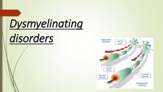
  • 4. Dys-myelination refers to the abnormal development or destruction of the myelin sheath, a fatty layer that surrounds and insulates nerve fibers in the brain and spinal cord. DEFINATION
  • 5. Cognitivedefecits an early presentation-hereditary diffuse leukoencephalopathy with axonal spheroids (HDLS) . Prominent features behavioural changes, mood changes and loss of realistic assessments of daily life experiences Bulbar symptoms at onset –Alexander disease Extrapyramidal signs and symptoms - dystonia and/or dyskinesias , are less frequent but might be a predominant manifestation in certain disorders, including progressive leukoencephalopathy with ovarian failure, various HLDs and AxD. Seizures – MLD , krabbes ,Alexander disease
  • 6. Adult Leukodystrophies: A Step by- Step Diagnostic Approach Based on imaging
  • 7. Step 1 Identify Symmetric White Matter Involvement  Symmetric white matter involvement at MRI is a typical finding in patients with leukodystrophies. (a) Axial T2-weighted MR image in a 23-year-old woman shows symmetric white matter involvement. (b) Axial FLAIR MR image shows an asymmetric pattern in a 46-year-old woman shows asymmetric white matter involvement
  • 8. Step 2: Look for a White Matter Involvement Pattern
  • 9. X- linked Adrenoleukodystrophy  Inherited disorder of peroxisomal metabolism.  Altered peroxisome metabolism in ALD results from absent or deficient acyl CoA synthetase leading to impaired β-oxidation of very-long-chain fatty acids (VLCFAs)  ABCD1 gene mutation
  • 10. Imaging  MRI : White matter abnormalities are usually first seen in occipital regions, with early involvement of the splenium of the corpus callosum and posterior limbs of the internal capsule.  The changes then progress to involve more anterior regions.  In cerebral ALD, contrast enhancement at the periphery of the signal abnormalities is said to be characteristic
  • 11.
  • 12.
  • 13.
  • 14.
  • 15. Axial T2-weighted MR image shows parieto-occipital symmetric white matter hyperintensity. (b) Contrast-enhanced axial T1-weighted MR image in the shows enhancement foci in the white (c) Sagittal T2-weighted MR image shows thoracic cord atrophy.
  • 16. The Loes Scoring System.  The Loes scoring system is universally applied to quantify the extent of cerebral involvement.  Regions of the brain (such as parietooccipital white matter, anterior temporal white matter, visual pathway, corpus callosum, auditory pathway, basal ganglia, projection fibers, and cerebellum) are subdivided and scored based on the extent of disease .  With a score of 0 if normal, 0.5 if unilateral involvement is present, and 1 if the lesion or atrophy is bilateral.  Global atrophy is also assessed.  A normal MRI scan has a score of 0, and the maximum severity score is 34.
  • 17. Abbreviation: MRI, magnetic resonance imaging. Each region is given a score of 0 for normal, 0.5 for unilateral involvement, and 1 for bilateral involvement or atrophy. The maximum score is 34. Parieto-occipital white matter (maximum 4) Basal ganglia (maximum 1) Anterior temporal white matter (maximum 4) •Frontal white matter (maximum 4) •Periventricular •Central •Subcortical •Local atrophy •Visual pathway (maximum 4)Optic radiation •Meyer’s loop •Lateral geniculate body •Optic tract •Corpus callosum (maximum 5) •Splenium •Genu •Body •Splenium atrophy •Genu atrophy •Auditory pathway (maximum 4)Medial geniculate body •Brachium of inferior colliculus •Lateral lemniscus •Pons •Global atrophy (maximum 4)Mild •Moderate •Severe •Brainstem •Cerebellum (maximum 2)White matter •Atrophy •Projection fibers (maximum 2)Internal capsule •Brain stem MRI severity scale scoring (Loes et al 5 )
  • 18.
  • 19. Metachromatic Leukodystrophy  AR lysosomal storage disorder  An enzyme deficiency of arylsulfatase A, which is encoded on the ARSA gene on chromosome 22 q/or pathogenic mutations in PSAP (which encodes prosaposin)  Sulfatides accumulate in the central and peripheral nervous systems, kidneys, testes, and visceral organs (gall bladder).  MLD is an adulthood leukodystrophy that is frequently misdiagnosed as early- onset dementia and/or a schizophrenic disorder, as the neurological symptoms can occur late in the disease course.
  • 20. A high index of suspicion for metachromatic leukodystrophy must exist for patients presenting with a peripheral neuropathy or gall bladder abnormality in the absence of other neurologic features. The MRI may not reveal pathology early in the disease course; therefore, a normal MRI is not sufficient to exclude metachromatic leukodystrophy.
  • 21.
  • 23.  A low arylsulfatase A level (less than 10% of normal values) is detected in white blood cells or cultured fibroblasts, confirmed by the detection of elevated sulfatides in the urine and ARSA sequencing for mutations.  Phase 1/II trial of intrathecal aryl sulfatase A .  Juvenile MLD – autologous HSCT .  Genetherapy under trial .
  • 24.
  • 25. Leukoencephalopathy with Axonal Spheroids and Pigmented Glia.— • Autosomal dominant • Colony-stimulating factor 1 receptor (CSF1R) gene mutation. • The gene defect affects the tyrosine kinase domain of macrophage colony-stimulating factor 1 receptor. • Unlike other leukodystrophies, this disease manifests exclusively in adults(20-50). • P/W Behavioral changes, dementia, motor impairment.
  • 26. (a) Luxol fast blue stain for myelin shows myelin loss and tissue vacuolation with axonal spheroids [arrows in (a) and (b)], that are immunopositive for amyloid precursor protein (b). Affected white matter has many lipid-laden macrophages immunohistochemistry-positive for HLA-DR (c)
  • 27.
  • 28. (a) Case 1 (MRI performed 1.2 years after start of symptoms); localized white matter lesions (arrow) in both frontal and parietal hemispheres involving the corpus callosum (arrow dashed). (b) Case 2 (MRI performed 1.9 years after start of symptoms); confluent white matter lesions in both frontal and parietal hemispheres with cortical atrophy in the affected areas. (c) Case 3 (MRI performed 3.5 years after start of symptoms); localized periventricular lesions (arrow) with corresponding frontoparietal atrophy and involvement of the corpus callosum (arrow dashed). (d) Case 4 (MRI performed 2.5 years after start of symptoms); bilateral frontoparietal white matter changes (arrow) extending into the corpus callosum (arrow dashed).
  • 29. Axial T2-weighted MRI with increased signal intensity in the frontoparietal white matter sparing arcuate U fibres (A) and in the corticospinal tracts (B, arrows). Sagittal proton density imaging with atrophy and increased signal intensity in the posterior body of the corpus callosum (C, arrow). Restricted diffusion on diffusion weighted imaging (D, arrows) that is dark on apparent diffusion coefficient mapping in the areas of T2 signal abnormality (E, arrows). Haematoxylin–eosin staining (F) demonstrates unpigmented macrophages and large neuroaxonal spheroids (arrows).
  • 30. leukoencephalopathy with axonal spheroids and pigmented glia in a 43-year-old woman. (a) Axial FLAIR MR image shows symmetric white matter involvement with diffuse hyperintensity and cysts (arrow). (b) Axial diffusion-weighted MR image shows areas of restricted diffusion, which was confirmed on the apparent diffusion coefficient map (not shown). (c) Nonenhanced CT image shows bilateral deep and periventricular cerebral calcification
  • 31. CT images in 40 year old male (A-C) show multiple symmetric small-sized discrete calcifications in the bilateral frontal and parietal subcortical and periventricular white matter.
  • 32. Small bilateral calcifications in the frontal white matter adjacent to the anterior horns of the lateral ventricles on an axial CT image (arrows). B, Sagittal view represents the symmetric and characteristic stepping stone appearance (arrows). C, Case 4. Small bilateral calcifications in the parietal subcortical white matter on an axial CT image (arrows), but there are no calcifications in the frontal area. D, Sagittal CT image displays subtle calcifications in the anterior pericallosal region bilaterally (arrows)
  • 33.
  • 34. Globoid Cell Leukodystrophy (Krabbe Disease)  Globoid cell leukodystrophy, or Krabbe disease, is an autosomal recessive lysosomal storage disorder caused by mutations in GALC on chromosome 14q31.  GALC encodes the enzyme galactosylceramidase, which is essential in the degradation of lipids (galactosylceramide and psychosine) during myelin turnover.  Adult form (10% of cases)  Presents with pyramidal tract dysfunction and spastic paraparesis.  Can also develop cognitive decline, seizures & cortical blindness.  20% of patients abnormal NCS (slowing of conduction velocity)
  • 35. Krabbe disease in a 48-year-oldman. (a) Axial FLAIR image shows bilateralparieto-occipital white matter involvement,extending to the splenium of the corpus callosum.(b) Coronal T2-weighted MR image also shows a posterior white matter involvement pattern extending to the corticospinal tracts bilaterally(arrows).
  • 36.
  • 37.
  • 38.
  • 39.
  • 40. Sjögren-Larsson Syndrome Sjögren-Larsson syndrome is a rare autosomal recessive disorder Sjögren-Larsson syndrome is caused by inactivating mutations in the aldehyde dehydrogenase 3 family member A2 gene (ALDH3A2), which encodes for fatty aldehyde dehydrogenase (FALDH) and results in abnormal metabolism of long-chain aliphatic aldehydes and alcohols.
  • 41. Magnetic resonance imaging of the brain in a 4year old child with Sjogren Larsson syndrome demonstrating hyperintense signal changes in the periventricular and deep white matter on FLAIR axial view (A and B) and hypointense signal changes on T1weighted axial image
  • 42. Late-phase SjögrenLarsson syndrome in a 27-yearold man. (a) Axial FLAIR MR image shows a periventricular white matter involvement pattern. (b) Proton spectroscopic image of the frontal white matter, obtained with a short echo time, shows abnormal peaks at 1.3 ppm (arrow), indicating lipid deposition. Cho = choline, Cr = creatine, mI = myoinositol, NAA = N-acetyl aspartate
  • 43.
  • 44. L-2-hydroxyglutaric aciduria • autosomal recessive inheritance [L2HGDH] gene • Cerebellar ataxia and intellectual decline. • Hearing loss is another possible symptom of the disease. • Increased L2-hydroxyglutaric acid in urine is diagnostic. MRI • shows predominant subcortical white matter involvement, initially focal and evolving to become confluent. • Periventricular white matter is spared. • Increased signal intensity on T2-weighted or FLAIR MR images may be observed in the globus pallidus, and less importantly, in the caudate nucleus and putamen, with symmetric distribution. These same signal intensity abnormalities also may be observed in the dentate nucleus .
  • 45. L-2-hydroxyglutaric aciduria in a 29-year-old woman. (a) Axial FLAIR image shows a subcortical pattern of white matter involvement. (b) Coronal T2-weighted MR image shows the same pattern and bilateral hyperintensities in the dentate nuclei (arrow), which are commonly observed in patients with this disease.
  • 46. Axial T2-weighted MRIs show (A) bilateral symmetrical white matter (WM) hyperintensity (red arrows) in the centripetal pattern involving subcortical and deep WM, with sparing of periventricular WM (white arrows). (B) Hyperintense basal ganglia (white arrows) with more hyperintensity along the outer rim of the putamen (outer rim sign, red arrow). (C) Hyperintense dentate nucleus (white arrows). (D) The fluid-attenuated inversion recovery image shows rarefaction (white arrows).
  • 47. CORONAL FLAIR SHOWS SUBCORTICAL T2/ FLAIR HYPERINTENSITY MRI shows hyperintensity of the basal ganglia with a higher signal intensity of the outer rim of the caudate nucleus (white arrow) and putamen (black arrow); the entire globus pallidus(open arrow) shows high signal intensity. (b) Axial T2- weighted shows a homogeneously affected caudate nucleus (black arrow), putamen (white arrow), and globus pallidus (open arrow) in an advanced stage of disease.
  • 48.
  • 49. Alexander disease Etiology & inheritance:  AD is sporadic leukoencephalopathy of unknown etiology.  GFAP gene  Mutations in the GFAP gene lead to the production of a structurally altered glial fibrillary acidic protein. The altered protein is thought to impair the formation of normal intermediate filaments.  No definitive diagnostic biochemical test for AD & the diagnosis is made by biopsy
  • 50.
  • 51.
  • 52.
  • 53. (a) Sagittal T1-weighted magnetic resonance image showing “tadpole” atrophy of brainstem; i.e., atrophy of medulla oblongata and cervical cord (arrowhead) with preserved basis pontis (arrow). (b) Axial T2 sequence showing marked atrophy of medulla oblongata (arrows)
  • 54. . T2-weighted MR image shows symmetric demyelination in the frontal lobe white matter.The internal and external capsules and parietal white matter are also involved. Alexander disease in a 5-year-old boy with macrocephaly
  • 55.
  • 56. Adult onset autosomal dominant leukodystrophy (ADLD)  ADLD is caused by duplications of the LMNB1 gene on chromosome 5q23, which result in overexpression of lamin B1 protein.  Overexpression of this protein leads to disruption of myelin homeostasis and slowly progressive, non-inflammatory demyelination, predominantly in deep white matter structures and cerebral peduncle, mistaken for chronic progressive MS.
  • 57.  Typically, patients present in 4th or 5th decade with autonomic abnormalities, followed by pyramidal symptoms, ataxia and cognitive deterioration MRI :  Diffuse white matter T2 hyperintensities involving the frontal lobe, parietal lobe and middle cerebellar peduncle.  Atrophy of the brainstem and corpus callosum
  • 58. FLAIR show symmetric hyperintense signals involving the deep white matter of both hemispheres with relative sparing of the periventricular regions (A, arrows) and hyperintensity of cerebellar peduncles and pontine nuclei (B, arrows). T1- weighted images show diffuse spinal cord atrophy (C).
  • 59. A, Frontoparietal changes are less severe in the periventricular region..C, Changes in the middle cerebellar peduncles and in the pontine nuclei.
  • 60. FIGURE 4. T1-weighted spin-echo image (T1W) (A), T2W (B), and cross- sectional (C) MR image T1W on sagittal surface and cross-sectional MRI showed diffuse shriking, with accompanying abnormal signal in white matter of thoracic one to four level spinal cord on T2WI.
  • 61.
  • 62. Cerebrotendinous Xanthomatosis  Autosomal recessive disorder caused by mutations in the CYP27A1 gene, which encodes sterol 27-hydroxylase.  The deficiency of this enzyme results in the accumulation of cholesterol and cholestanol leading to premature arteriosclerosis,neurotoxicity and the formation of xanthomas in tendons, CNS, skin, and other organs.
  • 63.  Adolescence with cataracts  In adulthood - spastic paraparesis, pyramidal tract signs, cerebellar ataxia, bulbar symptoms and peripheral neuropathy  Childhood history of diarrhoea or failure to thrive.
  • 64.  MRI : Non- specific supratentorial atrophy and deep periventricular white matter changes  Sparing of U fibres and corpus callosum are spared.  classic picture - high signal intensity within the cerebellar white matter and low signal intensity in the dentate nucleus on T2
  • 65. MRI brain of 20-year-old female, (a) T2 sagittal section showing mild cerebellar atrophy. (b and c) T2 and FLAIR axial section showing bilaterally symmetrical hyperintensities involving the dentate nuclei and the deep cerebellar white matter (arrow), which appears iso to hypointense on T1 (arrow in d).
  • 66. a) Achilles tendon xanthoma. Magnetic resonance imaging brain showing (b) symmetrical cerebellar atrophy and T2 hyperintensity of dendate nuclei, (c) fluid-attenuated inversion recovery hyperintensities in globus pallidus and white matter, (d) fluid-attenuated inversion recovery hyperintensity in substantia nigra
  • 67.
  • 68.
  • 69. Canavan disease  Etiology & inheritance: Synonym :Spongyform leukodystrophy, van Bogaert-Bertrand disease.  Caused by deficiency of N acetylaspartylase that results in the accumulation of N-acetyl aspartic acid in urine, plasma & brain.  Inheritance is autosomal recessive.
  • 70. Figure 1: (a) MRI of brain, T2 axial image, showing marked symmetrical hyperintensity of cerebral white matter with involvement of the subcortical arcuate fibres; (b) T2-weighted axial section at the level of cerebellum showing hyperintensities in dentate nuclei; (c) Axial section showing extensive symmetrical diffusion restriction in the subcortical white matter; (d) Single voxel MRS from left parietal white matter showing NAA peak with normal creatine and choline peaks CANAVAN
  • 71.
  • 72.
  • 73. Ashwin 1y/M Atrophic brainstem with diffuse white matter hyperintensities
  • 74. Adult polyglucosan body disease  APBD typically presents in the fifth to the sixth decade of life with a combination of upper and lower motor neuron impairment resembling amyotrophic lateral sclerosis,along with cerebellar ataxia or Parkinson disease-like symptoms with extrapyramidal movement disorders.  In some patients, polyneuropathic symptoms with hyporeflexia, distal symmetric sensory loss, muscle atrophy and fasciculations can be prominent, with slowed nerve conduction velocity and denervation potentials on electrophysiological testing.  Cognitive deficits, reflecting white matter involvement, tend to be very mild.
  • 75. The affected gene in APBD, GBE1, encodes a glycogen- branching enzyme (GBE1), dysfunction of which leads to accumulation of polyglucosan bodies in the central and peripheral nerves. Triheptanoin diet( 7 carbon triglyceride ) therapy under trial
  • 76. MRI  Diffuse periventricular white matter changes predominantly the occipital and temporal lobes and the mesencephalon and cerebellum.  Most prominent in the periventricular region, posterior limb of the internal capsule and external capsule .  Later stages - thinning of the corpus callosum diffuse cerebral, cerebellar and spinal cord atrophy
  • 77. Brain MRI (FLAIR) shows Symmetric hyperintense white matter lesions in the (A) periventricular area, (B) optic radiations, (C) mesencephalic white matter, (D) superior cerebellar peduncles, and (E) medulla. No contrast enhancement was noted (not shown).
  • 78. Cervical spine MRI(A) T2 fluid-attenuated inversion recovery and (B) T2-weighted images show marked atrophy of the medulla and cervical spinal cord. (C) T2- weighted images show hyperintensities in the corticospinal tracts and (D) increased signal in the dorsal columns (arrowhead).
  • 79. Differential diagnoses for adulthood leukodystrophies  Inherited vasculopathies with white matter involvement –CADASIL ,CARASIL  Inherited CNS diseases with grey and white matter involvement- fragile x ataxia syndrome ,DRPLA  Inborn errors of metabolism  Other disorders with white matter involvement like Wilson,s Disease  Acquired inflammatory, toxic, and traumatic white-matter predominant central nervous system disorders
  • 80.
  • 81. Assessment and Diagnosis  Initial assessment should focus on the exclusion of common acquired causes and severe small vessel disease(round 1).  If these initial tests are negative and the patient is suspected to have a genetic disorder, then the first line of testing should include white cell enzyme activities, a VLCFA profile(in men), plasma cholestanol and bile alcohols and plasma amino acids, to exclude the classical leukodystrophies which can present in adulthood
  • 82.
  • 83. 1)Hydrocortisone supplementation for Addison syndrome in adrenoleukodystrophy 2) cholecystectomy for gall bladder dysfunction, polyps and to prevent cancer in metachromatic leukodystrophy 3) avoid head trauma and ensure prompt treatment of fever and infection to avoid triggering deterioration Mangement
  • 84.
  • 85. Evolving Therapeutic Approach Multimodal therapy approaches have the highest potential not only of halting but also repairing the complex and multifactorial pathology of leukodystrophies. The CRISPR (clustered regularly interspaced palindromic repeats)-Cas (CRISPR-associated protein) approach is a promising method for precise gene editing,