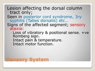
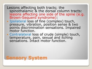
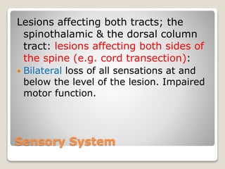
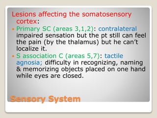
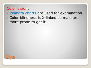
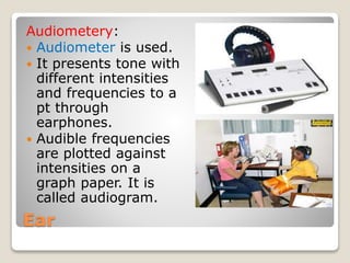
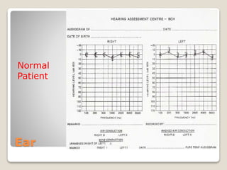
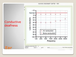
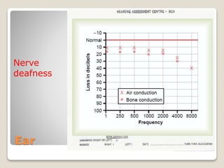
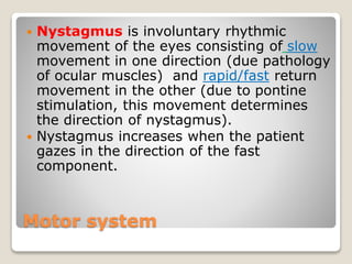
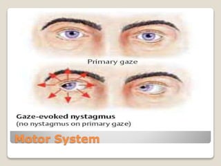
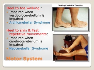
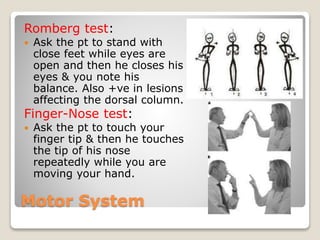
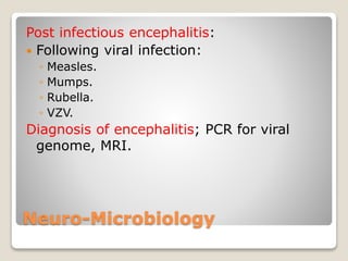
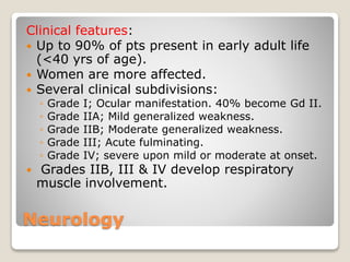
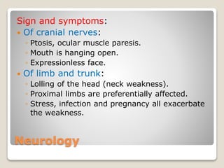
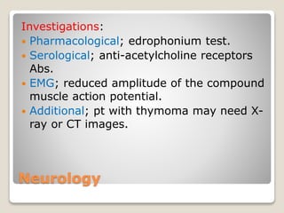
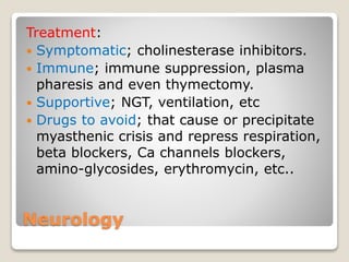
Applied Neurosciences
Done by:
Ahmed Khalafalla Mohammed
Ahmed Abdalla Alamin
Topics
 Sensory System.
 Special senses:
◦ Eye.
◦ Ear.
 Motor System.
 Cranial nerves.
 Nerve conduction
study.
 EEG.
 Neuro-microbiology.
 Imaging.
 Neurology.
 Neuro-surgery.
Sensory System
Lesion affecting the spinothalamic
tract only; Syringomyelia:
 Signs: Dissociated sensory loss of
the affected segment;
◦ Loss of pain & temperature.
◦ Intact touch (fine touch).
◦ Intact motor function.
Sensory System
Lesion affecting the spinothalamic
tract with concomitant motor
impairment; Anterior cord
syndrome:
 Signs of the affected segment;
◦ Loss of pain & temperature.
◦ Intact touch (fine touch).
◦ Loss of motor function.
Sensory System
Lesion affecting the dorsal column
tract only;
Seen in posterior cord syndrome, 3ry
syphilis (Tabes dorsalis) etc…
Signs of the affected segment; sensory
ataxia:
◦ Loss of vibratory & positional sense. +ve
Romberg sign.
◦ Intact pain & temperature.
◦ Intact motor function.
Sensory System
Lesions affecting both tracts; the
spinothalamic & the dorsal column tracts:
lesions affecting one side of the spine (e.g.
Brown-Sequerd syndrome):
 Ipsilateral loss of fine (complex) touch,
pressure, vibration, position sense & two
points discrimination sensations. Impaired
motor function.
 Contralateral loss of crude (simple) touch,
temperature, pain, sexual and itching
sensations. Intact motor function.
Sensory System
Lesions affecting both tracts; the
spinothalamic & the dorsal column
tract: lesions affecting both sides of
the spine (e.g. cord transection):
 Bilateral loss of all sensations at and
below the level of the lesion. Impaired
motor function.
Sensory System
Lesions affecting both tracts; the
spinothalamic & the dorsal column
tracts: lesions affecting the
thalamus:
 One thalamus: contralateral loss of
sensation but the pt still can smell.
 Both thalami: impaired sensation sparing
the ability to smell.
Sensory System
Lesions affecting the somatosensory
cortex:
 Primary SC (areas 3,1,2): contralateral
impaired sensation but the pt still can feel
the pain (by the thalamus) but he can’t
localize it.
 S association C (areas 5,7): tactile
agnosia; difficulty in recognizing, naming
& memorizing objects placed on one hand
while eyes are closed.
Eye
Light reflex:
 Direct on the examined eye.
 Indirect (consensual) on the other one.
 Abnormalities:
◦ Bilaterally: Argyll Robertson pupil; impaired
light reflex with intact accommodation. Caused
by syphilis.
◦ Unilaterally: Holmes-Adie (myotonic) pupil;
impaired light reflex with sluggish
accommodation.
Eye
Eye
Fundoscopy (Ophthalmoscopy):
 For examination of the retina (macula,
optic disc and blood vessels).
 For detection of Papilledema (swelling of
the optic disc) which indicates increased
ICP so lumbar puncture will be
contraindicated.
Eye
Ophthalmoscopes:
Eye
Ophthalmoscopy:
Eye
Retina:
Eye
Visual Acuity:
 Snellen’s charts
are used.
 Normal ratio is
6/6.
Eye
Color vision:
 Ishihara charts are used for examination.
 Color blindness is X-linked so male are
more prone to get it.
Eye
Ishihara charts:
Eye
Visual Field:
 Using no machines; confrontation
method.
 Using special machines; Perimetry.
Eye
Goldmann kinetic Perimeter
Eye
Automated Perimetry:
Ear
 Weber’s test.
 Rinne’s test.
Tunning Fork
Ear
Weber’s test:
 Normally; pt hears
equally on both sides.
 Conductive deafness;
pt hears better on
the affected side (due
to loss of masking
effect of the
environment).
 Nerve deafness; pt
hears better on the
normal side.
Ear
Rinne’s test:
 Normally; air conduction
is better than bone
conduction.
 Conductive deafness; pt
hears bone conduction
but not the air
conduction.
 Nerve deafness; pt does
not hear any sound
whether it is through
bone or air conduction.
Ear
Audiometery:
 Audiometer is used.
 It presents tone with
different intensities
and frequencies to a
pt through
earphones.
 Audible frequencies
are plotted against
intensities on a
graph paper. It is
called audiogram.
Ear
Normal
Patient
Ear
Conductive
deafness
Ear
Nerve
deafness
Ear
Mixed
deafness
Motor System
Examination of deep reflexes:
 Based on the stretch reflex.
 The “Hammer” is used.
 Hyper-reflexia in UMNL.
 Hypo-reflexia in LMNL.
Motor System
 Knee reflex; (L1,2,3 ).
 Biceps reflex; (C5,6).
 Ankle reflex; (S1).
Motor System
Lesions affecting the pyramidal system:
 At the cortex → contralateral flaccid weakness that is
limited to a particular area (e.g. monoparesis).
 At the internal capsule → contralateral paralysis or
weakness (hemiplegia).
 At the brain stem → contralateral paralysis or
weakness (hemiplegia) + ipsilateral cranial nerve palsy.
 At the spinal cord:
◦ Complete transection → quadriplegia (cervical lesion below C4)
or paraplegia (thoracic lesion).
◦ Hemisection → ipsilateral hemiplegia (cervical lesion below C4)
or ipsilateral monoplegia (thoracic lesion).
Motor System
Lesions affecting the extra-pyramidal
system:
 At the basal ganglia.
 At the cerebellum.
Motor System
Disorders of the basal ganglia:
 Hyperkinetic disorders; Tics, Chorea,
Athetosis, Ballismus and Dystonia.
 Hypokinetic disorders; Parkinson’s
disease.
Motor System
Tics:
 Repeated
involuntary
movements.
 Mostly in the face.
Motor System
Chorea:
Dancing movement.
Impaired caudate
nucleas.
Motor System
Athetosis:
Slow writhing, pronounced in hands, fingers
and may be in the neck.
Motor System
Balismus:
Violent movement in
one side of the
body (hemi-
balismaus).
Motor System
Parkinson’s Disease:
Resting tremor.
Slow shuffling gate.
Akinesia.
Bradykinesia.
Motor System
Lesions affecting the cerebellum:
 Cerebellar ataxia:
•Delay in movement initiation.
•Dysmetria: alterations in the rate and force of a
movement.
•Asynergia: decomposition of movement.
•Past Pointing.
•Intention Tremor.
•Dysarthria.
•Dysdiadochokinesis.
•hypotonia.
Motor system
 Nystagmus is involuntary rhythmic
movement of the eyes consisting of slow
movement in one direction (due pathology
of ocular muscles) and rapid/fast return
movement in the other (due to pontine
stimulation, this movement determines
the direction of nystagmus).
 Nystagmus increases when the patient
gazes in the direction of the fast
component.
Motor System
Motor System
Heel to toe walking :
 Impaired when
vestibulocerebellum is
impaired
 Archicerebellar Syndrome
Heel to shin & Fast
repetitive movements:
 Impaired when
cerebrocerebellum is
impaired
 Neocerebellar Syndrome
Motor System
Romberg test:
 Ask the pt to stand with
close feet while eyes are
open and then he closes his
eyes & you note his
balance. Also +ve in lesions
affecting the dorsal column.
Finger-Nose test:
 Ask the pt to touch your
finger tip & then he touches
the tip of his nose
repeatedly while you are
moving your hand.
Cranial Nerves
Olfactory Nerve lesion;
 Anosmia.
Cranial Nerves
Optic nerve:
Right nasal hemianopiaLoss of right eye visionBitemporal heteronymus
hemianopia
Left homonymus
hemianopia
with macular sparing
(NB: the macula has a
duel blood supply.)
Cranial Nerves
Oculomotor nerve:
 Ptosis.
 Able to abduct his
eye.
 According to the
impaired part of the
nerve, ocular motor
deficit occurs.
Cranial Nerve
Trochlear nerve:
 Eye is upward and
medially rotated
due to un opposed
action of the SR
muscle.
 Impaired
abduction.
 Paralyzed SO
muscle.
Cranial nerves
Trigeminal nerve:
 Numb face.
 Atrophied muscles of
mastication.
Cranial Nerves
Abducent nerve:
 Eye is medially
rotated due to
unopposed action of
MR.
 Impaired abduction
of the affected eye.
Cranial Nerves
Facial nerve:
 Upper part of the face receives duel motor
supply from both cerebral hemispheres.
 Lower part of the face receives single motor
supply from the contralateral cerebral
hemisphere.
 UMNL; Contralateral paralysis of the lower
half of the face + Contralateral hemiplegia
(Nb: hemiplegia is ipsilateral to the paralyzed site of the face
& contralateral to the lesion site).
 LMNL at the level of pons; Ipsilateral
paralysis of half of the face (upper & lower) +
Contralateral hemiplegia.
 LMNL at the nerve level; Ipsilateral paralysis
of half of the face only.
Cranial Nerves
 UMNL
 LMNL (e.g. bell’s palsy)
Cranial Nerves
Vestibulo-cochlear nerve:
 Vestibular nerve; vertigo (not as fainting).
 Cochlear nerve; deafness.
Cranial Nerves
Glossopharyngeal and vagus nerves:
 Gag reflex (for both).
 Unilateral vagus lesion; deviated uvula to
the opposite site.
 Bilateral vagus lesion; uvula does not lift.
Cranial Nerves
Accessory nerve:
 Bilaterally; ask the pt to flex his neck and
resist the movement.
 Unilaterally; ask the pt to turn his head to
one side and resist him.
 Ask the to shrug his shoulder.
Cranial Nerve
Hypoglossal nerve:
 Ask the pt to put out his tongue:
◦ UMNL; tongue deviates to the side opposite to
the lesion.
◦ LMNL; tongue deviates to the same side of the
lesion, fasciculation & atrophy.
Cranial Nerves
Test:
Nerve Conduction Studies
 They are:
1. Sensory studies : nerves are studied by
stimulating them at one point and
recording at a distant site along those
nerves.
2. Motor studies : nerves are studied by
stimulating them at two points
(proximal + distal) and recording the
muscle action potentials or stimulating
them at one point & recording them at
two points.
Nerve Conduction Studies
Parameters for nerve studies:
1. Amplitude → reduced due axonal neuropathy
e.g. Diabetic axonal neuropathy.
2. Velocity (can be calculated from both distance
between the cathodes and latency period
between stimulation and wave appearance) →
slow in demyelination neuropathy (e.g.
Guillian-Barré syndrome).
• These parameters are normal in Myasthenia
gravis.
• Both parameters are found defective in severe
cases of local nerve entrapment (e.g. Carpel
tunnel syndrome).
Nerve Conduction Studies
Sensory studies:
1. Amplitude (10 µV)
2. Velocity (70-90 m/s)
Nerve Conduction Studies
Recording electrodes
Stimulating electrodes
Nerve Conduction Studies
Motor studies:
1. Amplitude (2-3 mV).
2. Velocity (50-70 m/s).
Nerve Conduction Studies
Recording electrodes
Stimulating electrode
EEG
The Electro-Encephalo-Gram:
 It is a record of the
electrical activity of the
brain using electrodes
placed on the scalp or
directly on the surface of
the cortex.
 It can be unipolar or bipolar.
EEG
EEG
Epilepsy
 Epilepsy is a chronic brain disorder
characterized by recurrent synchronous
discharges of neuronal groups in the cortex.
 The discharges, which are also called
seizures, may be localized or
generalized.
 Seizures have been classified as partial or
generalized, depending on the spread, and
medication is selected on the basis of the
classification.
EEG
Classification of epilepsy:
 Partial; simple or complex.
 Generalized;
◦ Tonic clonic (grand mal) seizures.
◦ Absence (petit mal) seizures.
◦ Other types; myoclonic, febrile & status
epilepticus.
EEG
EEG
 In generalized, or non-focal, seizures, there is a
massive spread of electrical activity over both
hemispheres.
 Petit mal (absence) seizures, which begin in
childhood, involve a transient loss of consciousness
without a loss of muscle tone, so those patients rarely
fall down.
 In grand mal (tonic-clonic) seizures, the patient
suddenly loses consciousness and falls down.
Convulsions consist of increased muscle tone (tonic)
periods alternating with jerking movements (clonic).
After the convulsions, loss of consciousness may
persist.
 Status epilepticus is a dangerous, uninterrupted
series of seizures that requires urgent intravenous
administration of drugs.
EEG
 Partial (focal) seizures result from localized
discharges that spread from the focus to adjacent
brain areas. The patient may remain conscious, and
the nature of the seizure depends on the area of the
brain affected. Discharges in the motor cortex may
involve first the seemingly purposeful movement of
extremities (fingers), with spread up the arms to the
face, and down to the legs as neuronal groups are
progressively recruited into the discharge.
 Motor seizures are also called jacksonian motor
seizures.
 In simple partial seizures, the patient remains
conscious, but if activity spreads to the other
hemisphere, the patient may lose consciousness and
the seizure is termed complex or psychomotor,
since there may be hallucinatory experiences.
EEG
On EEG
 A focal seizure produces a characteristic EEG
spike, whereas a generalized seizure
produces a series of spikes that are picked
up all over the skull simultaneously. The EEG
also distinguishes between the tonic and
clonic phases of the generalized
discharge.
 During absence seizures, there are
characteristic spike & wave patterns of
discharge.
EEG
EEG
EEG
Treatment; anti-epileptics:
 Partial and grand mal seizures; Na
valproate, phenytoin & carbamazepine.
 For pregnant ladies; lamotrigine
 Absent seizures; ethosuximide.
 Status epilepticus; emergency tx:
lorazepam, diazepam.
Neuro-Microbiology
Medically important microbial diseases of the
Nervous System:
 Meningitis.
 Encephalitis.
 Brain abscess.
 Pott’s disease.
 Botulism.
 Tetanus.
 Poliomyelitis.
 Rabies.
 AIDS opportunistic infections.
 Sleeping sickness.
 Post infection Guillian-Barré syndrome.
Neuro-Microbiology
Meningitis:
 Inflammation of the meninges which it is
characterized by headache, fever, neck
stiffness, photophobia, rash
(meningococcal rash), etc..
 Forms:
◦ Acute meningitis.
◦ Chronic meningitis.
Neuro-Microbiology
Acute meningitis:
 Pyogenic (usually bacterial); which is
generally caused by:
◦ S. pneumoniae, N. menigitidis & H. influenzae
cause meningitis in childern.
◦ E. coli & S. agalactae cause neonatal meningitis.
◦ S. pneumoniae, N. menigitidis & L. monocytogenes
cause meningitis in adults.
 Aseptic (usually viral); caused by
enteroviruses, mumps, HSV-2, varicella-
zoster virus, HIV, Epstein-Barr virus, LCMV,
etc...
Neuro-Microbiology
Chronic meningitis:
 Tuberculous meningitis.
 Meningiovascular syphilis.
 Lyme disease.
 Cryptococcus neoformans.
Neuro-Microbiology
Causative
agent
WBCs Differential RBCs Protein Glucose
Bacterial >200/µl Polymorphs Few High Low
Viral <200/µl Lymphocytes - Slight
high
Normal
Fungal <50/µl Lymphocytes - Not high Low
TB 50-500/µl Lymphocytes - usually Low
Typical changes in the CSF in different types of meningitis
Neuro-Microbiology
Encephalitis:
 Infection of the brain parenchyma.
 Characterized by; headache, fever,
personality change, hallucinations, aphasia,
signs of meningism.
 Caused by:
◦ Bacterial; bartonella, mycoplasma, listeria.
◦ Parasitic; malaria, toxoplasma, sleeping sickness,
naegleria.
◦ Viral (commonest); HSV (most common cause, Mx:
acyclovir), enteroviruses, rabies, paramyxoviruses,
arboviruses, CMV.
Neuro-Microbiology
Post infectious encephalitis:
 Following viral infection:
◦ Measles.
◦ Mumps.
◦ Rubella.
◦ VZV.
Diagnosis of encephalitis; PCR for viral
genome, MRI.
Neuro-Microbiology
CNS abscesses:
 Focal pyogenic infection.
 Exerted effects by:
◦ Brain/spine damage.
◦ Paranchymal compression.
◦ Raised ICP.
◦ Interfering with blood/CSF flow.
 Include:
◦ Brain abscess.
◦ Subdural empyema.
◦ Intracranial epidural abscess.
◦ Spinal epidural abscess.
◦ Spinal cord abscesses.
Neuro-Microbiology
Brain abscess:
 Often polymicrobial (strep., enteric,
anaerobes); S. aureus may causes abscess
associated with IE.
 Less common etiologies; nocardia, fungi, T.
gondii & neurocystocercosis.
 Clinical features; headache, fever, mental
status changes, hemiparesis, papilledema.
 Diagnosis; neuroimaging, lumbar puncture is
contraindicted.
 Treatment; surgico-medical, prophlyctic
antiepileptic agents, measures to lower ICP.
Neuro-Microbiology
Ring-shaped enhancement following
contrast administration on CT (Abscess):
Neuro-Microbiology
Pott’s disease:
 Often preceded by pulmonary TB. It
attacks the vertebral bodies of the spinal
column making kyphosis.
 Charachterized by; signs and symptoms
of TB (e.g. night sweating), back pain,
weakness.
 Diagnosis; imaging of the chest for former
TB infection, spinal MRI, PCR, ELISA,
culture.
 Treatment; TB regimen.
Neuro-Microbiology
Guillian-Barré syndrome:
 Immuno-mediated acute inflammatory
demyelinating polyneuropathy. Characterized by
progressive weakness & areflexia.
 Differential diagnosis can be mixed with polio
(acute flaccid paralysis) but the incidence of polio
is low.
 Associated infections:
◦ Viral; inflenza, EBV, HSV, CMV, HIV, WestNV.
◦ Bacterial; C. jejuni, Mycoplasma.
◦ Parasitic; malaria (P. falciparum).
◦ Vaccination; inflenza (swine).
 Diagnosis; typical clinical features, high CSF
protein, recovering from febrile illness, immuno-
diagnosis.
 Treatment; plasmapharesis, immunoglobulins.
Neuro-Microbiology
AIDS opportunistic infections:
 Many causative agents but mainly:
◦ Toxoplasmosis; cause focal calcified or ring
enhancing masses lesions with focal
neurological symptoms.
◦ CMV; cause encephalitis, myelitis, treated with
ganciclovir.
◦ Cryptococcal meningitis.
◦ JC virus; cause progressive multifocal
leucoencephalopathy.
Imaging
Imaging modalities of the CNS:
 Spiral Computed Tomography scanning (CT)
for brain imaging mainly (it is the primary
modality). It can be used in spinal imaging in
certain cases & when MRI is contraindicated.
 Magnetic Resonance Imaging (MRI) for brain
and spinal imaging.
 Ultrasonograghy is used for infants by placing
the probe on the fontanels before they close.
 X rays are used also.
Imaging
CT MRI
 Quicker.
 CT scan is superior in
head trauma.
 CT measures densities.
 Bright = Hyperdense.
 Black = Hypodense.
 Bone is hyperdense.
 CSF & water are
hypodense.
 Axial (horizontal) images
only.
 Slow.
 MRI is superior in
detecting strokes.
 MRI measures signal
intensities.
 Bright = hyperintense
 Black = hypointense.
 Bone is hypointense.
 CSF & water are
hyperintense in T2 &
hypointense in T1.
 Multi-axial images.
Imaging
Type of
hemorrhage
Time course Mass effect
Acute ≈ 8 to 72 hrs +++
Early subacute ≈ 3 days to 1 wk +++/++
Late subacute ≈ 1 wk to months ±
Chronic Months to years ‫ــ‬
Imaging
CT:
 It is primary modality of brain imaging even
though it is not as sensitive as the MRI in
detecting strokes. That is because it very quick &
shows blood & other secondary causes of
strokes.
 With or without contrast.
 Bone and calcifications are hyperdense.
 Acute hemorrhage is hyperdense.
 Early sub-acute hemorrhage is hyperdense.
 Late sub-acute hemorrhage is isodense to brain.
 Chronic hemorrhage is hypodense.
 Infarcts are hypodense.
Imaging
Spiral CT machine
Imaging CT images showing
base of the skull
Imaging CT images showing
pons & cerebellum
Imaging CT images showing
the midbrain
Imaging CT images showing
Lateral ventricles
Imaging CT with contrast images
showing circle of Willis
Imaging
MRI
 Two major types of images.
◦ T1-wighted images.
◦ T2-wighted images.
Imaging
T1W images T2W images
 With or without contrast
(gadolinium)
 CSF & water are
hypointense.
 Bone and calcifications are
hypointense.
 Acute hemmorrhage is
iso/hypo-intense.
 Subacute hemmorrhage is
hyperintense.
 Chronic hemmorrhage is
hypointense.
 Infarcts are hypointense in
general.
 Without contrast.
 CSF & water are
hyperintense.
 Bone and calcifications are
hypointense.
 Acute hemmorrhage is
hypointense.
 Early subacute
hemmorrhage is
hypointense. Late SH is
hyperintense + low signal.
 Chronic hemmorrhage is
hypointense.
 Infarcts are hyperintense in
general.
Imaging
T2-weighted imageT1-weighted image
Imaging
Neurology
 Stroke.
 Myasthenia Gravis.
Neurology
Stroke
 Two types:
1. Ischemic.
2. Hemorrhagic.
 They are not distinguishable clinically but
there are pointers;
◦ Hemorrhage (meningism, severe headache &
coma) .
◦ Ischemia (carotid bruit, Atrial fibrillation, past
TIA, IHD).
Neurology
Causes of cerebrovascular diseases:
 Infarction – 71 - 85%
 Hemorrhage (intracerebral) – 10 - 26%
 Others (CST, SAH) – 2.9 - 5%
Neurology
Risk factors for Stroke:
 Hypertension; remains the main
modifiable risk factor for stroke.
 Diabetes Mellitus.
 Hypercholesterolemia, IHD.
 Sickle cell disease.
 Smoking
 Physical inactivity, Obesity & alcohol.
 TIA.
 Others.
Neurology
On Examination:
 Atrial fibrillation (AF) - emboli.
 Heart valve disease – cardio-embolic
stroke.
 Carotid bruits are not predictive of
severity of carotid stenosis.
 Cerebral bruits may suggest arterio-
venous malformation.
Neurology
Transient ischemic attack (TIA or Mini
Stroke):
 TIA is a very small stroke that is caused by a
temporary blocked blood vessel and leaves no
permanent brain damage.
 A TIA is a warning that there is something seriously
wrong with the blood flow to the brain and that the
individual is at risk of having a full-blown stroke.
 Most symptoms of a TIA disappear within an hour,
although they may persist for up to 24 hours.
 About one-third of those who have a TIA will have an
acute stroke sometime in the future. 5-10 times risk
of subsequent stroke.
 As many as 20% may sustain a small infarct
visible on CT.
 Only 15% of strokes are preceded by TIAs.
Neurology
Neurology
ACA territory
infarct on CT
Neurology
ACA territory
infarct on MRI
Neurology
MCA territory
infarct on CT
Neurology
MCA territory infarct
With occluded MCA on CT
Neurology
MCA territory
infarct on MR
Neurology
PCA territory
infarct on CT
Neurology
PCA territory
infarct on MRI
Neurology Young infarct on MR
Neurology
Intra-cerebral
hemorrhage
Note the fluid/fluid level
between CSF (up) & blood
(below) by the effect of gravity
Neurology
Subarachnoid hemorrhage:
 It is spontaneous bleeding into the subarachnoid
space.
 Caused by; rupture of aneurysms, arterio-venous
malformations, etc..
 Symptoms & signs:
◦ Sudden acute ‘worst ever’ headache which is often occipital.
◦ Neck stiffness due to meningeal irritation that should
differentiated from meningitis.
◦ Vomiting, collapse, seizures and coma may follow.
 Diagnosis; finding blood stained CSF in lumbar
puncture & on CT.
 Management; supportive, surgery, etc..
 Complications; rebleeding, ischemia, hydrocephalus,
hyponatermia.
Neurology
CT image shows
Subarachnoid
hemorrhage
NB: The presence of CSF/blood
mixture makes CSF hyperdense
Neurology
Myasthenia Gravis:
 It is a disorder of the neuromuscular
transmission characterized by variable
muscle weakness (hypotonic) and
fatiguing that is relieved by rest.
 Caused by; an organ-specific autoimmune
reaction with antibodies directed against
acetylcholine receptors (nicotinic
receptors).
Neurology
Clinical features:
 Up to 90% of pts present in early adult life
(<40 yrs of age).
 Women are more affected.
 Several clinical subdivisions:
◦ Grade I; Ocular manifestation. 40% become Gd II.
◦ Grade IIA; Mild generalized weakness.
◦ Grade IIB; Moderate generalized weakness.
◦ Grade III; Acute fulminating.
◦ Grade IV; severe upon mild or moderate at onset.
 Grades IIB, III & IV develop respiratory
muscle involvement.
Neurology
Sign and symptoms:
 Of cranial nerves:
◦ Ptosis, ocular muscle paresis.
◦ Mouth is hanging open.
◦ Expressionless face.
 Of limb and trunk:
◦ Lolling of the head (neck weakness).
◦ Proximal limbs are preferentially affected.
◦ Stress, infection and pregnancy all exacerbate
the weakness.
Neurology
Investigations:
 Pharmacological; edrophonium test.
 Serological; anti-acetylcholine receptors
Abs.
 EMG; reduced amplitude of the compound
muscle action potential.
 Additional; pt with thymoma may need X-
ray or CT images.
Neurology
Treatment:
 Symptomatic; cholinesterase inhibitors.
 Immune; immune suppression, plasma
pharesis and even thymectomy.
 Supportive; NGT, ventilation, etc
 Drugs to avoid; that cause or precipitate
myasthenic crisis and repress respiration,
beta blockers, Ca channels blockers,
amino-glycosides, erythromycin, etc..
Neurology
Other forms of Myasthenias:
 Neonatal myasthenia:
◦ Born to mothers with MG.
◦ Due to autoimmune Abs (IgG) attacking NMJ.
◦ Starts within 48 hrs & ends 2-12 wks.
 Congenital myasthenia:
◦ Non-autoimmune.
◦ Due to structural abnormalities of the receptors
themselves caused by gene mutations.
◦ Does not respond well to MG treatment.
Neuro-Surgery
 Head injury.
 Raised intracranial pressure.
 Hydrocephalus.
Neuro-Surgery
Head injury:
 Brain damage may occur from primary
injury or the secondary effects of that
injury.
Neuro-Surgery
Primary brain damage:
 Cortical contusion and lacerations; occur
under the site of the injury or opposite
(contre-coup)it.
 Diffuse white matter lesions; as a result of
shearing following deceleration and
damaging the axons.
Neuro-Surgery
Secondary brain damage:
 Hemorrhage:
◦ Epidural (extradural) hematoma.
◦ Subdural hemorrhage.
 Brain swelling (edema).
 Brain shift & tentorial/tonsillar herniation.
 Ischemia.
 Infection.
Neuro-Surgery
Examination:
 Lacerations and bruising; indicate the
presence of fractures.
 Basal skull fracture signs.
 Conscious level; Glasgow coma scale.
 Pupil reflex; tests optic & oculomotor nerves
but the latter is important regarding
herniations & expanding masses.
 Limb weakness; can be contralateral &
ipsilateral (due to indentation of the
contralateral cerebral peduncle by the edge
of the tentorium cerebelli).
 Eye movement; prognostic guide.
Neuro-Surgery
Basal skull fracture signs:
 Anterior fossa fracture:
◦ CSF rhinorrhoea (nasal CSF discharge).
◦ Bilateral periorbital hematoma.
◦ Subconjuncival hemorrhage.
 Petrious fracture:
◦ Bleeding from external meatus.
◦ CSF otorrhoea (CSF from external meatus).
◦ Battle’s sign; bruising over the mastoid.
Neuro-Surgery
Epidural hematoma:
 It often occurs due to a fractured temporal or
parietal bones lacerating the middle
meningeal artery & vein. It can be also due
to any tear in dural venous sinus.
 Signs and symptoms; due raised ICP causing
deteriorating consciousness level,
hypertension+bradycardia, focal neurological
signs (e.g. hemiparesis).
 Diagnosis; lens shaped collection on CT,
lumbar puncture is contraindicated.
 Management; ‘horse shoe’ craniotomy flap,
burr hole, ligation of the bleeding vessel,
measures to lower ICP.
Neuro-Surgery
Epidural
hematoma on CT
Neuro-Surgery
Epidural
hematoma on MRI
Neuro-Surgery
Subdural hematoma:
 There may be history of trauma or not
because it may be forgotten because it was
so minor or so long ago.
 The elderly are most susceptible, as brain
atrophy makes bridging veins vulnerable.
 Signs & symptoms; headache, personality
change, physical & intellectual slowing,
seizures, neurological symptoms, etc…
 Diagnosis; finding crescent-shaped collection
over hemisphere on CT.
 Management; irrigation/evacuation, via burr
twist drill and burr hole craniostomy.
Neuro-Surgery
Subdural
hematoma on CT
Neuro-Surgery
Subdural
hematoma on MRI
Neuro-Surgery
Raised Intracranial pressure:
 MONRO KELLIE DOCTRINE (An increase in
one constituent or expanding mass within
the skull results in an increase in
intracranial pressure).
 Causes:
◦ Expanding mass.
◦ Increase in brain water content.
◦ Increase in cerebral blood volume.
◦ Increase in CSF.
Neuro-Surgery
 Features:
◦ Headache.
◦ Vomiting.
◦ Papilloedema.
 Complications; brain shifts:
◦ Tentorial herniation.
◦ Subfalcine herniation.
◦ Tonsilar herniation.
 Investigations; CT, MRI, etc...
Neuro-Surgery
Treatment:
 Rest
 Position
 Sedation
 Steroids
 Hyper-osmotic solutions
 Hyperventilation
 Surgery, CSF drainage.
Neuro-Surgery
Hydrocephalus:
 Classification:
◦ Obstructive; CSF flow obstruction within the
ventricular system.
◦ Communicating; CSF flow obstruction out/with
ventricular system (with subarachnoid
communication).
 Signs & symptoms; mainly due to raised ICP.
 Investigations; CT, ultrasound, skull X-ray,
isotope cisternography, etc…
 Management; drainage, shunts.

Applied neurosciences

  • 1.
    Applied Neurosciences Done by: AhmedKhalafalla Mohammed Ahmed Abdalla Alamin
  • 2.
    Topics  Sensory System. Special senses: ◦ Eye. ◦ Ear.  Motor System.  Cranial nerves.  Nerve conduction study.  EEG.  Neuro-microbiology.  Imaging.  Neurology.  Neuro-surgery.
  • 3.
    Sensory System Lesion affectingthe spinothalamic tract only; Syringomyelia:  Signs: Dissociated sensory loss of the affected segment; ◦ Loss of pain & temperature. ◦ Intact touch (fine touch). ◦ Intact motor function.
  • 4.
    Sensory System Lesion affectingthe spinothalamic tract with concomitant motor impairment; Anterior cord syndrome:  Signs of the affected segment; ◦ Loss of pain & temperature. ◦ Intact touch (fine touch). ◦ Loss of motor function.
  • 5.
    Sensory System Lesion affectingthe dorsal column tract only; Seen in posterior cord syndrome, 3ry syphilis (Tabes dorsalis) etc… Signs of the affected segment; sensory ataxia: ◦ Loss of vibratory & positional sense. +ve Romberg sign. ◦ Intact pain & temperature. ◦ Intact motor function.
  • 6.
    Sensory System Lesions affectingboth tracts; the spinothalamic & the dorsal column tracts: lesions affecting one side of the spine (e.g. Brown-Sequerd syndrome):  Ipsilateral loss of fine (complex) touch, pressure, vibration, position sense & two points discrimination sensations. Impaired motor function.  Contralateral loss of crude (simple) touch, temperature, pain, sexual and itching sensations. Intact motor function.
  • 7.
    Sensory System Lesions affectingboth tracts; the spinothalamic & the dorsal column tract: lesions affecting both sides of the spine (e.g. cord transection):  Bilateral loss of all sensations at and below the level of the lesion. Impaired motor function.
  • 8.
    Sensory System Lesions affectingboth tracts; the spinothalamic & the dorsal column tracts: lesions affecting the thalamus:  One thalamus: contralateral loss of sensation but the pt still can smell.  Both thalami: impaired sensation sparing the ability to smell.
  • 9.
    Sensory System Lesions affectingthe somatosensory cortex:  Primary SC (areas 3,1,2): contralateral impaired sensation but the pt still can feel the pain (by the thalamus) but he can’t localize it.  S association C (areas 5,7): tactile agnosia; difficulty in recognizing, naming & memorizing objects placed on one hand while eyes are closed.
  • 10.
    Eye Light reflex:  Directon the examined eye.  Indirect (consensual) on the other one.  Abnormalities: ◦ Bilaterally: Argyll Robertson pupil; impaired light reflex with intact accommodation. Caused by syphilis. ◦ Unilaterally: Holmes-Adie (myotonic) pupil; impaired light reflex with sluggish accommodation.
  • 11.
  • 12.
    Eye Fundoscopy (Ophthalmoscopy):  Forexamination of the retina (macula, optic disc and blood vessels).  For detection of Papilledema (swelling of the optic disc) which indicates increased ICP so lumbar puncture will be contraindicated.
  • 13.
  • 14.
  • 15.
  • 16.
    Eye Visual Acuity:  Snellen’scharts are used.  Normal ratio is 6/6.
  • 17.
    Eye Color vision:  Ishiharacharts are used for examination.  Color blindness is X-linked so male are more prone to get it.
  • 18.
  • 19.
    Eye Visual Field:  Usingno machines; confrontation method.  Using special machines; Perimetry.
  • 20.
  • 21.
  • 22.
    Ear  Weber’s test. Rinne’s test. Tunning Fork
  • 23.
    Ear Weber’s test:  Normally;pt hears equally on both sides.  Conductive deafness; pt hears better on the affected side (due to loss of masking effect of the environment).  Nerve deafness; pt hears better on the normal side.
  • 24.
    Ear Rinne’s test:  Normally;air conduction is better than bone conduction.  Conductive deafness; pt hears bone conduction but not the air conduction.  Nerve deafness; pt does not hear any sound whether it is through bone or air conduction.
  • 25.
    Ear Audiometery:  Audiometer isused.  It presents tone with different intensities and frequencies to a pt through earphones.  Audible frequencies are plotted against intensities on a graph paper. It is called audiogram.
  • 26.
  • 27.
  • 28.
  • 29.
  • 30.
    Motor System Examination ofdeep reflexes:  Based on the stretch reflex.  The “Hammer” is used.  Hyper-reflexia in UMNL.  Hypo-reflexia in LMNL.
  • 31.
    Motor System  Kneereflex; (L1,2,3 ).  Biceps reflex; (C5,6).  Ankle reflex; (S1).
  • 32.
    Motor System Lesions affectingthe pyramidal system:  At the cortex → contralateral flaccid weakness that is limited to a particular area (e.g. monoparesis).  At the internal capsule → contralateral paralysis or weakness (hemiplegia).  At the brain stem → contralateral paralysis or weakness (hemiplegia) + ipsilateral cranial nerve palsy.  At the spinal cord: ◦ Complete transection → quadriplegia (cervical lesion below C4) or paraplegia (thoracic lesion). ◦ Hemisection → ipsilateral hemiplegia (cervical lesion below C4) or ipsilateral monoplegia (thoracic lesion).
  • 33.
    Motor System Lesions affectingthe extra-pyramidal system:  At the basal ganglia.  At the cerebellum.
  • 34.
    Motor System Disorders ofthe basal ganglia:  Hyperkinetic disorders; Tics, Chorea, Athetosis, Ballismus and Dystonia.  Hypokinetic disorders; Parkinson’s disease.
  • 35.
  • 36.
  • 37.
    Motor System Athetosis: Slow writhing,pronounced in hands, fingers and may be in the neck.
  • 38.
    Motor System Balismus: Violent movementin one side of the body (hemi- balismaus).
  • 39.
    Motor System Parkinson’s Disease: Restingtremor. Slow shuffling gate. Akinesia. Bradykinesia.
  • 40.
    Motor System Lesions affectingthe cerebellum:  Cerebellar ataxia: •Delay in movement initiation. •Dysmetria: alterations in the rate and force of a movement. •Asynergia: decomposition of movement. •Past Pointing. •Intention Tremor. •Dysarthria. •Dysdiadochokinesis. •hypotonia.
  • 41.
    Motor system  Nystagmusis involuntary rhythmic movement of the eyes consisting of slow movement in one direction (due pathology of ocular muscles) and rapid/fast return movement in the other (due to pontine stimulation, this movement determines the direction of nystagmus).  Nystagmus increases when the patient gazes in the direction of the fast component.
  • 42.
  • 43.
    Motor System Heel totoe walking :  Impaired when vestibulocerebellum is impaired  Archicerebellar Syndrome Heel to shin & Fast repetitive movements:  Impaired when cerebrocerebellum is impaired  Neocerebellar Syndrome
  • 44.
    Motor System Romberg test: Ask the pt to stand with close feet while eyes are open and then he closes his eyes & you note his balance. Also +ve in lesions affecting the dorsal column. Finger-Nose test:  Ask the pt to touch your finger tip & then he touches the tip of his nose repeatedly while you are moving your hand.
  • 45.
    Cranial Nerves Olfactory Nervelesion;  Anosmia.
  • 46.
    Cranial Nerves Optic nerve: Rightnasal hemianopiaLoss of right eye visionBitemporal heteronymus hemianopia Left homonymus hemianopia with macular sparing (NB: the macula has a duel blood supply.)
  • 47.
    Cranial Nerves Oculomotor nerve: Ptosis.  Able to abduct his eye.  According to the impaired part of the nerve, ocular motor deficit occurs.
  • 48.
    Cranial Nerve Trochlear nerve: Eye is upward and medially rotated due to un opposed action of the SR muscle.  Impaired abduction.  Paralyzed SO muscle.
  • 49.
    Cranial nerves Trigeminal nerve: Numb face.  Atrophied muscles of mastication.
  • 50.
    Cranial Nerves Abducent nerve: Eye is medially rotated due to unopposed action of MR.  Impaired abduction of the affected eye.
  • 51.
    Cranial Nerves Facial nerve: Upper part of the face receives duel motor supply from both cerebral hemispheres.  Lower part of the face receives single motor supply from the contralateral cerebral hemisphere.  UMNL; Contralateral paralysis of the lower half of the face + Contralateral hemiplegia (Nb: hemiplegia is ipsilateral to the paralyzed site of the face & contralateral to the lesion site).  LMNL at the level of pons; Ipsilateral paralysis of half of the face (upper & lower) + Contralateral hemiplegia.  LMNL at the nerve level; Ipsilateral paralysis of half of the face only.
  • 52.
    Cranial Nerves  UMNL LMNL (e.g. bell’s palsy)
  • 53.
    Cranial Nerves Vestibulo-cochlear nerve: Vestibular nerve; vertigo (not as fainting).  Cochlear nerve; deafness.
  • 54.
    Cranial Nerves Glossopharyngeal andvagus nerves:  Gag reflex (for both).  Unilateral vagus lesion; deviated uvula to the opposite site.  Bilateral vagus lesion; uvula does not lift.
  • 55.
    Cranial Nerves Accessory nerve: Bilaterally; ask the pt to flex his neck and resist the movement.  Unilaterally; ask the pt to turn his head to one side and resist him.  Ask the to shrug his shoulder.
  • 56.
    Cranial Nerve Hypoglossal nerve: Ask the pt to put out his tongue: ◦ UMNL; tongue deviates to the side opposite to the lesion. ◦ LMNL; tongue deviates to the same side of the lesion, fasciculation & atrophy.
  • 57.
  • 58.
    Nerve Conduction Studies They are: 1. Sensory studies : nerves are studied by stimulating them at one point and recording at a distant site along those nerves. 2. Motor studies : nerves are studied by stimulating them at two points (proximal + distal) and recording the muscle action potentials or stimulating them at one point & recording them at two points.
  • 59.
    Nerve Conduction Studies Parametersfor nerve studies: 1. Amplitude → reduced due axonal neuropathy e.g. Diabetic axonal neuropathy. 2. Velocity (can be calculated from both distance between the cathodes and latency period between stimulation and wave appearance) → slow in demyelination neuropathy (e.g. Guillian-Barré syndrome). • These parameters are normal in Myasthenia gravis. • Both parameters are found defective in severe cases of local nerve entrapment (e.g. Carpel tunnel syndrome).
  • 60.
    Nerve Conduction Studies Sensorystudies: 1. Amplitude (10 µV) 2. Velocity (70-90 m/s)
  • 61.
    Nerve Conduction Studies Recordingelectrodes Stimulating electrodes
  • 62.
    Nerve Conduction Studies Motorstudies: 1. Amplitude (2-3 mV). 2. Velocity (50-70 m/s).
  • 63.
    Nerve Conduction Studies Recordingelectrodes Stimulating electrode
  • 64.
    EEG The Electro-Encephalo-Gram:  Itis a record of the electrical activity of the brain using electrodes placed on the scalp or directly on the surface of the cortex.  It can be unipolar or bipolar.
  • 66.
  • 67.
    EEG Epilepsy  Epilepsy isa chronic brain disorder characterized by recurrent synchronous discharges of neuronal groups in the cortex.  The discharges, which are also called seizures, may be localized or generalized.  Seizures have been classified as partial or generalized, depending on the spread, and medication is selected on the basis of the classification.
  • 68.
    EEG Classification of epilepsy: Partial; simple or complex.  Generalized; ◦ Tonic clonic (grand mal) seizures. ◦ Absence (petit mal) seizures. ◦ Other types; myoclonic, febrile & status epilepticus.
  • 69.
  • 70.
    EEG  In generalized,or non-focal, seizures, there is a massive spread of electrical activity over both hemispheres.  Petit mal (absence) seizures, which begin in childhood, involve a transient loss of consciousness without a loss of muscle tone, so those patients rarely fall down.  In grand mal (tonic-clonic) seizures, the patient suddenly loses consciousness and falls down. Convulsions consist of increased muscle tone (tonic) periods alternating with jerking movements (clonic). After the convulsions, loss of consciousness may persist.  Status epilepticus is a dangerous, uninterrupted series of seizures that requires urgent intravenous administration of drugs.
  • 71.
    EEG  Partial (focal)seizures result from localized discharges that spread from the focus to adjacent brain areas. The patient may remain conscious, and the nature of the seizure depends on the area of the brain affected. Discharges in the motor cortex may involve first the seemingly purposeful movement of extremities (fingers), with spread up the arms to the face, and down to the legs as neuronal groups are progressively recruited into the discharge.  Motor seizures are also called jacksonian motor seizures.  In simple partial seizures, the patient remains conscious, but if activity spreads to the other hemisphere, the patient may lose consciousness and the seizure is termed complex or psychomotor, since there may be hallucinatory experiences.
  • 72.
    EEG On EEG  Afocal seizure produces a characteristic EEG spike, whereas a generalized seizure produces a series of spikes that are picked up all over the skull simultaneously. The EEG also distinguishes between the tonic and clonic phases of the generalized discharge.  During absence seizures, there are characteristic spike & wave patterns of discharge.
  • 73.
  • 74.
  • 75.
    EEG Treatment; anti-epileptics:  Partialand grand mal seizures; Na valproate, phenytoin & carbamazepine.  For pregnant ladies; lamotrigine  Absent seizures; ethosuximide.  Status epilepticus; emergency tx: lorazepam, diazepam.
  • 76.
    Neuro-Microbiology Medically important microbialdiseases of the Nervous System:  Meningitis.  Encephalitis.  Brain abscess.  Pott’s disease.  Botulism.  Tetanus.  Poliomyelitis.  Rabies.  AIDS opportunistic infections.  Sleeping sickness.  Post infection Guillian-Barré syndrome.
  • 77.
    Neuro-Microbiology Meningitis:  Inflammation ofthe meninges which it is characterized by headache, fever, neck stiffness, photophobia, rash (meningococcal rash), etc..  Forms: ◦ Acute meningitis. ◦ Chronic meningitis.
  • 78.
    Neuro-Microbiology Acute meningitis:  Pyogenic(usually bacterial); which is generally caused by: ◦ S. pneumoniae, N. menigitidis & H. influenzae cause meningitis in childern. ◦ E. coli & S. agalactae cause neonatal meningitis. ◦ S. pneumoniae, N. menigitidis & L. monocytogenes cause meningitis in adults.  Aseptic (usually viral); caused by enteroviruses, mumps, HSV-2, varicella- zoster virus, HIV, Epstein-Barr virus, LCMV, etc...
  • 79.
    Neuro-Microbiology Chronic meningitis:  Tuberculousmeningitis.  Meningiovascular syphilis.  Lyme disease.  Cryptococcus neoformans.
  • 80.
    Neuro-Microbiology Causative agent WBCs Differential RBCsProtein Glucose Bacterial >200/µl Polymorphs Few High Low Viral <200/µl Lymphocytes - Slight high Normal Fungal <50/µl Lymphocytes - Not high Low TB 50-500/µl Lymphocytes - usually Low Typical changes in the CSF in different types of meningitis
  • 81.
    Neuro-Microbiology Encephalitis:  Infection ofthe brain parenchyma.  Characterized by; headache, fever, personality change, hallucinations, aphasia, signs of meningism.  Caused by: ◦ Bacterial; bartonella, mycoplasma, listeria. ◦ Parasitic; malaria, toxoplasma, sleeping sickness, naegleria. ◦ Viral (commonest); HSV (most common cause, Mx: acyclovir), enteroviruses, rabies, paramyxoviruses, arboviruses, CMV.
  • 82.
    Neuro-Microbiology Post infectious encephalitis: Following viral infection: ◦ Measles. ◦ Mumps. ◦ Rubella. ◦ VZV. Diagnosis of encephalitis; PCR for viral genome, MRI.
  • 83.
    Neuro-Microbiology CNS abscesses:  Focalpyogenic infection.  Exerted effects by: ◦ Brain/spine damage. ◦ Paranchymal compression. ◦ Raised ICP. ◦ Interfering with blood/CSF flow.  Include: ◦ Brain abscess. ◦ Subdural empyema. ◦ Intracranial epidural abscess. ◦ Spinal epidural abscess. ◦ Spinal cord abscesses.
  • 84.
    Neuro-Microbiology Brain abscess:  Oftenpolymicrobial (strep., enteric, anaerobes); S. aureus may causes abscess associated with IE.  Less common etiologies; nocardia, fungi, T. gondii & neurocystocercosis.  Clinical features; headache, fever, mental status changes, hemiparesis, papilledema.  Diagnosis; neuroimaging, lumbar puncture is contraindicted.  Treatment; surgico-medical, prophlyctic antiepileptic agents, measures to lower ICP.
  • 85.
  • 86.
    Neuro-Microbiology Pott’s disease:  Oftenpreceded by pulmonary TB. It attacks the vertebral bodies of the spinal column making kyphosis.  Charachterized by; signs and symptoms of TB (e.g. night sweating), back pain, weakness.  Diagnosis; imaging of the chest for former TB infection, spinal MRI, PCR, ELISA, culture.  Treatment; TB regimen.
  • 87.
    Neuro-Microbiology Guillian-Barré syndrome:  Immuno-mediatedacute inflammatory demyelinating polyneuropathy. Characterized by progressive weakness & areflexia.  Differential diagnosis can be mixed with polio (acute flaccid paralysis) but the incidence of polio is low.  Associated infections: ◦ Viral; inflenza, EBV, HSV, CMV, HIV, WestNV. ◦ Bacterial; C. jejuni, Mycoplasma. ◦ Parasitic; malaria (P. falciparum). ◦ Vaccination; inflenza (swine).  Diagnosis; typical clinical features, high CSF protein, recovering from febrile illness, immuno- diagnosis.  Treatment; plasmapharesis, immunoglobulins.
  • 88.
    Neuro-Microbiology AIDS opportunistic infections: Many causative agents but mainly: ◦ Toxoplasmosis; cause focal calcified or ring enhancing masses lesions with focal neurological symptoms. ◦ CMV; cause encephalitis, myelitis, treated with ganciclovir. ◦ Cryptococcal meningitis. ◦ JC virus; cause progressive multifocal leucoencephalopathy.
  • 89.
    Imaging Imaging modalities ofthe CNS:  Spiral Computed Tomography scanning (CT) for brain imaging mainly (it is the primary modality). It can be used in spinal imaging in certain cases & when MRI is contraindicated.  Magnetic Resonance Imaging (MRI) for brain and spinal imaging.  Ultrasonograghy is used for infants by placing the probe on the fontanels before they close.  X rays are used also.
  • 90.
    Imaging CT MRI  Quicker. CT scan is superior in head trauma.  CT measures densities.  Bright = Hyperdense.  Black = Hypodense.  Bone is hyperdense.  CSF & water are hypodense.  Axial (horizontal) images only.  Slow.  MRI is superior in detecting strokes.  MRI measures signal intensities.  Bright = hyperintense  Black = hypointense.  Bone is hypointense.  CSF & water are hyperintense in T2 & hypointense in T1.  Multi-axial images.
  • 91.
    Imaging Type of hemorrhage Time courseMass effect Acute ≈ 8 to 72 hrs +++ Early subacute ≈ 3 days to 1 wk +++/++ Late subacute ≈ 1 wk to months ± Chronic Months to years ‫ــ‬
  • 92.
    Imaging CT:  It isprimary modality of brain imaging even though it is not as sensitive as the MRI in detecting strokes. That is because it very quick & shows blood & other secondary causes of strokes.  With or without contrast.  Bone and calcifications are hyperdense.  Acute hemorrhage is hyperdense.  Early sub-acute hemorrhage is hyperdense.  Late sub-acute hemorrhage is isodense to brain.  Chronic hemorrhage is hypodense.  Infarcts are hypodense.
  • 93.
  • 94.
    Imaging CT imagesshowing base of the skull
  • 95.
    Imaging CT imagesshowing pons & cerebellum
  • 96.
    Imaging CT imagesshowing the midbrain
  • 97.
    Imaging CT imagesshowing Lateral ventricles
  • 98.
    Imaging CT withcontrast images showing circle of Willis
  • 99.
    Imaging MRI  Two majortypes of images. ◦ T1-wighted images. ◦ T2-wighted images.
  • 100.
    Imaging T1W images T2Wimages  With or without contrast (gadolinium)  CSF & water are hypointense.  Bone and calcifications are hypointense.  Acute hemmorrhage is iso/hypo-intense.  Subacute hemmorrhage is hyperintense.  Chronic hemmorrhage is hypointense.  Infarcts are hypointense in general.  Without contrast.  CSF & water are hyperintense.  Bone and calcifications are hypointense.  Acute hemmorrhage is hypointense.  Early subacute hemmorrhage is hypointense. Late SH is hyperintense + low signal.  Chronic hemmorrhage is hypointense.  Infarcts are hyperintense in general.
  • 101.
  • 102.
  • 103.
  • 104.
    Neurology Stroke  Two types: 1.Ischemic. 2. Hemorrhagic.  They are not distinguishable clinically but there are pointers; ◦ Hemorrhage (meningism, severe headache & coma) . ◦ Ischemia (carotid bruit, Atrial fibrillation, past TIA, IHD).
  • 105.
    Neurology Causes of cerebrovasculardiseases:  Infarction – 71 - 85%  Hemorrhage (intracerebral) – 10 - 26%  Others (CST, SAH) – 2.9 - 5%
  • 106.
    Neurology Risk factors forStroke:  Hypertension; remains the main modifiable risk factor for stroke.  Diabetes Mellitus.  Hypercholesterolemia, IHD.  Sickle cell disease.  Smoking  Physical inactivity, Obesity & alcohol.  TIA.  Others.
  • 107.
    Neurology On Examination:  Atrialfibrillation (AF) - emboli.  Heart valve disease – cardio-embolic stroke.  Carotid bruits are not predictive of severity of carotid stenosis.  Cerebral bruits may suggest arterio- venous malformation.
  • 108.
    Neurology Transient ischemic attack(TIA or Mini Stroke):  TIA is a very small stroke that is caused by a temporary blocked blood vessel and leaves no permanent brain damage.  A TIA is a warning that there is something seriously wrong with the blood flow to the brain and that the individual is at risk of having a full-blown stroke.  Most symptoms of a TIA disappear within an hour, although they may persist for up to 24 hours.  About one-third of those who have a TIA will have an acute stroke sometime in the future. 5-10 times risk of subsequent stroke.  As many as 20% may sustain a small infarct visible on CT.  Only 15% of strokes are preceded by TIAs.
  • 109.
  • 110.
  • 111.
  • 112.
  • 113.
  • 114.
  • 115.
  • 116.
  • 117.
  • 118.
    Neurology Intra-cerebral hemorrhage Note the fluid/fluidlevel between CSF (up) & blood (below) by the effect of gravity
  • 119.
    Neurology Subarachnoid hemorrhage:  Itis spontaneous bleeding into the subarachnoid space.  Caused by; rupture of aneurysms, arterio-venous malformations, etc..  Symptoms & signs: ◦ Sudden acute ‘worst ever’ headache which is often occipital. ◦ Neck stiffness due to meningeal irritation that should differentiated from meningitis. ◦ Vomiting, collapse, seizures and coma may follow.  Diagnosis; finding blood stained CSF in lumbar puncture & on CT.  Management; supportive, surgery, etc..  Complications; rebleeding, ischemia, hydrocephalus, hyponatermia.
  • 120.
    Neurology CT image shows Subarachnoid hemorrhage NB:The presence of CSF/blood mixture makes CSF hyperdense
  • 121.
    Neurology Myasthenia Gravis:  Itis a disorder of the neuromuscular transmission characterized by variable muscle weakness (hypotonic) and fatiguing that is relieved by rest.  Caused by; an organ-specific autoimmune reaction with antibodies directed against acetylcholine receptors (nicotinic receptors).
  • 122.
    Neurology Clinical features:  Upto 90% of pts present in early adult life (<40 yrs of age).  Women are more affected.  Several clinical subdivisions: ◦ Grade I; Ocular manifestation. 40% become Gd II. ◦ Grade IIA; Mild generalized weakness. ◦ Grade IIB; Moderate generalized weakness. ◦ Grade III; Acute fulminating. ◦ Grade IV; severe upon mild or moderate at onset.  Grades IIB, III & IV develop respiratory muscle involvement.
  • 123.
    Neurology Sign and symptoms: Of cranial nerves: ◦ Ptosis, ocular muscle paresis. ◦ Mouth is hanging open. ◦ Expressionless face.  Of limb and trunk: ◦ Lolling of the head (neck weakness). ◦ Proximal limbs are preferentially affected. ◦ Stress, infection and pregnancy all exacerbate the weakness.
  • 124.
    Neurology Investigations:  Pharmacological; edrophoniumtest.  Serological; anti-acetylcholine receptors Abs.  EMG; reduced amplitude of the compound muscle action potential.  Additional; pt with thymoma may need X- ray or CT images.
  • 125.
    Neurology Treatment:  Symptomatic; cholinesteraseinhibitors.  Immune; immune suppression, plasma pharesis and even thymectomy.  Supportive; NGT, ventilation, etc  Drugs to avoid; that cause or precipitate myasthenic crisis and repress respiration, beta blockers, Ca channels blockers, amino-glycosides, erythromycin, etc..
  • 126.
    Neurology Other forms ofMyasthenias:  Neonatal myasthenia: ◦ Born to mothers with MG. ◦ Due to autoimmune Abs (IgG) attacking NMJ. ◦ Starts within 48 hrs & ends 2-12 wks.  Congenital myasthenia: ◦ Non-autoimmune. ◦ Due to structural abnormalities of the receptors themselves caused by gene mutations. ◦ Does not respond well to MG treatment.
  • 127.
    Neuro-Surgery  Head injury. Raised intracranial pressure.  Hydrocephalus.
  • 128.
    Neuro-Surgery Head injury:  Braindamage may occur from primary injury or the secondary effects of that injury.
  • 129.
    Neuro-Surgery Primary brain damage: Cortical contusion and lacerations; occur under the site of the injury or opposite (contre-coup)it.  Diffuse white matter lesions; as a result of shearing following deceleration and damaging the axons.
  • 130.
    Neuro-Surgery Secondary brain damage: Hemorrhage: ◦ Epidural (extradural) hematoma. ◦ Subdural hemorrhage.  Brain swelling (edema).  Brain shift & tentorial/tonsillar herniation.  Ischemia.  Infection.
  • 131.
    Neuro-Surgery Examination:  Lacerations andbruising; indicate the presence of fractures.  Basal skull fracture signs.  Conscious level; Glasgow coma scale.  Pupil reflex; tests optic & oculomotor nerves but the latter is important regarding herniations & expanding masses.  Limb weakness; can be contralateral & ipsilateral (due to indentation of the contralateral cerebral peduncle by the edge of the tentorium cerebelli).  Eye movement; prognostic guide.
  • 132.
    Neuro-Surgery Basal skull fracturesigns:  Anterior fossa fracture: ◦ CSF rhinorrhoea (nasal CSF discharge). ◦ Bilateral periorbital hematoma. ◦ Subconjuncival hemorrhage.  Petrious fracture: ◦ Bleeding from external meatus. ◦ CSF otorrhoea (CSF from external meatus). ◦ Battle’s sign; bruising over the mastoid.
  • 133.
    Neuro-Surgery Epidural hematoma:  Itoften occurs due to a fractured temporal or parietal bones lacerating the middle meningeal artery & vein. It can be also due to any tear in dural venous sinus.  Signs and symptoms; due raised ICP causing deteriorating consciousness level, hypertension+bradycardia, focal neurological signs (e.g. hemiparesis).  Diagnosis; lens shaped collection on CT, lumbar puncture is contraindicated.  Management; ‘horse shoe’ craniotomy flap, burr hole, ligation of the bleeding vessel, measures to lower ICP.
  • 134.
  • 135.
  • 136.
    Neuro-Surgery Subdural hematoma:  Theremay be history of trauma or not because it may be forgotten because it was so minor or so long ago.  The elderly are most susceptible, as brain atrophy makes bridging veins vulnerable.  Signs & symptoms; headache, personality change, physical & intellectual slowing, seizures, neurological symptoms, etc…  Diagnosis; finding crescent-shaped collection over hemisphere on CT.  Management; irrigation/evacuation, via burr twist drill and burr hole craniostomy.
  • 137.
  • 138.
  • 139.
    Neuro-Surgery Raised Intracranial pressure: MONRO KELLIE DOCTRINE (An increase in one constituent or expanding mass within the skull results in an increase in intracranial pressure).  Causes: ◦ Expanding mass. ◦ Increase in brain water content. ◦ Increase in cerebral blood volume. ◦ Increase in CSF.
  • 140.
    Neuro-Surgery  Features: ◦ Headache. ◦Vomiting. ◦ Papilloedema.  Complications; brain shifts: ◦ Tentorial herniation. ◦ Subfalcine herniation. ◦ Tonsilar herniation.  Investigations; CT, MRI, etc...
  • 141.
    Neuro-Surgery Treatment:  Rest  Position Sedation  Steroids  Hyper-osmotic solutions  Hyperventilation  Surgery, CSF drainage.
  • 142.
    Neuro-Surgery Hydrocephalus:  Classification: ◦ Obstructive;CSF flow obstruction within the ventricular system. ◦ Communicating; CSF flow obstruction out/with ventricular system (with subarachnoid communication).  Signs & symptoms; mainly due to raised ICP.  Investigations; CT, ultrasound, skull X-ray, isotope cisternography, etc…  Management; drainage, shunts.