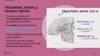
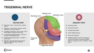
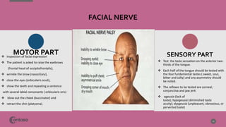
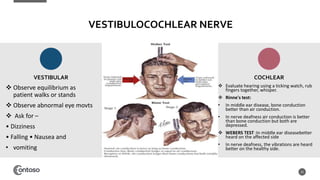
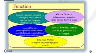
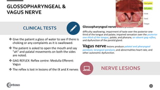
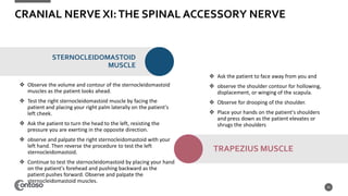
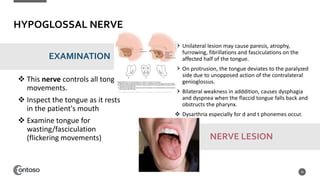
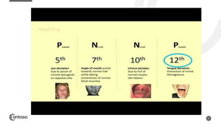
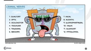
CRANIAL
NERVE
EXAMINATION
DR SAI PRIYANKA NERUSU (PT)
PRACTICAL
WEBINAR
3
CRANIAL NERVES
4
5
PRE EXAMINATION
• Wash hands
• Introduce yourself
• Confirm patient details – name
/ DOB Explain the examination
• Gain consent
• Keep your kit ready
• Remember to check on both
sides
• Ask patient if they have pain
anywhere before you begin!
6
7
OLFACTORY NERVE
 Type : Sensory Nerve
 Function : Detection of Smell
 Ask subject to close the eyes and occlude
untested nostril
 Check each nostril independently
 Don’t use irritating substances as they
stimulate sensory part of trigeminal nerve –
False results
 Don’t touch patient as it give cue when test
object is being presented
 Smell Intact – If patient reports detection of
odor
 Recognition of odor – Involves olfactory
memory which is higher cortical function
 Hyposmia is diminished olfactory acuity.
 Anosmia, the inability to recognize odors, may
be unilateral or bilateral.
 Dysosmia is an abnormal sense of smell.
8
OPTIC NERVE
Specific testing of the optic nerve
involves
• Visual acuity Assessment
• Color perception
• Visual fields Assessment, and
• Pupillary light reflexes Assessment
• Accommodation Assessment and
• Funduscopic Examination
9
OPTIC NERVE
• Examine the ability of brain to
interpret images , gives an idea of
optical integrity of eyes
• Seated 6 meters away from snellers
chart
• 6/5- 6 denotes distance between chart
and subject and 5 denotes the last line
read by subject – person able to read
at 5 mts which can be read by healthy
person at 6m
• 6/6- worse to normal vision
• 6/5 – Normal vision
• CF Vision, HM vision, LP Vision
10
VISUAL ACUITYTEST
• Tested by using Psuedoisochromatic
plates of ishihara
• Assess to understand whether
patient able to differentiaite colors
• Ask patient to identify numbers /
letters in plates
• Color vision more in optic neuritis
and RED RESATURATION – First
feature of optic nerve injury
• RED GREEN Deficiency
11
COLOR PERCEPTIONTEST
CONFRONTATION METHOD
12
VISUAL FIELDTESTS
HOMONYMOUS VISUAL FIELD
DEFECTS
PERIPHERAL VISUAL FIELD DEFECTS
CENTRAL VISUAL FIELD DEFECTS
• Afferent:
1. Optic nerve
2. Proprioceptive fibers from
extraocular muscles
• Centre: Nucleus of Perlia
• Efferent: Oculomotor nerve
• The actions of CN III will result in
convergence of the eyeballs by contracting
the medial recti during accommodation.
The PARA SYMPATHETIC FIBERS result in
thickening of the LENS (via CILIARY MUSCLE
contraction) and pupillary constriction (by
activation of the constrictor pupillae muscles
of the iris) 13
ACCOMMODATION REFLEX
Reflexes
14
PUPILLARY LIGHT REFLEX
DIRECT LIGHT REFLEX
CONSENSUAL LIGHT
REFLEX
Bilateral Activation
of Edinger
westphatl nucleus
• The fundus examination is conducted
to assess the retina, optic disc,
and retinal vessels.
• The examination is performed using
an ophthalmoscope.
• It is a handheld illuminated lens
apparatus that allows the examiner
to view a magnified version of the
retina.
15
FUNDOSCOPY
EXAMINATION
OCCULOMOTOR NERVE
TROCHLEAR NERVE
ABDUCENS NERVE
The third, fourth and sixth cranial
nerves are responsible for
movements of the eyeball and
hence if they are affected singly
or together they cause defective
ocular movements.
16
EXAMINATION
Look for
• Pursuit Movements
• Saccades
• H test
Note the range & movement of eye
balls , look for action of yoke muscles
17
IDENTIFY
THE
CONDITION
18
TRIGEMINAL NERVE (5
CRANIAL NERVE)
• The sensory portion of
the trigeminal supplies touch–pain–
temperature to the face.
• The nerve has three divisions: the
ophthalmic, maxillary, and
mandibular nerves
• The mandibular division carries the
motor portion.
• The motor portion conveys
proprioceptive impulses from the
temporomandibular joint.
19
TRIGEMINAL NERVE
MOTOR PART
 Temporalis muscle : clench teeth + palpate
muscle
 Masseters : clench teeth + palpate muscle,
holding its ant. & post. Borers.
 Pterygoids: Fixed head ; open mouth + open
mouth against resistance to test tone
 In Unilateral pterygoid paralysis : The jaw is
deviated to the diseased side
 In Bilateral pterygoid paralysis : inability to
open mouth
 JAW JERK : Normal/Hypetrclonus/Absent
Afferent: Trigeminal nerve; Efferent :
Trigeminal nerve
SENSORY PART
 Test with cotton
 Test with pin prick
 Compare both sides
 Ask for differentiation
 Check all 3 divisions
 CORNEAL REFLEX: check for blinking of
muscle and look for Consensual Reflex
Afferent: Trigeminal nerve ; Efferent : Facial
Nerve
20
FACIAL NERVE
21
 Test the taste sensation on the anterior two-
thirds of the tongue.
 Each half of the tongue should be tested with
the four fundamental tastes ( sweet, sour,
bitter and salty) and any asymmetry should
be noted.
 The reflexes to be tested are corneal,
conjunctiva and jaw jerk
 ageusia (lack of
taste); hypogeusia (diminished taste
acuity); dysgeusia (unpleasant, obnoxious, or
perverted taste)
MOTOR PART SENSORY PART
 Inspection of facial expression
 The patient is asked to raise the eyebrows
(frontal head of occipitofrontalis),
 wrinkle the brow (nasociliary),
 close the eyes (orbicularis oculi),
 show the teeth and repeating a sentence
with several labial consonants ( orbicularis oris)
 blow out the cheek (buccinator) and
 retract the chin (platysma).
VESTIBULOCOCHLEAR NERVE
 Observe equilibrium as
patient walks or stands
 Observe abnormal eye movts
 Ask for –
• Dizziness
• Falling • Nausea and
• vomiting
22
 Evaluate hearing using a ticking watch, rub
fingers together, whisper.
 Rinne's test:
• In middle ear disease, bone conduction
better than air conduction.
• In nerve deafness air conduction is better
than bone conduction but both are
depressed.
 WEBERS TEST :In middle ear diseasebetter
heard on the affected side
• In nerve deafness, the vibrations are heard
better on the healthy side.
VESTIBULAR COCHLEAR
23
GLOSSOPHARYNGEAL &
VAGUS NERVE
CLINICALTESTS
 Give the patient a glass of water to see if there is
choking or any complaints as it is swallowed.
 The patient is asked to open the mouth and say
"ah" and palatal movements on both the sides
are noted.
 GAG REFLEX: Reflex centre: Medulla Efferent:
Vagus
 The reflex is lost in lesions of the IX and X nerves
Glossopharyngeal nerve lesions produce
difficulty swallowing; impairment of taste over the posterior one-
third of the tongue and palate; impaired sensation over the posterior
one-third of the tongue, palate, and pharynx; an absent gag reflex;
and dysfunction of the parotid gland.
Vagus nervelesions produce palatal and pharyngeal
paralysis; laryngeal paralysis; and abnormalities heart rate; and
other autonomic dysfunction.
NERVE LESIONS
24
CRANIAL NERVE XI:THE SPINAL ACCESSORY NERVE
 Observe the volume and contour of the sternocleidomastoid
muscles as the patient looks ahead.
 Test the right sternocleidomastoid muscle by facing the
patient and placing your right palm laterally on the patient's
left cheek.
 Ask the patient to turn the head to the left, resisting the
pressure you are exerting in the opposite direction.
 observe and palpate the right sternocleidomastoid with your
left hand. Then reverse the procedure to test the left
sternocleidomastoid.
 Continue to test the sternocleidomastoid by placing your hand
on the patient's forehead and pushing backward as the
patient pushes forward. Observe and palpate the
sternocleidomastoid muscles.
25
STERNOCLEIDOMASTOID
MUSCLE
 Ask the patient to face away from you and
 observe the shoulder contour for hollowing,
displacement, or winging of the scapula.
 Observe for drooping of the shoulder.
 Place your hands on the patient's shoulders
and press down as the patient elevates or
shrugs the shoulders
TRAPEZIUS MUSCLE
HYPOGLOSSAL NERVE
 This nerve controls all tongue
movements.
 Inspect the tongue as it rests
in the patient's mouth
 Examine tongue for
wasting/fasciculation
(flickering movements)
26
EXAMINATION
 Unilateral lesion may cause paresis, atrophy,
furrowing, fibrillations and fasciculations on the
affected half of the tongue.
 On protrusion, the tongue deviates to the paralyzed
side due to unopposed action of the contralateral
genioglossus.
 Bilateral weakness in adddition, causes dysphagia
and dyspnea when the flaccid tongue falls back and
obstructs the pharynx.
 Dysarthria especially for d and t phonemes occur.
NERVE LESION
27
28
REFERENCES
29
 PJ Mehta – Practical Book of medicine
 Clinical Methods: The History, Physical, and Laboratory Examinations. 3rd
edition by Walker HK, Hall WD, Hurst JW
 OSCE And Clinical skills handbook: Hurley KF, second edition.Elsevier Canada
2011
 Online osceskills website. www.osceskills.com
 http://geekymedics.com/eye-examination-osce-guide/
 Tim Hall: PACES for the MRCP with 250 cases .Third edition.
 Google images
THANKYOU !
Follow me on
https://instagram.com/physiotherapyposts?igs
hid=OGQ5ZDc2ODk2ZA
nspphysio1879@gmail.com

CRANIAL NERVE EXAMINATION.pptx

  • 1.
  • 2.
  • 3.
  • 4.
  • 5.
  • 6.
    PRE EXAMINATION • Washhands • Introduce yourself • Confirm patient details – name / DOB Explain the examination • Gain consent • Keep your kit ready • Remember to check on both sides • Ask patient if they have pain anywhere before you begin! 6
  • 7.
    7 OLFACTORY NERVE  Type: Sensory Nerve  Function : Detection of Smell  Ask subject to close the eyes and occlude untested nostril  Check each nostril independently  Don’t use irritating substances as they stimulate sensory part of trigeminal nerve – False results  Don’t touch patient as it give cue when test object is being presented  Smell Intact – If patient reports detection of odor  Recognition of odor – Involves olfactory memory which is higher cortical function  Hyposmia is diminished olfactory acuity.  Anosmia, the inability to recognize odors, may be unilateral or bilateral.  Dysosmia is an abnormal sense of smell.
  • 8.
  • 9.
    Specific testing ofthe optic nerve involves • Visual acuity Assessment • Color perception • Visual fields Assessment, and • Pupillary light reflexes Assessment • Accommodation Assessment and • Funduscopic Examination 9 OPTIC NERVE
  • 10.
    • Examine theability of brain to interpret images , gives an idea of optical integrity of eyes • Seated 6 meters away from snellers chart • 6/5- 6 denotes distance between chart and subject and 5 denotes the last line read by subject – person able to read at 5 mts which can be read by healthy person at 6m • 6/6- worse to normal vision • 6/5 – Normal vision • CF Vision, HM vision, LP Vision 10 VISUAL ACUITYTEST
  • 11.
    • Tested byusing Psuedoisochromatic plates of ishihara • Assess to understand whether patient able to differentiaite colors • Ask patient to identify numbers / letters in plates • Color vision more in optic neuritis and RED RESATURATION – First feature of optic nerve injury • RED GREEN Deficiency 11 COLOR PERCEPTIONTEST
  • 12.
    CONFRONTATION METHOD 12 VISUAL FIELDTESTS HOMONYMOUSVISUAL FIELD DEFECTS PERIPHERAL VISUAL FIELD DEFECTS CENTRAL VISUAL FIELD DEFECTS
  • 13.
    • Afferent: 1. Opticnerve 2. Proprioceptive fibers from extraocular muscles • Centre: Nucleus of Perlia • Efferent: Oculomotor nerve • The actions of CN III will result in convergence of the eyeballs by contracting the medial recti during accommodation. The PARA SYMPATHETIC FIBERS result in thickening of the LENS (via CILIARY MUSCLE contraction) and pupillary constriction (by activation of the constrictor pupillae muscles of the iris) 13 ACCOMMODATION REFLEX
  • 14.
    Reflexes 14 PUPILLARY LIGHT REFLEX DIRECTLIGHT REFLEX CONSENSUAL LIGHT REFLEX Bilateral Activation of Edinger westphatl nucleus
  • 15.
    • The fundusexamination is conducted to assess the retina, optic disc, and retinal vessels. • The examination is performed using an ophthalmoscope. • It is a handheld illuminated lens apparatus that allows the examiner to view a magnified version of the retina. 15 FUNDOSCOPY EXAMINATION
  • 16.
    OCCULOMOTOR NERVE TROCHLEAR NERVE ABDUCENSNERVE The third, fourth and sixth cranial nerves are responsible for movements of the eyeball and hence if they are affected singly or together they cause defective ocular movements. 16
  • 17.
    EXAMINATION Look for • PursuitMovements • Saccades • H test Note the range & movement of eye balls , look for action of yoke muscles 17
  • 18.
  • 19.
    TRIGEMINAL NERVE (5 CRANIALNERVE) • The sensory portion of the trigeminal supplies touch–pain– temperature to the face. • The nerve has three divisions: the ophthalmic, maxillary, and mandibular nerves • The mandibular division carries the motor portion. • The motor portion conveys proprioceptive impulses from the temporomandibular joint. 19
  • 20.
    TRIGEMINAL NERVE MOTOR PART Temporalis muscle : clench teeth + palpate muscle  Masseters : clench teeth + palpate muscle, holding its ant. & post. Borers.  Pterygoids: Fixed head ; open mouth + open mouth against resistance to test tone  In Unilateral pterygoid paralysis : The jaw is deviated to the diseased side  In Bilateral pterygoid paralysis : inability to open mouth  JAW JERK : Normal/Hypetrclonus/Absent Afferent: Trigeminal nerve; Efferent : Trigeminal nerve SENSORY PART  Test with cotton  Test with pin prick  Compare both sides  Ask for differentiation  Check all 3 divisions  CORNEAL REFLEX: check for blinking of muscle and look for Consensual Reflex Afferent: Trigeminal nerve ; Efferent : Facial Nerve 20
  • 21.
    FACIAL NERVE 21  Testthe taste sensation on the anterior two- thirds of the tongue.  Each half of the tongue should be tested with the four fundamental tastes ( sweet, sour, bitter and salty) and any asymmetry should be noted.  The reflexes to be tested are corneal, conjunctiva and jaw jerk  ageusia (lack of taste); hypogeusia (diminished taste acuity); dysgeusia (unpleasant, obnoxious, or perverted taste) MOTOR PART SENSORY PART  Inspection of facial expression  The patient is asked to raise the eyebrows (frontal head of occipitofrontalis),  wrinkle the brow (nasociliary),  close the eyes (orbicularis oculi),  show the teeth and repeating a sentence with several labial consonants ( orbicularis oris)  blow out the cheek (buccinator) and  retract the chin (platysma).
  • 22.
    VESTIBULOCOCHLEAR NERVE  Observeequilibrium as patient walks or stands  Observe abnormal eye movts  Ask for – • Dizziness • Falling • Nausea and • vomiting 22  Evaluate hearing using a ticking watch, rub fingers together, whisper.  Rinne's test: • In middle ear disease, bone conduction better than air conduction. • In nerve deafness air conduction is better than bone conduction but both are depressed.  WEBERS TEST :In middle ear diseasebetter heard on the affected side • In nerve deafness, the vibrations are heard better on the healthy side. VESTIBULAR COCHLEAR
  • 23.
  • 24.
    GLOSSOPHARYNGEAL & VAGUS NERVE CLINICALTESTS Give the patient a glass of water to see if there is choking or any complaints as it is swallowed.  The patient is asked to open the mouth and say "ah" and palatal movements on both the sides are noted.  GAG REFLEX: Reflex centre: Medulla Efferent: Vagus  The reflex is lost in lesions of the IX and X nerves Glossopharyngeal nerve lesions produce difficulty swallowing; impairment of taste over the posterior one- third of the tongue and palate; impaired sensation over the posterior one-third of the tongue, palate, and pharynx; an absent gag reflex; and dysfunction of the parotid gland. Vagus nervelesions produce palatal and pharyngeal paralysis; laryngeal paralysis; and abnormalities heart rate; and other autonomic dysfunction. NERVE LESIONS 24
  • 25.
    CRANIAL NERVE XI:THESPINAL ACCESSORY NERVE  Observe the volume and contour of the sternocleidomastoid muscles as the patient looks ahead.  Test the right sternocleidomastoid muscle by facing the patient and placing your right palm laterally on the patient's left cheek.  Ask the patient to turn the head to the left, resisting the pressure you are exerting in the opposite direction.  observe and palpate the right sternocleidomastoid with your left hand. Then reverse the procedure to test the left sternocleidomastoid.  Continue to test the sternocleidomastoid by placing your hand on the patient's forehead and pushing backward as the patient pushes forward. Observe and palpate the sternocleidomastoid muscles. 25 STERNOCLEIDOMASTOID MUSCLE  Ask the patient to face away from you and  observe the shoulder contour for hollowing, displacement, or winging of the scapula.  Observe for drooping of the shoulder.  Place your hands on the patient's shoulders and press down as the patient elevates or shrugs the shoulders TRAPEZIUS MUSCLE
  • 26.
    HYPOGLOSSAL NERVE  Thisnerve controls all tongue movements.  Inspect the tongue as it rests in the patient's mouth  Examine tongue for wasting/fasciculation (flickering movements) 26 EXAMINATION  Unilateral lesion may cause paresis, atrophy, furrowing, fibrillations and fasciculations on the affected half of the tongue.  On protrusion, the tongue deviates to the paralyzed side due to unopposed action of the contralateral genioglossus.  Bilateral weakness in adddition, causes dysphagia and dyspnea when the flaccid tongue falls back and obstructs the pharynx.  Dysarthria especially for d and t phonemes occur. NERVE LESION
  • 27.
  • 28.
  • 29.
    REFERENCES 29  PJ Mehta– Practical Book of medicine  Clinical Methods: The History, Physical, and Laboratory Examinations. 3rd edition by Walker HK, Hall WD, Hurst JW  OSCE And Clinical skills handbook: Hurley KF, second edition.Elsevier Canada 2011  Online osceskills website. www.osceskills.com  http://geekymedics.com/eye-examination-osce-guide/  Tim Hall: PACES for the MRCP with 250 cases .Third edition.  Google images
  • 30.
    THANKYOU ! Follow meon https://instagram.com/physiotherapyposts?igs hid=OGQ5ZDc2ODk2ZA nspphysio1879@gmail.com

Editor's Notes

  • #15 Light falling on the retina is conveyed via optic nerve, optic chiasma and then through both optic tracks to both lateral geniculate bodies. Fibers subserving light reflex are relayed via peri aqueduct to both EdingerWestphal nuclei. Hence, light falling on either eye, constricts both pupils (basis of consensual light reflex) When both medial rectus muscles are activated to converge the eyes, Edinger-Westphal nuclei are activated and constrict the pupils (basis of accommodation reflex). The final relay of the pathway is in the ciliary ganglion in the posterior orbit from where it reaches the constrictor muscle of the pupil. This completes the light reflex pathway.