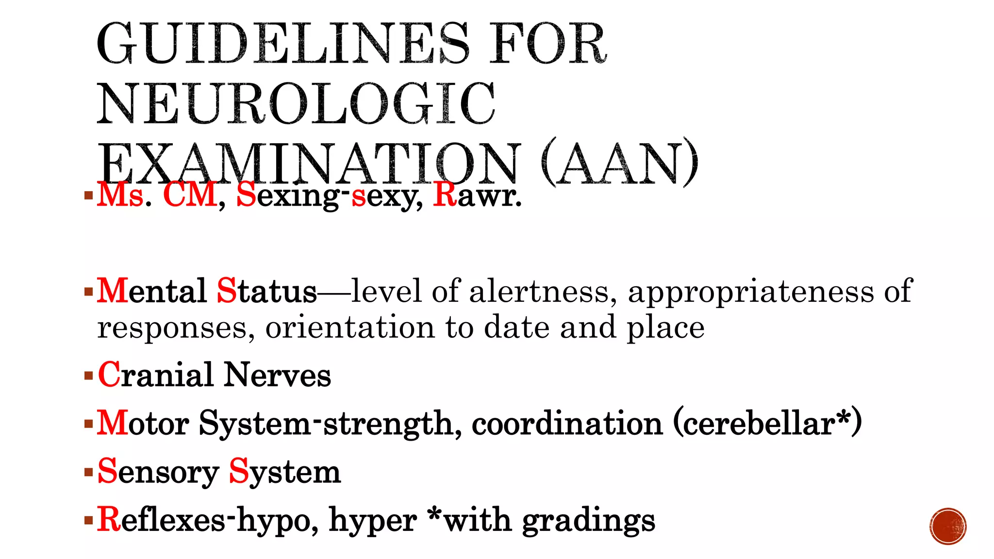
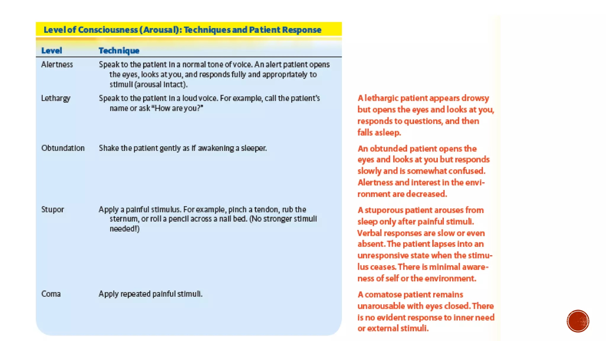
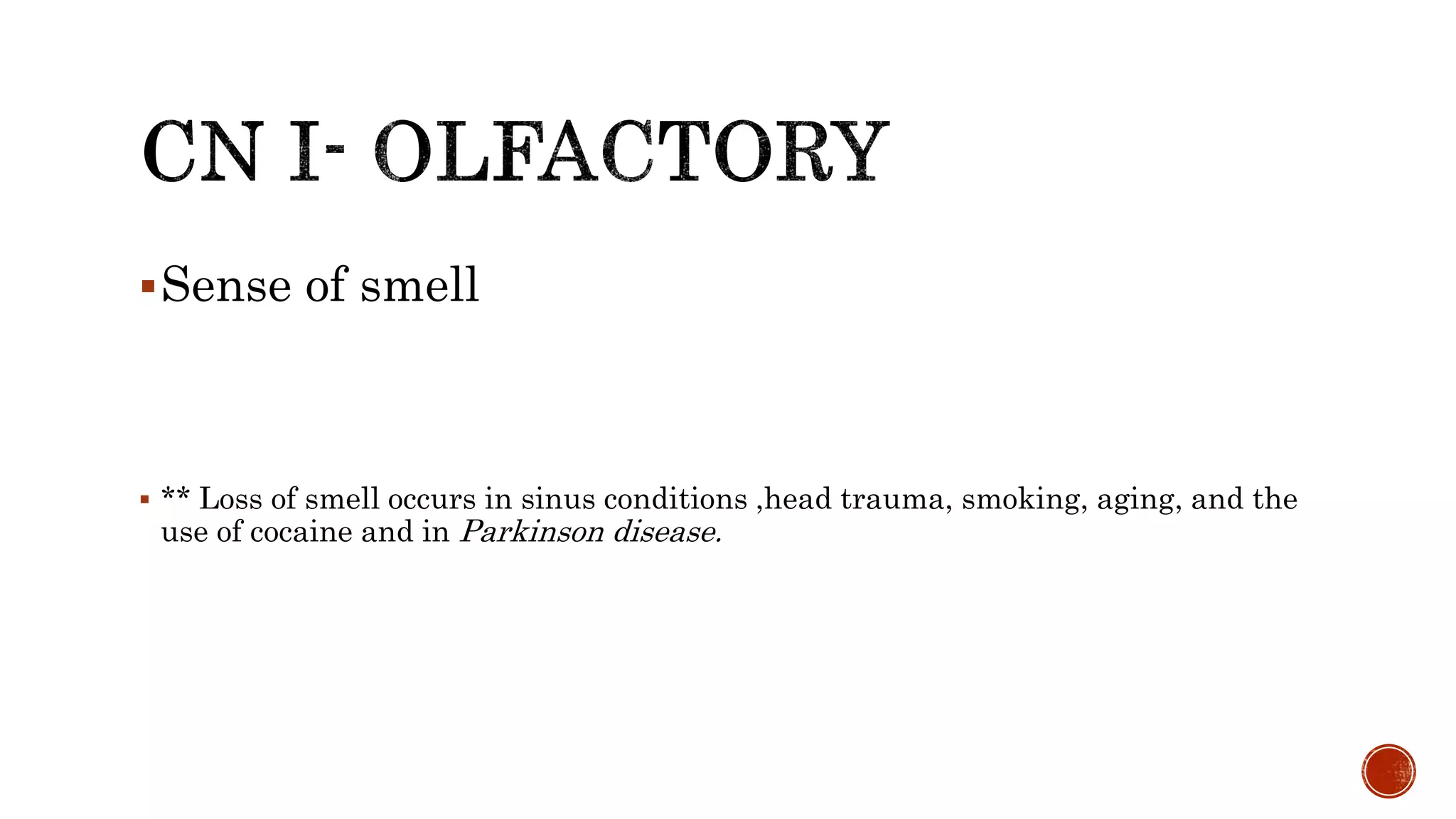
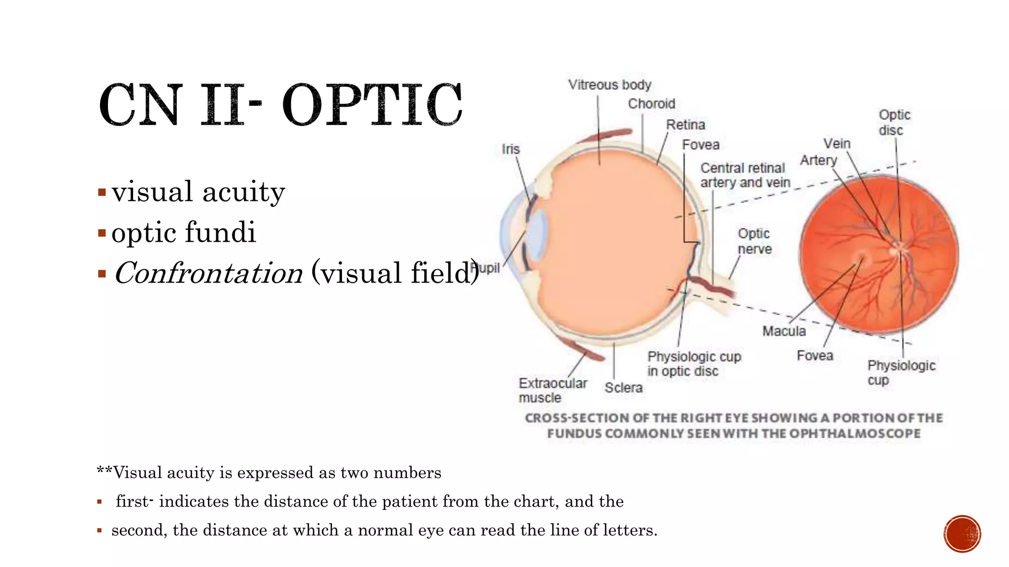
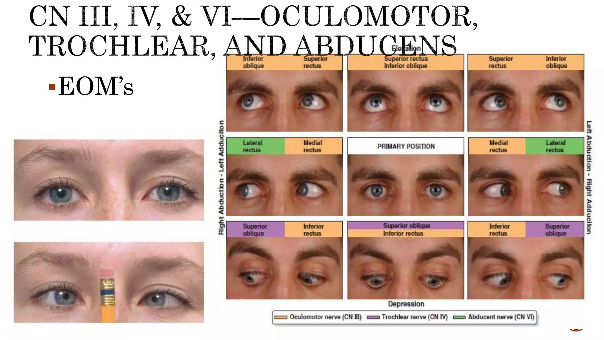
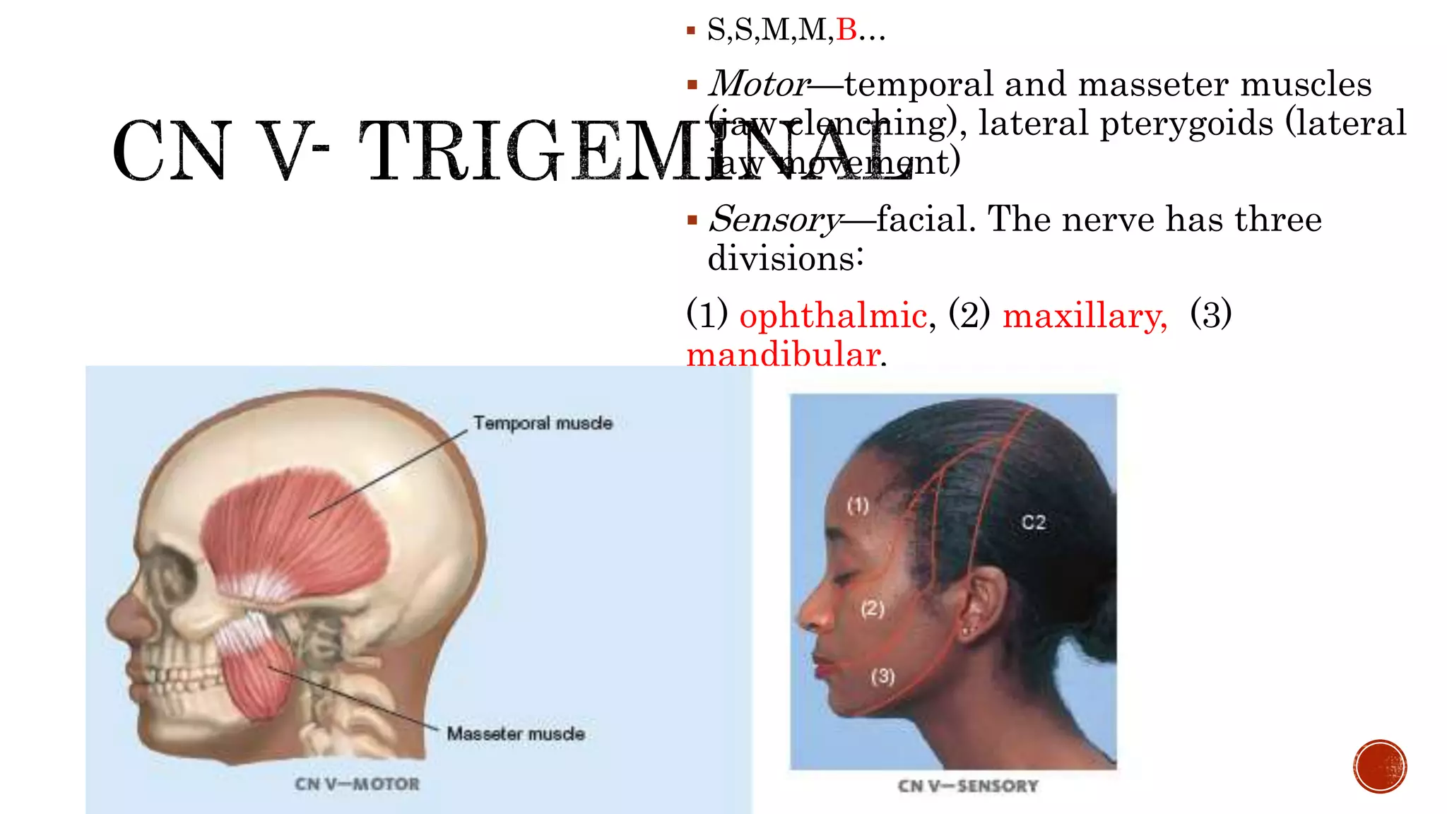
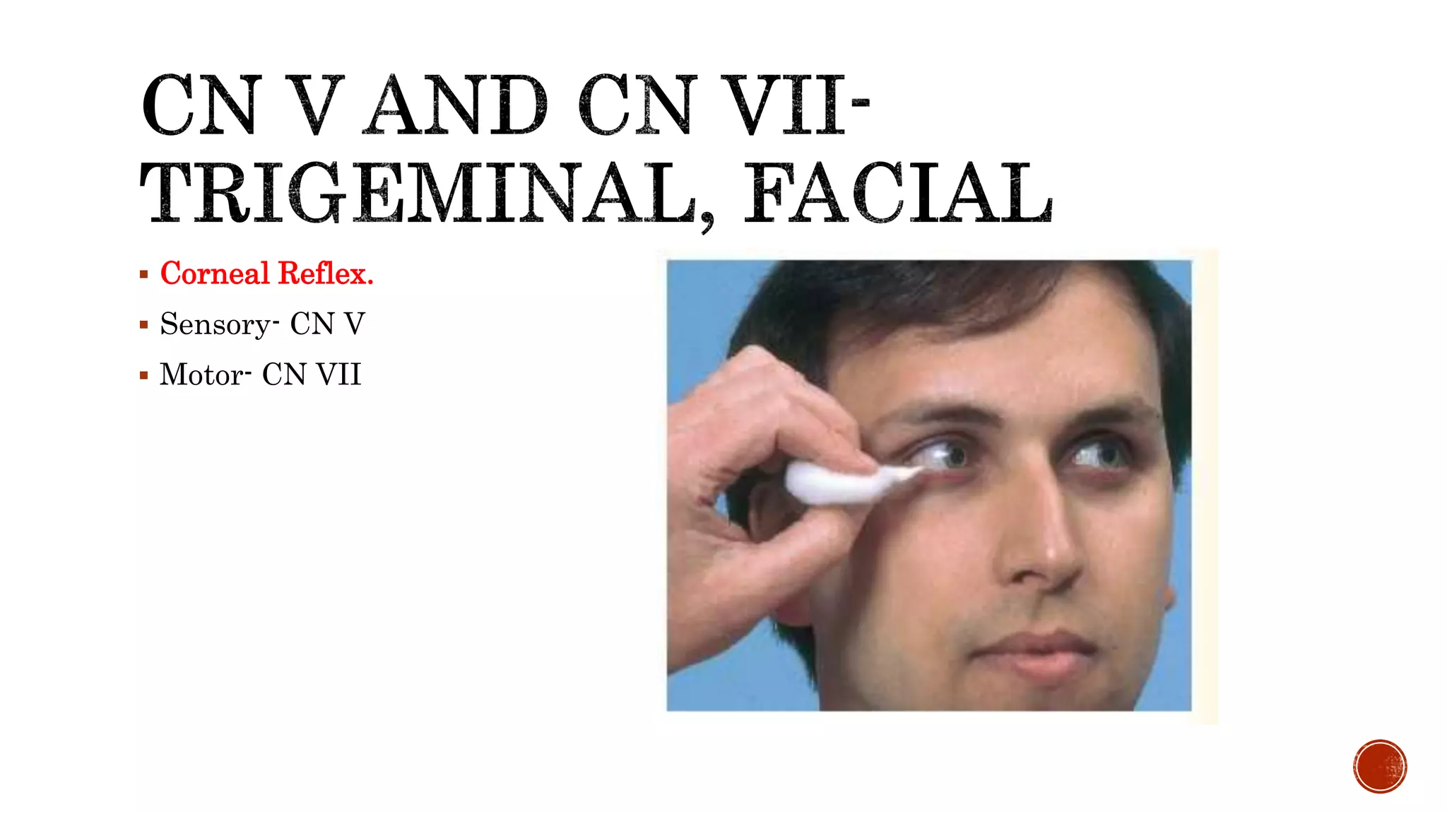
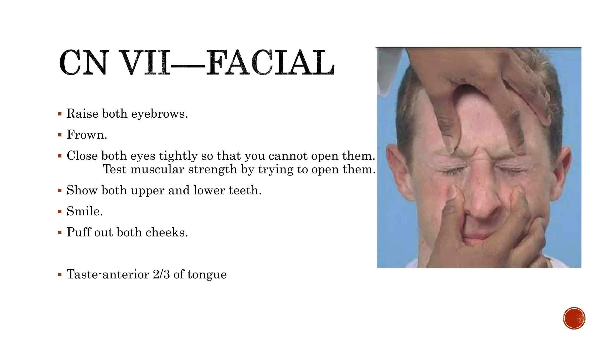
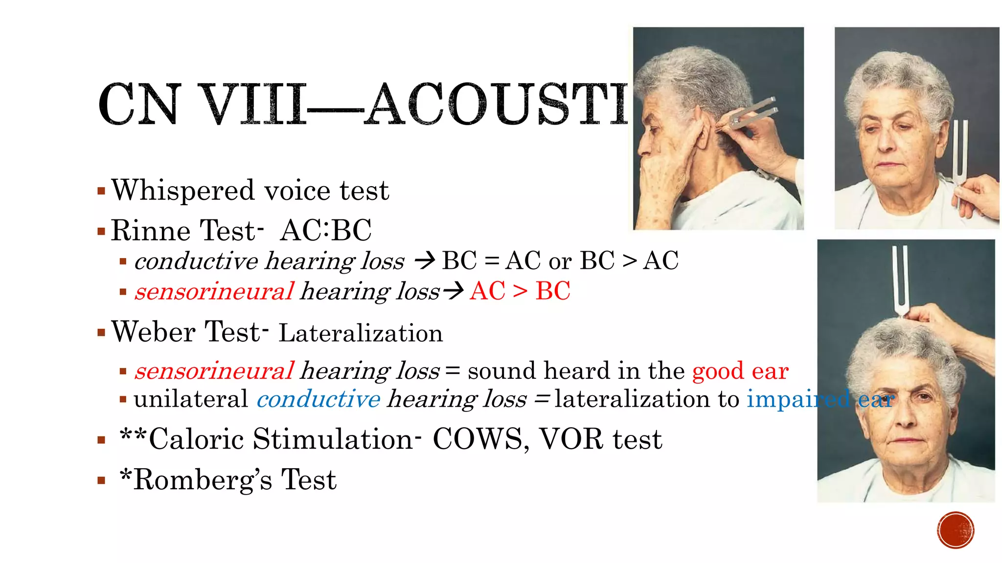
This document provides information about Baymax, who introduces himself as a personal healthcare provider. It then provides details on performing a physical exam, including preparing for the exam, establishing rapport with the patient, ensuring privacy and comfort, and explaining findings. Common symptoms that may warrant examination are listed. The physical exam components covered include vital signs, skin, head, eyes, ears, nose, mouth, neck, lungs, heart, abdomen, back, extremities, neurologic exam and mental status exam.
3. Prepare equipment/materials ahead.
Universal precaution.
Establish rapport.
Explain the procedure.
Ensure privacy and comfort.
Comprehensive, ergonomic PE.
Explain result/findings.
4. Common or Concerning Symptoms
Headache
Dizziness or vertigo
Generalized, proximal, or distal weakness
Numbness, abnormal or loss of sensations
Loss of consciousness, syncope, or near-syncope
Seizures
Tremors or involuntary movements
5. Sudden numbness or weakness of the face, arm, or leg
Sudden confusion, trouble speaking or understanding
Sudden trouble seeing in one or both eyes
Sudden trouble walking, dizziness, or loss of balance or
coordination
Sudden severe headache
6. Facial drooping: A section of the face, usually only on one
side, that is drooping and hard to move
Arm weakness: The inability to raise one's arm fully
Speech difficulties: An inability or difficulty to understand
or produce speech
Time: Time is of the essence when having a stroke, and an
immediate call to emergency services or trip to the
hospital is recommended.[
7. Ms. CM, Sexing-sexy, Rawr.
Mental Status—level of alertness, appropriateness of
responses, orientation to date and place
Cranial Nerves
Motor System-strength, coordination (cerebellar*)
Sensory System
Reflexes-hypo, hyper *with gradings
8.
9. Appearance and behavior
LOC
Posture and Motor behavior
Dress, grooming, hygiene
Facial Expression
Manner, affect
Speech and language
Quantity, Rate, Volume, Articulation, Fluency
Mood
Thoughts and perceptions
Cognition, including memory, attention,
information and vocabulary, calculations, abstract thinking, and constructional
ability -higher cortical fx
10. patient’s capacity for arousal, or wakefulness.
determined by the level of activity that the patient can be
aroused to perform in response to escalating stimuli from
the examiner.
11.
12.
13.
14. Mnemonics CN Sensory/Motor /Both
Oh I Olfactory Some
Oh II Optic Say
Oh III Occular Many
To IV Trochlear (“2”) Money
Touch V Trigeminal (“3”) But
And VI Abducens My
Feel VII Facial Brother
A/ Very VIII Acoustic/Vestibulocochlear Says
Good IX Glossopharyhngeal Big
Velvet X Vagus Brains
So XI Spinal Accessory Matter
Heavenly XII Hypoglossal Most
15.
16. Sense of smell
** Loss of smell occurs in sinus conditions ,head trauma, smoking, aging, and the
use of cocaine and in Parkinson disease.
17. visual acuity
optic fundi
Confrontation (visual field)
**Visual acuity is expressed as two numbers
first- indicates the distance of the patient from the chart, and the
second, the distance at which a normal eye can read the line of letters.
21. CN III Oculomotor Pupillary constriction, opening the
eye (lid elevation), and most extraocular
movements
CN IV Trochlear Downward, internal rotation of the
eye
CN VI Abducens Lateral deviation of the eye
22. S,S,M,M,B…
Motor—temporal and masseter muscles
(jaw clenching), lateral pterygoids (lateral
jaw movement)
Sensory—facial. The nerve has three
divisions:
(1) ophthalmic, (2) maxillary, (3)
mandibular.
24. Raise both eyebrows.
Frown.
Close both eyes tightly so that you cannot open them.
Test muscular strength by trying to open them.
Show both upper and lower teeth.
Smile.
Puff out both cheeks.
Taste-anterior 2/3 of tongue
25. Whispered voice test
Rinne Test- AC:BC
conductive hearing loss BC = AC or BC > AC
sensorineural hearing loss AC > BC
Weber Test- Lateralization
sensorineural hearing loss = sound heard in the good ear
unilateral conductive hearing loss = lateralization to impaired ear
**Caloric Stimulation- COWS, VOR test
*Romberg’s Test
27. Voice?
Dysphagia?
Say “aaaahhh”
Gag reflex
*The palate fails to rise with a bilateral lesion of CN X. In unilateral paralysis, one side of
the palate fails to rise and, together with the uvula, is pulled toward the normal side.
28. Observe Trapezius muscle
* Trapezius muscle paralysis, the shoulder droops, and the scapula is displaced
downward and laterally.
Sternocleidomastoid
Trapezius weakness with atrophy and fasciculations- peripheral nerve disorder.
A supine patient with bilateral weakness of the sternomastoids has difficulty
raising the head off the pillow.
29. articulation of words- CN V, VII, and X, as well as XII
Tongue- atrophy or fasciculations (relaxed)
asymmetry, atrophy, or deviation from the midline (protruded)
move from side to side- note the symmetry of the movement
push against the inside of each cheek in turn as you palpate for strength
**Tongue atrophy and fasciculations-ALS; polio
**unilateral cortical lesion-protruded tongue deviates transiently
away from the side of the cortical lesion, toward the side of
weakness.
30.
31. body position -during movement and at rest.
involuntary movements- tremors, tics, or fasciculations.
Note location, quality, rate, rhythm, and amplitude and relation
to posture, activity, fatigue, emotion, and other factors.
characteristics of the muscles (bulk, tone, and strength)
coordination
If you see an abnormality, identify the muscle(s) involved.
Determine whether the abnormality is central or peripheral in origin, and begin to learn which nerves
innervate the affected muscles.
32.
33. ELBOW
FLEXION and EXTENSION @
by having the patient pull and
push against your hand
Flexion (C5, C6—biceps)
extension (C6, C7, C8—triceps)
34. extension (C6, C7, C8,
radial nerve—
extensor carpi radialis
longus and brevis)
make a fist and resist
your pulling it down.
35.
36.
37. (C7, C8, T1)
squeeze two of your fingers as hard
as possible and not let them go
Weak grip
cervical radiculopathy,
de Quervain’s tenosynovitis,
carpal tunnel syndrome,
arthritis,
epicondylitis.
38. (C8, T1, ulnar nerve).
palm down and fingers spread
not not to let you examiner
move
the fingers as he forces them
together
40. Strength, tremors, movement
Flexion, extension, and lateral bending of the spine, and
Thoracic expansion and diaphragmatic excursion during respiration.
41. extension (S1—gluteus maximus) Have the patient
push the posterior thigh down against your hand.
Flexion-(L2, L3, L4— iliopsoas)
your hand on the patient’s thigh
Ask patient to raise the leg against
adduction (L2, L3, L4—adductors).
Ask the patient to bring both legs together
Again, with resistance
Abduction (L4, L5, S1—gluteus medius and
minimus).
patient to spread both legs against your hands
*Symmetric weakness of the proximal muscles -myopathy;
*symmetric weakness of distal muscles -a polyneuropathy, or
disorder of peripheral nerves.
42.
43. Extension (L2, L3, L4—quadriceps).
knee in flexion; ask to straighten the leg against
your hand
quadriceps - strongest muscle in the body
Flexion (L4, L5, S1, S2—hamstrings)
knee flexed with the foot resting on the bed.
Tell patient to keep the foot down as you try to
straighten the leg.
45. for rhythmic movement and steady posture
Rapid alternating movements
Upper, Lower Ext.
Point-to-point movements
Arms—Finger-to-Nose Test
Legs—Heel-to-Shin Test
Gait and stance, other related body movements
Heel-to-toe, heel, toe
Hopping in place
Romberg’s
Pronator Drift
46.
47. In cerebellar disease one movement cannot be followed quickly by its
opposite and movements are slow, irregular, and clumsy.
48. Gait that lacks coordination, with reeling and instability –ATAXIC
May be due to cerebellar disease, loss of position sense, or
intoxication.
**Inability to heel-walk is a sensitive test for corticospinal tract
damage.
49. Dorsal column disease ataxia, vision compensates for the
sensory loss.
(+) if loses balance when eyes are closed, a positive
Cerebellar ataxia- difficulty standing with feet together –
with yes open or closed.
50. Pronation of one forearm.
Sensitive and specific for a corticospinal tract lesion
originating in the contralateral hemisphere.
51.
52.
53.
54. Pain and temperature (spinothalamic tracts)
Position and vibration (posterior columns)
Light touch (both of these pathways)
Discriminative sensations, which depend on some of the above sensations but also
involve the cortex
Stereognosis
Graphestesia
2-point-discrimination
Point localization
55. Compare symmetric areas on the two sides of the body.
When testing pain, temperature, and touch sensation, also compare the distal with the proximal areas.
Scatter the stimuli to sample most of the dermatomes and major peripheral nerves
both shoulders (C4)
inner and outer aspects of the forearms (C6 and T1)
thumbs and little fingers (C6 and C8),
fronts of both thighs (L2),
medial and lateral aspects of both calves (L4 and L5)
little toes (S1)
medial aspect of each buttock (S3).
When testing vibration and position sensation, first test the fingers and toes. If these are normal, you may
safely assume that more proximal areas will also be normal.
●● Vary the pace of your testing. This is important so that the patient does not merely respond to your
repetitive rhythm.
●● When you detect an area of sensory loss or hypersensitivity, map out its boundaries in detail. Stimulate
first at a point of reduced sensation, and move by progressive steps until the patient detects the change.
56.
57. pointed end- small areas (i.e finger ), broad-end- larger areas
Encourage the patient to relax; position the limbs properly and symmetrically.
Hold reflex hammer loosely between your thumb and index finger so that it
swings freely in an arc within the limits set by your palm and other fingers.
With wrist relaxed, strike tendon briskly using a rapid wrist movement.
Strike should be quick and direct, not glancing.
Note the speed, force, and amplitude of the reflex response and grade the response
using the scale below.
Always compare the response of one side with the other. Reflexes are usually
graded on a 0 to 4+ scale.
58. Hyperactive reflexes
(hyperreflexia) - seen in
CNS lesions along the
descending corticospinal
tract.
Look for associated
upper motor neuron
findings of
weakness, spasticity, or a
positive Babinski sign.
Hypoactive or absent reflexes
(hyporeflexia)
- seen in diseases of spinal nerve
roots, spinal nerves, plexuses, or
peripheral nerves.
-Look for associated findings of
lower motor unit disease, namely
weakness, atrophy, and fasciculations.
**MgSO4- decreases DTR
Fasciculation- a brief, spontaneous contraction affecting a small number of muscle fibers, often causing a flicker of movement under the skin. It can be a symptom of disease of the motor neurons.
Tic- a habitual spasmodic contraction of the muscles, most often in the face.
Stereognosis. Stereognosis refers
to the ability to identify an object
by feeling it. Place in the patient’s
hand a familiar object such as a coin,
paper clip, key, pencil, or cotton
ball, and ask the patient to tell you
what it is. Normally a patient will
manipulate it skillfully and identify
it correctly within 5 seconds.
Asking the patient to distinguish
“heads” from “tails” on a coin is a
sensitive test of stereognosis.
Number identification (graphesthesia).
When motor impairment,
arthritis, or other conditions prevent
the patient from manipulating
an object well enough to identify
it, test the ability to identify numbers.
With the blunt end of a pen or
pencil, draw a large number in the
patient’s palm. A normal person
can identify most such numbers.
●● Two-point discrimination. Using
the two ends of an opened paper
clip, or the sides of two pins, touch
a finger pad in two places simultaneously.
Alternate the double stimulus
irregularly with a one-point
touch. Be careful not to cause pain.
Find the minimal distance at which patient can discriminate one from
two points (normally <5 mm on the finger pads). This test may be used
on other parts of the body, but normal distances vary widely from one
body region to another.