Apoptosis is a controlled, programmed cell death process that is essential for normal development and homeostasis. During apoptosis, cells actively trigger intracellular events that lead to cell fragmentation and phagocytosis without causing inflammation. Apoptosis is distinct from necrosis, which is unregulated cell death caused by external cellular injuries. Key aspects of apoptosis include activation of caspases, DNA fragmentation, and changes to cell membranes that mark cells for phagocytosis. Apoptosis pathways can be triggered by extracellular signals or internal cell damage and are important in development, tissue homeostasis, and diseases like cancer when the process goes awry.


![Apoptosis and Cancer
• Apoptosis does not occur in Cancer
• Cancerous cells trick and skip Apoptosis in number of ways
Inactivation of p53 [shooting the guard]
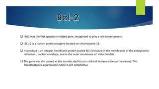
Produce Bcl-2 or a protein which mimics Bcl-2
Inhibits expression of Apaf-1](https://image.slidesharecdn.com/apop-140921044210-phpapp01/85/Apoptosis-42-320.jpg)
![Apoptosis and Autoimmune Disease
Autoimmune
Lymph Proliferative
Syndrome[ALPS]
Apoptosis doesnot
occur in self
reactive T & B cells
RBC
Hemolytic
Anemia
Neutrophil
Neutropenia
Platelets
Thrombocyto
penia](https://image.slidesharecdn.com/apop-140921044210-phpapp01/85/Apoptosis-43-320.jpg)

![Overview
Apoptosis is a good thing
Too little of a good thing is
bad [Cancer]
Too much of a good thing is
also bad [HIV]](https://image.slidesharecdn.com/apop-140921044210-phpapp01/85/Apoptosis-45-320.jpg)
![Sources
Textbook of Medical Physiology –Guyton & Hall [12th edition]
Review of Medical Physiology-William F Ganong [24th edition]
Internet Sources :
• www.ncbi.nlm.nih.gov/books/NBK26873
• http://en.wikipedia.org/wiki/Apoptosis
Articles :
Apoptosis- Molecular mechanisms and Pathogenicity
http://link.springer.com/article/10.1023/A:1009616228304#pg2
http://www.excli.de/vol8/Rastogi_08_2009/Rastogi_030809_proof.pdf](https://image.slidesharecdn.com/apop-140921044210-phpapp01/85/Apoptosis-46-320.jpg)