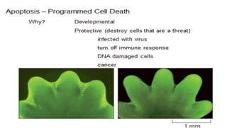
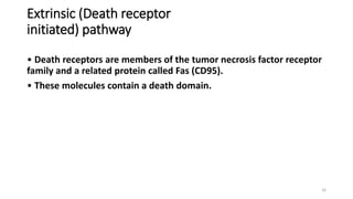
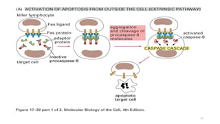
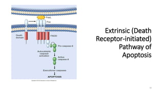
APOPTOSIS
1
Mohammed Jazeel M.
2017-11-111
Necrosis
• Morphologic expression of cell death
• Progressive disintegration of cell structure
• Initiated by overwhelming stress
• Usually elicits an acute inflammatory cell response (neutrophils
may be present).
2
Apoptosis
• Pathway of cell death induced by a tightly regulated suicide
program.
• Controlled by specific genes.
• Fragmentation of DNA.
• Fragmentation of nucleus.
• Apoptotic bodies are phagocytized.
• No neutrophils.
3
Consequences of Cell Death
Necrosis
• Loss of functional tissue
• Impaired organ function
Apoptosis
• Removal of damaged or unnecessary cells
4
5
Causes of Apoptosis
• Physiologic
• Pathologic
6
Physiologic
Embryogenesis and fetal development.
• Hormone dependent involution.
Prostate glandular epithelium after castration
Regression of lactating breast after weaning
• Cell loss in proliferating cell populations.
• Immature lymphocytes
• Epithelial cells in the GI tract
• Death of cells that have served their function.
7
Apoptosis in Pathologic
Conditions
• DNA damage due to radiation, chemotherapy.
• Accumulation of misfolded proteins leads to ER stress which ends
with apoptosis.
• Cell death in viral infections that induce
apoptosis such as HIV and Adenovirus or by the host immune
response such as hepatitis.
• Organ atrophy after duct obstruction.
8
General Characteristics
NECROSIS
• Usually affects large areas
of contiguous cells
• Control of intracellular
environment is lost early
• Cells swell and
organelles swell
APOPTOSIS
• Usually affects scattered
individual cells
• Control of intracellular
environment maintained in early
stages
• Cells contract
9
• NECROSIS
• Nuclear chromatin marginates
early,
• When DNA is cleaved, which is
usually a late event, fragments are
random in size
APOPTOSIS
• Nuclear chromatin marginates and
chromatin condenses, becoming very
compact
• Chromatin condensation and DNA
fragmentation occur together;
DNA cleaved into multiples of 200
base pair units
10
• A - early apoptosis; chromatin
margination & condensation
• B - later in apoptosis; nucleus
is fragmented
• C - phagocytosis of apoptotic
cellular remnants by adjacent cell
• D - swollen, necrotic cell for
Comparison
11
Mechanisms of Apoptosis
• Death receptor (Extrinsic) pathway
• Mitochrondrial (Intrinsic) pathway
• Execution Phase
• Removal of dead cells
12
Mechanisms of Apoptosis
• Cells contain intrinsic death and survival signals that are genetically
regulated.
• Genes are highly conserved across species and are homologous to
ced (cell death abnormal) genes in nematodes that initiate or inhibit
apoptosis.
13
Intrinsic -Mitochrondrial pathway
• Increased mitochondrial permeability with release of pro-apoptotic
molecules into the cytoplasm (cytochrome c).
• Synthesis of anti-apoptotic molecules (Bcl-2) promoted by Growth
factors.
• When cells are deprived of growth factors or subjected to stress anti
apoptotic molecules (Bcl-2) are lost.
Bcl-2 is over expressed in most follicular B-cell lymphomas – allowing
abnormal cells to proliferate.
• Mitochondrial membrane becomes permeable and proteins that activate
caspase leak out.
14
Intrinsic
(Mitochondrial)
Pathway of
Apoptosis
15
Extrinsic (Death receptor
initiated) pathway
• Death receptors are members of the tumor necrosis factor receptor
family and a related protein called Fas (CD95).
• These molecules contain a death domain.
16
17
Extrinsic (Death
Receptor-initiated)
Pathway of
Apoptosis
18
Removal of Dead Cells
• Dying cells secrete factors the recruit phagocytes.
• This facilitates prompt clearance before they undergo secondary
necrosis.
• Dead cells disappear without a trace and do not produce
inflammation
19
Apotosis- Summary
• “Programmed cell death” can be activated by moderate stress which
has damaged the cell beyond its ability to recover fully or by viral
infection.
• This has the desirable effect of removing damaged or infected cells.
• Selective manipulation of apoptotic pathways may be an important
approach for treating cancer in the future.
20
21

Apoptosis

  • 1.
  • 2.
    Necrosis • Morphologic expressionof cell death • Progressive disintegration of cell structure • Initiated by overwhelming stress • Usually elicits an acute inflammatory cell response (neutrophils may be present). 2
  • 3.
    Apoptosis • Pathway ofcell death induced by a tightly regulated suicide program. • Controlled by specific genes. • Fragmentation of DNA. • Fragmentation of nucleus. • Apoptotic bodies are phagocytized. • No neutrophils. 3
  • 4.
    Consequences of CellDeath Necrosis • Loss of functional tissue • Impaired organ function Apoptosis • Removal of damaged or unnecessary cells 4
  • 5.
  • 6.
    Causes of Apoptosis •Physiologic • Pathologic 6
  • 7.
    Physiologic Embryogenesis and fetaldevelopment. • Hormone dependent involution. Prostate glandular epithelium after castration Regression of lactating breast after weaning • Cell loss in proliferating cell populations. • Immature lymphocytes • Epithelial cells in the GI tract • Death of cells that have served their function. 7
  • 8.
    Apoptosis in Pathologic Conditions •DNA damage due to radiation, chemotherapy. • Accumulation of misfolded proteins leads to ER stress which ends with apoptosis. • Cell death in viral infections that induce apoptosis such as HIV and Adenovirus or by the host immune response such as hepatitis. • Organ atrophy after duct obstruction. 8
  • 9.
    General Characteristics NECROSIS • Usuallyaffects large areas of contiguous cells • Control of intracellular environment is lost early • Cells swell and organelles swell APOPTOSIS • Usually affects scattered individual cells • Control of intracellular environment maintained in early stages • Cells contract 9
  • 10.
    • NECROSIS • Nuclearchromatin marginates early, • When DNA is cleaved, which is usually a late event, fragments are random in size APOPTOSIS • Nuclear chromatin marginates and chromatin condenses, becoming very compact • Chromatin condensation and DNA fragmentation occur together; DNA cleaved into multiples of 200 base pair units 10
  • 11.
    • A -early apoptosis; chromatin margination & condensation • B - later in apoptosis; nucleus is fragmented • C - phagocytosis of apoptotic cellular remnants by adjacent cell • D - swollen, necrotic cell for Comparison 11
  • 12.
    Mechanisms of Apoptosis •Death receptor (Extrinsic) pathway • Mitochrondrial (Intrinsic) pathway • Execution Phase • Removal of dead cells 12
  • 13.
    Mechanisms of Apoptosis •Cells contain intrinsic death and survival signals that are genetically regulated. • Genes are highly conserved across species and are homologous to ced (cell death abnormal) genes in nematodes that initiate or inhibit apoptosis. 13
  • 14.
    Intrinsic -Mitochrondrial pathway •Increased mitochondrial permeability with release of pro-apoptotic molecules into the cytoplasm (cytochrome c). • Synthesis of anti-apoptotic molecules (Bcl-2) promoted by Growth factors. • When cells are deprived of growth factors or subjected to stress anti apoptotic molecules (Bcl-2) are lost. Bcl-2 is over expressed in most follicular B-cell lymphomas – allowing abnormal cells to proliferate. • Mitochondrial membrane becomes permeable and proteins that activate caspase leak out. 14
  • 15.
  • 16.
    Extrinsic (Death receptor initiated)pathway • Death receptors are members of the tumor necrosis factor receptor family and a related protein called Fas (CD95). • These molecules contain a death domain. 16
  • 17.
  • 18.
  • 19.
    Removal of DeadCells • Dying cells secrete factors the recruit phagocytes. • This facilitates prompt clearance before they undergo secondary necrosis. • Dead cells disappear without a trace and do not produce inflammation 19
  • 20.
    Apotosis- Summary • “Programmedcell death” can be activated by moderate stress which has damaged the cell beyond its ability to recover fully or by viral infection. • This has the desirable effect of removing damaged or infected cells. • Selective manipulation of apoptotic pathways may be an important approach for treating cancer in the future. 20
  • 21.