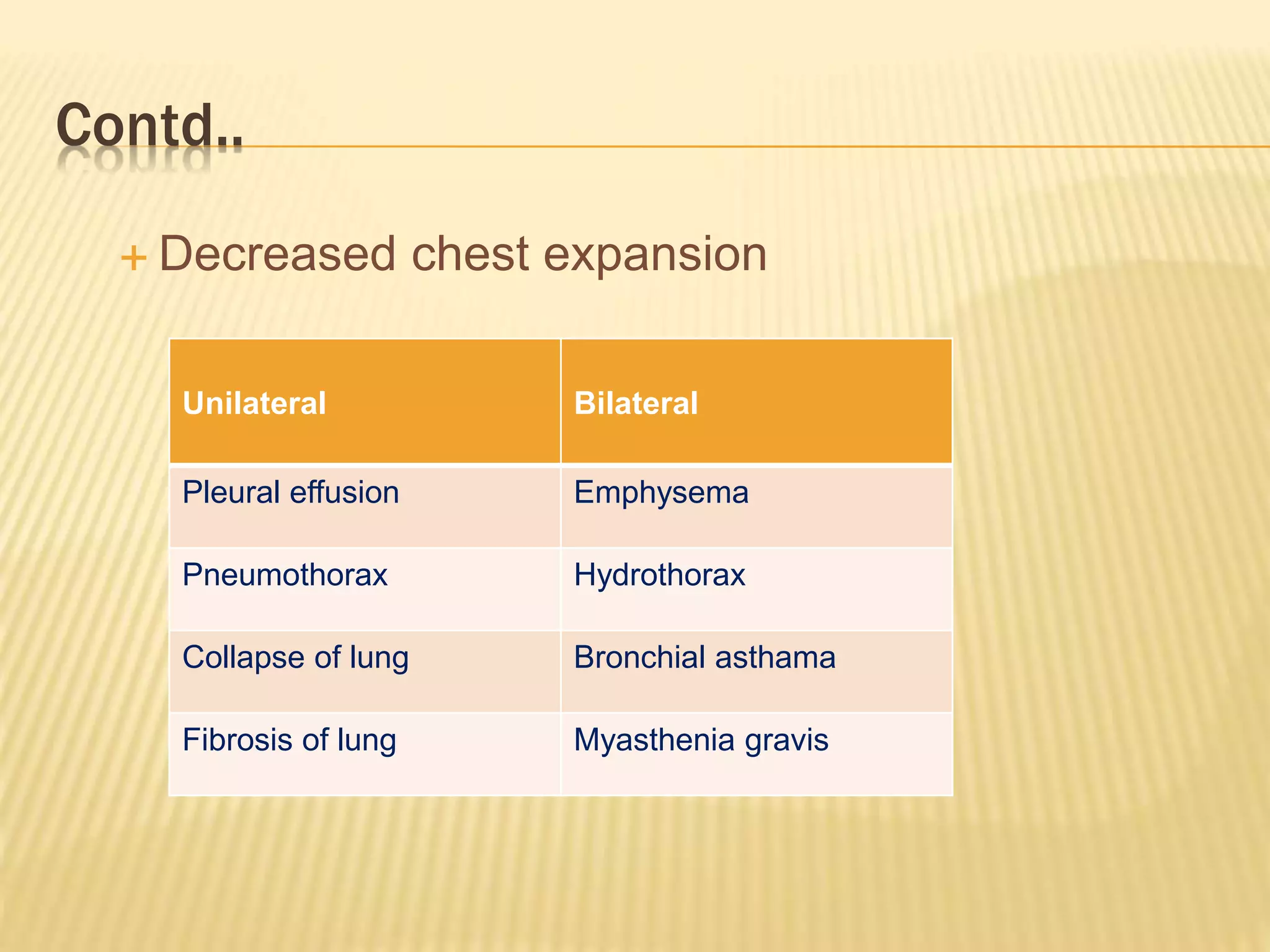
INSPECTION
1. Shape of the chest
 Normal: Bilaterally symmetrical and elliptical in cross
section
AP:Trans=5:7
 Chest deformity
 Flat chest: AP:Trans=1:2
 Pulmonary TB
 Fibrothorax
Contd..
 Barrel shaped chest: AP:Trans=1:1
 Physiological: Infancy,old age
 Pathological: COPD
 Pigeon chest(Pectus carinatus): Forward protrusion of
sternum and adjacent costal cartilages
 Rickets
 Childhood asthma
 Marfan’s syndrome
Contd..
 Funnel chest(Pectus excavatum): Exaggeration of the
normal hollowness on the lower end of sternum
 Development defect: Apex beat is shifted further to the left and the
vital capacity is restricted
 Marfan’s syndrome
 Scorbutic rosary: Sharpangulation with or without beading or
rosary formation of the ribs
D/t backward displacement of sternum
 Vit C deficiency
Contd..
 Harrison’s sulcus: D/t indrawing of the ribs to form
symmetrical horizontal groove above the costal
margin,along the line of attachment of the diaphragm d/t
hyperinflamation of the lung and reapeated strong
contraction of the diaphragm
 Chronic respiratory Dzs in childhood
 Childhood asthma
 Rickets
 Rickety rosary: Bead like enlargement of costochondral
junction
 Rickets
2. Movement of the chest:
 Movement of the chest with respiration
 Movement of the chest equally on both side
a) Unilateral: b) Bilateral
• Pleural effusion • Emphysema
• Chest trauma • Hydrothorax
• Pneumothorax • Obesity
• Hydropneumothorax • Bronchial asthma
• Consolidation • Diffuse interstitial fibrosis
• Fibrosis of lung • Myasthenia gravis
Contd..
3. Apical Impulse:
 Stand on the right side of the patient and look tangentially
over the precordium
 Helps to note the precordial shift
4. Tracheal deviation:
 Ask the patient to look forward an look for any deviation
Contd..
5. Respiration:
 Rate
 Rhythm
 Type
6. Venous Prominance:
 Superior venacava syndrome: Presence of distended vein
over the chest wall
Contd..
7. Retraction/Fullness of intercostal space:
 Abnormal retraction:
 Severe asthma
 COPD
 Upper airway obstruction
 Fullness of intercostal space:
 Pleural effusion
 Haemothorax
 pneumothorax
Contd..
8. Level of nipple:
 Whether both the nipple are at the same level or not
9. Skin over the chest:
• Cold abscess
• Ulcer
• Swelling
• Scar mark
Contd..
10. Accessory muscle: Whether accesory muscles of respiration are
working or not
 Inspiration:Active process d/t contraction of the intercostal
muscles and diaphragm
Muscles:
 Scalene
 Sternocleidomastoid
 Platysma
 Pectoralis
 Serratus anterior
 Expiration: Passive process d/t elastic recoil of the lung
Muscles
 Abdominal recti muscles
 latissimus dorsi
PALPATION
1. Surface temperature
2. Tenderness:
• Rib tenderness: Trauma, fracture
• Intercostal tenderness: Liver abscess, empyema thoracis
3. Corroboration of inspetory findings
4. Spinal deformity
Contd..
5. Position of trachea and apex beat
 Palpate in the standing or sitting position with arm placed symmetrically
on two sides.
 Flex the neck with left hand so that chin remain in same side
 Insert the tip of index finger in suprasternal notch
 Feel the tracheal ring
 Now side the index finger in the angle between sternocleidomastoid
muscles and trachea on both side
 On the deviated side angle is narrowed and feel resistant
Contd..
• Shift of trachea:
To the same side To the opposite
side
Fibrosis of lung Massive pleural effusion
Collapse of lung Pneumothorax
pneumonectomy Hydropneumothorax
Contd..
6. Movement of chest:
 Upper part of thorax:
 Face the patient’s back
 Place both hands over the patient’s supraclavicular
fossa.
 Compare on both sides the extent of upward movement
of the hands during quiet respiration
Contd..
 Anterior thoracic movement:
 Face the patient
 Keep the finger tip of both the hands on either side of patients rib
cage so that the tip of thumbs approximate each other in midline
without touching the chest wall
 Ask the patient to take deep breath
 Compare the movement of thumbs on both sides away from
midline
 It can also be assessed by holding a loose fold of skin between
the thumbs and noting their separation
Contd..
 Posterior thoracic movement
 Perform at the infrascapular region
Contd..
7. Chest expansion:
 Done using inch tape
 In male: measure at the level of nipple
 In female: measure just below breast
 Measure normal circumference of chest
 Ask to take deep inspiration, again measure the chest circumference
 Difference between the two is known as chest expansion
 Normal expansion=5-8cm
Contd..
 Decreased chest expansion
Unilateral Bilateral
Pleural effusion Emphysema
Pneumothorax Hydrothorax
Collapse of lung Bronchial asthama
Fibrosis of lung Myasthenia gravis
Contd..
7. Vocal fremitus: palpation of laryngeal vibration on the
chest wall when patient is asked to repeat 9-9 or 1-1-1
• Place the flat of hand or ulnar border of the right hand over the
intercostal space
• Compare the patient to tell 9-9 or 1-1-1
• Compare on both side
Increased: Consolidation
Decreased: Pleural effusion
PERCUSSION
 Cardinal rules:
Method:
 Place the middle finger of the left hand(pleximeter) of the
examiner firmly over the chest wall over the ICS such that other
finger don’t touch the chest wall
 Then strike the centre of middle phalanx of the pleximeter finger
with the tip of middle finger of right hand(plexor)
 The finger should be moved immediately after the striking action
in tapping movement. The percussion finger is bent to make its
terminal phalanx right angled so that it strikes the other finger
perpendicularly
Contd..
 The percussion movement should be sudden originating from the
wrist
 Always percuss the opposite side of chest on the equivalent
position and compare with notes on other side
Contd..
 Position of the patient:
Sitting position is the best for percussion
Supine position is not desirable because of the
alteration of percussion note by the underlying
structure in which patient lies
Contd..
i. For anterior percussion:
Patients should sit erect with hands by his side
ii. For posterior percussion:
patient should bend his head forward and keep his
hands over the shoulder.This position keep the two
scapula away so that more lung field is available for
percussion
iii. Lateral percussion:
The patient should sit with his hand held over the head
 Area of percussion
i. Anterior chest wall:
a) Clavicle:
 Direct percussion
 Percussion is done within middle 1/3rd of clavicle
b) Supraclavicular region
 It is a band of resonance 5-7cm size over the supraclavicular
fossa
 Boundaries:
 Medially: Scalenus muscle of neck
 Laterally: Acromian process of scapula
 Anteriorly: Clavicle
 Posteriorly: Trapezius
The percussion is done by standing behind the patient and
resonance of the lung apices is assessed
 Hyper resonance: Emphysema
 Impaired resonance: Pulmonary TB
c) Infraclavicular:
2nd to 6th ICS; however
the percussion note
cannot be compared
due to relative cardiac
dullness on the left side
Contd..
ii. Lateral chest wall
Percuss from 4th to 8th ICS in mid axillary line
Contd..
iii. Posterior chest wall
a) Suprascapular
b) Interscaular
c) Infrascapular region upto the 11th ICS
Types of
percussion note
Lesion
1. Tympanitic Hollow viscus
2. Sub tympanitic Above the level of pleural effusion
3. Hyper-resonant Pneumotharax
4. Resonant Normal lung
5. Impaired Pulmonary fibrosis
6. Dull Consolidation,collapse
7. Stony dull Pleural effusion,haemothorax
AUSCULTATION
Preliminaries
 Auscultation is carried out with diaphragm of stethoscope as
most respiratory sound are high pitched
 Listen with the patient relaxed and breathing deeply through
an open mouth.
 Instruct the patient to turn the face to one side, ask to breath
regularly and deeply through open mouth
Contd..
 Auscultate the both sides alternately
 Avoid auscultation within 3cm of the midline anteriorly and
posteriorly as these area may transmit sounds directly from
the trachea or main bronchi
 Listen anteriorly from above the clavicle down to the 6th rib,
laterally from axilla to the 8th rib and posteriorly down to the
level of the 11th rib
 In each area listen to the quality and amplitude of breath
sound
Contd..
Position of the patient: Sitting position
Auscultatory area:
i. Anterior: From an area above the clavicle down to
6th rib
ii. Axilla: Area upto 8th rib
iii. Posterior: Above the level of spine of scapula down
to 11th rib
Contd..
1. Breath sounds
Breath sounds are produced by vibration of vocal cord
due to turbulent air flow in larger airways which is
conducted by the overlying lung tissue to the chest wall
Contd..
i. Vesicular breath sound:
 Vesicular breathing. Respiratory sounds known
as vesicular breathing arise due to vibration of the
elastic elements of the alveolar walls during their
filling with air in inspiration.
 The alveoli are filled with air in sequence.
Therefore, the summation of the great number
of sounds produced during vibration of the alveolar
walls gives a long soft (blowing) noise that can
be heard during the entire inspiration phase, its
intensity gradually increasing.
Contd..
 Normal vesicular breathing is better heard over the
anterior surface of the chest, below the 2nd rib, laterally
of the parasternal line, and also in the axillary regions
and below the scapular angle, i.e. at points where the
largest masses of the pulmonary tissue are located.
 Vesicular breathing is heard worse at the apices of the
lungs and their lowermost parts, where the masses of the
pulmonary tissue are less abundand. While carrying out
comparative auscultation, it should be remembered that
the expiration sounds are louder and longer in the right
lung due to a better conduction of the laryngeal sounds
by the right main bronchus, which is shorter and wider.
Contd..
Condition with diminished vesicular breath sound:
 Bronchial asthma
 Tumor
 Pleural effusion
 Pleural thickeing
 Emphysema
Contd..
ii. Bronchial breath sound:
 Respiratory sounds known as bronchial or tubular
breathing arise in the larynx and the trachea as air
passes through the vocal slit.
 As air is inhaled, it passes through the vocal slit to enter
wider trachea where it is set in vortex-type motion. Sound
waves thus generated propagate along the air column
throughout the entire bronchial tree. Sounds generated
by the vibration of these waves are harsh.
Contd..
 During expiration, air also passes through the vocal slit
to enter a wider space of the larynx where it is set in a
vortex motion.
 But since the vocal slit is narrower during expiration, the
respiratory sound becomes louder, harsher and longer.
This type of breathing is called laryngotracheal (by the
site of its generation).
Contd..
 Bronchial breathing is well heard in physiological
cases over the larynx, the trachea, and at points of
projection of the tracheal bifurcation (anteriorly, over
the manubrium sterni, at the point of its junction with
the sternum, and posteriorly in the interscapular
space, at the level of the 3rd and 4th thoracic
vertebrae).
 Bronchial breathing is not heard over the other parts
of the chest because of large masses of the
pulmonary tissue found between the bronchi and the
chest wall.
Contd..
Types of bronchial breathing:
a. Tubular:
They are high pitched and present in:
 pneumonic consolidation
 collapse lung
b. Cavernous:
They are low pitched and heard in the presence of thick
walled cavity with a communicating bronchus
c. Amphoric:
They are low pitched, with a high tone and metallic
quality and present in:
 Bronchopleural fistula
 Tension pneumothorax
Contd..
Causes of absent breath sound:
 Pleural effusion(massive)
 Thickned pleura
 Pneumothorax
 Collapsed lung
Contd..
2. Added sounds
i. Crackles:
They are non musical, interrupted added sounds of short
duration. They are explosive in nature
Types:
 Fine: less loud,short,arise from alveoli
 Coarse: Low pitched,loud nd arise frombronchi and
bronchioles
Contd..
Crackles may be:
 Early inspiratory: Chronic bronchitis
 Mid inspiratory: Bronchiectasis
 Late inspiratory: Asbestosis,pulmonary fibrosis,pneumonitis
 Expiratory: Chronic bronchitis
Mechanism of crackles:
 Bubbling or flow of air through secretion in the bronchial
level
Contd..
ii. Ronchi
They are musical,continuous added sounds.
They may be:
 Low pitched: arising from large airways
 High pitched: arising from small airways
Eg.
 Tumors
 Foreign body
 Bronchial asthma
 Emphysema
Contd..
iii. Wheeze:
 Wheezing is a high-pitched whistling sound made while
breathing. Most commonly wheezing occurs during breathing out
(expiration), but it can sometimes be related to breathing in
(inspiration)
 Wheezing results from a narrowing of the airways and typically
indicates some difficulty breathing. The narrowing of the airways can
be caused by inflammation from asthma, an infection, an allergic
reaction, or by a physical obstruction, such as a tumor or a foreign
object that's been inhaled.
Contd..
The most common cause of recurrent wheezing is asthma.
Possible causes of wheezing include:
 Allergies
 Anaphylaxis (a severe allergic reaction, such as to an insect bite or medication)
 Asthma
 Bronchiectasis
 Bronchiolitis (especially in young children)
 Pneumonia
 Respiratory syncytial virus (RSV)
Contd..
Causes(contd..)
 Bronchitis
 COPD(chronic obstructive pulmonary disease) and other lung diseases
 Emphysema
 Foreign object inhaled: First aid
 GERD(gastroesophageal reflux disease)
 Heart failure
 Lung cancer
Contd..
3. Vocal resonance:
It is a voice sound heard with the chest piece of
stethoscope
i. Increased vocal resonance:
 Consolidation
 Collapse with patent bronchus
 Open pneumothorax
ii. Decreased vocal resonance
 Pleural effusion
 Pneumothorax
 Emphysema
Contd..
4. Aegophony
The voice may sound nasal or bleating; heard over the
level of a pleural effusion,or in some cases over an area
of consolidation
5. Pleural rub
It is superficial localized grating sound best heard with
pressure of stethoscope
It is produced when inflamed parietal and visceral pleura
move over one another
Not altered by coughing and usually associated with pleuritic
pain

Systemic examination of respiratory system

  • 2.
    INSPECTION 1. Shape ofthe chest  Normal: Bilaterally symmetrical and elliptical in cross section AP:Trans=5:7  Chest deformity  Flat chest: AP:Trans=1:2  Pulmonary TB  Fibrothorax
  • 3.
    Contd..  Barrel shapedchest: AP:Trans=1:1  Physiological: Infancy,old age  Pathological: COPD  Pigeon chest(Pectus carinatus): Forward protrusion of sternum and adjacent costal cartilages  Rickets  Childhood asthma  Marfan’s syndrome
  • 4.
    Contd..  Funnel chest(Pectusexcavatum): Exaggeration of the normal hollowness on the lower end of sternum  Development defect: Apex beat is shifted further to the left and the vital capacity is restricted  Marfan’s syndrome  Scorbutic rosary: Sharpangulation with or without beading or rosary formation of the ribs D/t backward displacement of sternum  Vit C deficiency
  • 5.
    Contd..  Harrison’s sulcus:D/t indrawing of the ribs to form symmetrical horizontal groove above the costal margin,along the line of attachment of the diaphragm d/t hyperinflamation of the lung and reapeated strong contraction of the diaphragm  Chronic respiratory Dzs in childhood  Childhood asthma  Rickets  Rickety rosary: Bead like enlargement of costochondral junction  Rickets
  • 6.
    2. Movement ofthe chest:  Movement of the chest with respiration  Movement of the chest equally on both side a) Unilateral: b) Bilateral • Pleural effusion • Emphysema • Chest trauma • Hydrothorax • Pneumothorax • Obesity • Hydropneumothorax • Bronchial asthma • Consolidation • Diffuse interstitial fibrosis • Fibrosis of lung • Myasthenia gravis
  • 7.
    Contd.. 3. Apical Impulse: Stand on the right side of the patient and look tangentially over the precordium  Helps to note the precordial shift 4. Tracheal deviation:  Ask the patient to look forward an look for any deviation
  • 8.
    Contd.. 5. Respiration:  Rate Rhythm  Type 6. Venous Prominance:  Superior venacava syndrome: Presence of distended vein over the chest wall
  • 9.
    Contd.. 7. Retraction/Fullness ofintercostal space:  Abnormal retraction:  Severe asthma  COPD  Upper airway obstruction  Fullness of intercostal space:  Pleural effusion  Haemothorax  pneumothorax
  • 10.
    Contd.. 8. Level ofnipple:  Whether both the nipple are at the same level or not 9. Skin over the chest: • Cold abscess • Ulcer • Swelling • Scar mark
  • 11.
    Contd.. 10. Accessory muscle:Whether accesory muscles of respiration are working or not  Inspiration:Active process d/t contraction of the intercostal muscles and diaphragm Muscles:  Scalene  Sternocleidomastoid  Platysma  Pectoralis  Serratus anterior  Expiration: Passive process d/t elastic recoil of the lung Muscles  Abdominal recti muscles  latissimus dorsi
  • 12.
    PALPATION 1. Surface temperature 2.Tenderness: • Rib tenderness: Trauma, fracture • Intercostal tenderness: Liver abscess, empyema thoracis 3. Corroboration of inspetory findings 4. Spinal deformity
  • 13.
    Contd.. 5. Position oftrachea and apex beat  Palpate in the standing or sitting position with arm placed symmetrically on two sides.  Flex the neck with left hand so that chin remain in same side  Insert the tip of index finger in suprasternal notch  Feel the tracheal ring  Now side the index finger in the angle between sternocleidomastoid muscles and trachea on both side  On the deviated side angle is narrowed and feel resistant
  • 15.
    Contd.. • Shift oftrachea: To the same side To the opposite side Fibrosis of lung Massive pleural effusion Collapse of lung Pneumothorax pneumonectomy Hydropneumothorax
  • 16.
    Contd.. 6. Movement ofchest:  Upper part of thorax:  Face the patient’s back  Place both hands over the patient’s supraclavicular fossa.  Compare on both sides the extent of upward movement of the hands during quiet respiration
  • 17.
    Contd..  Anterior thoracicmovement:  Face the patient  Keep the finger tip of both the hands on either side of patients rib cage so that the tip of thumbs approximate each other in midline without touching the chest wall  Ask the patient to take deep breath  Compare the movement of thumbs on both sides away from midline  It can also be assessed by holding a loose fold of skin between the thumbs and noting their separation
  • 19.
    Contd..  Posterior thoracicmovement  Perform at the infrascapular region
  • 20.
    Contd.. 7. Chest expansion: Done using inch tape  In male: measure at the level of nipple  In female: measure just below breast  Measure normal circumference of chest  Ask to take deep inspiration, again measure the chest circumference  Difference between the two is known as chest expansion  Normal expansion=5-8cm
  • 22.
    Contd..  Decreased chestexpansion Unilateral Bilateral Pleural effusion Emphysema Pneumothorax Hydrothorax Collapse of lung Bronchial asthama Fibrosis of lung Myasthenia gravis
  • 23.
    Contd.. 7. Vocal fremitus:palpation of laryngeal vibration on the chest wall when patient is asked to repeat 9-9 or 1-1-1 • Place the flat of hand or ulnar border of the right hand over the intercostal space • Compare the patient to tell 9-9 or 1-1-1 • Compare on both side Increased: Consolidation Decreased: Pleural effusion
  • 24.
    PERCUSSION  Cardinal rules: Method: Place the middle finger of the left hand(pleximeter) of the examiner firmly over the chest wall over the ICS such that other finger don’t touch the chest wall  Then strike the centre of middle phalanx of the pleximeter finger with the tip of middle finger of right hand(plexor)  The finger should be moved immediately after the striking action in tapping movement. The percussion finger is bent to make its terminal phalanx right angled so that it strikes the other finger perpendicularly
  • 25.
    Contd..  The percussionmovement should be sudden originating from the wrist  Always percuss the opposite side of chest on the equivalent position and compare with notes on other side
  • 26.
    Contd..  Position ofthe patient: Sitting position is the best for percussion Supine position is not desirable because of the alteration of percussion note by the underlying structure in which patient lies
  • 27.
    Contd.. i. For anteriorpercussion: Patients should sit erect with hands by his side ii. For posterior percussion: patient should bend his head forward and keep his hands over the shoulder.This position keep the two scapula away so that more lung field is available for percussion iii. Lateral percussion: The patient should sit with his hand held over the head
  • 28.
     Area ofpercussion i. Anterior chest wall: a) Clavicle:  Direct percussion  Percussion is done within middle 1/3rd of clavicle b) Supraclavicular region  It is a band of resonance 5-7cm size over the supraclavicular fossa
  • 29.
     Boundaries:  Medially:Scalenus muscle of neck  Laterally: Acromian process of scapula  Anteriorly: Clavicle  Posteriorly: Trapezius The percussion is done by standing behind the patient and resonance of the lung apices is assessed  Hyper resonance: Emphysema  Impaired resonance: Pulmonary TB
  • 30.
    c) Infraclavicular: 2nd to6th ICS; however the percussion note cannot be compared due to relative cardiac dullness on the left side
  • 31.
    Contd.. ii. Lateral chestwall Percuss from 4th to 8th ICS in mid axillary line
  • 32.
    Contd.. iii. Posterior chestwall a) Suprascapular b) Interscaular c) Infrascapular region upto the 11th ICS Types of percussion note Lesion 1. Tympanitic Hollow viscus 2. Sub tympanitic Above the level of pleural effusion 3. Hyper-resonant Pneumotharax 4. Resonant Normal lung 5. Impaired Pulmonary fibrosis 6. Dull Consolidation,collapse 7. Stony dull Pleural effusion,haemothorax
  • 34.
    AUSCULTATION Preliminaries  Auscultation iscarried out with diaphragm of stethoscope as most respiratory sound are high pitched  Listen with the patient relaxed and breathing deeply through an open mouth.  Instruct the patient to turn the face to one side, ask to breath regularly and deeply through open mouth
  • 35.
    Contd..  Auscultate theboth sides alternately  Avoid auscultation within 3cm of the midline anteriorly and posteriorly as these area may transmit sounds directly from the trachea or main bronchi  Listen anteriorly from above the clavicle down to the 6th rib, laterally from axilla to the 8th rib and posteriorly down to the level of the 11th rib  In each area listen to the quality and amplitude of breath sound
  • 36.
    Contd.. Position of thepatient: Sitting position Auscultatory area: i. Anterior: From an area above the clavicle down to 6th rib ii. Axilla: Area upto 8th rib iii. Posterior: Above the level of spine of scapula down to 11th rib
  • 37.
    Contd.. 1. Breath sounds Breathsounds are produced by vibration of vocal cord due to turbulent air flow in larger airways which is conducted by the overlying lung tissue to the chest wall
  • 38.
    Contd.. i. Vesicular breathsound:  Vesicular breathing. Respiratory sounds known as vesicular breathing arise due to vibration of the elastic elements of the alveolar walls during their filling with air in inspiration.  The alveoli are filled with air in sequence. Therefore, the summation of the great number of sounds produced during vibration of the alveolar walls gives a long soft (blowing) noise that can be heard during the entire inspiration phase, its intensity gradually increasing.
  • 39.
    Contd..  Normal vesicularbreathing is better heard over the anterior surface of the chest, below the 2nd rib, laterally of the parasternal line, and also in the axillary regions and below the scapular angle, i.e. at points where the largest masses of the pulmonary tissue are located.  Vesicular breathing is heard worse at the apices of the lungs and their lowermost parts, where the masses of the pulmonary tissue are less abundand. While carrying out comparative auscultation, it should be remembered that the expiration sounds are louder and longer in the right lung due to a better conduction of the laryngeal sounds by the right main bronchus, which is shorter and wider.
  • 40.
    Contd.. Condition with diminishedvesicular breath sound:  Bronchial asthma  Tumor  Pleural effusion  Pleural thickeing  Emphysema
  • 41.
    Contd.. ii. Bronchial breathsound:  Respiratory sounds known as bronchial or tubular breathing arise in the larynx and the trachea as air passes through the vocal slit.  As air is inhaled, it passes through the vocal slit to enter wider trachea where it is set in vortex-type motion. Sound waves thus generated propagate along the air column throughout the entire bronchial tree. Sounds generated by the vibration of these waves are harsh.
  • 42.
    Contd..  During expiration,air also passes through the vocal slit to enter a wider space of the larynx where it is set in a vortex motion.  But since the vocal slit is narrower during expiration, the respiratory sound becomes louder, harsher and longer. This type of breathing is called laryngotracheal (by the site of its generation).
  • 43.
    Contd..  Bronchial breathingis well heard in physiological cases over the larynx, the trachea, and at points of projection of the tracheal bifurcation (anteriorly, over the manubrium sterni, at the point of its junction with the sternum, and posteriorly in the interscapular space, at the level of the 3rd and 4th thoracic vertebrae).  Bronchial breathing is not heard over the other parts of the chest because of large masses of the pulmonary tissue found between the bronchi and the chest wall.
  • 44.
    Contd.. Types of bronchialbreathing: a. Tubular: They are high pitched and present in:  pneumonic consolidation  collapse lung b. Cavernous: They are low pitched and heard in the presence of thick walled cavity with a communicating bronchus c. Amphoric: They are low pitched, with a high tone and metallic quality and present in:  Bronchopleural fistula  Tension pneumothorax
  • 45.
    Contd.. Causes of absentbreath sound:  Pleural effusion(massive)  Thickned pleura  Pneumothorax  Collapsed lung
  • 46.
    Contd.. 2. Added sounds i.Crackles: They are non musical, interrupted added sounds of short duration. They are explosive in nature Types:  Fine: less loud,short,arise from alveoli  Coarse: Low pitched,loud nd arise frombronchi and bronchioles
  • 47.
    Contd.. Crackles may be: Early inspiratory: Chronic bronchitis  Mid inspiratory: Bronchiectasis  Late inspiratory: Asbestosis,pulmonary fibrosis,pneumonitis  Expiratory: Chronic bronchitis Mechanism of crackles:  Bubbling or flow of air through secretion in the bronchial level
  • 48.
    Contd.. ii. Ronchi They aremusical,continuous added sounds. They may be:  Low pitched: arising from large airways  High pitched: arising from small airways Eg.  Tumors  Foreign body  Bronchial asthma  Emphysema
  • 49.
    Contd.. iii. Wheeze:  Wheezingis a high-pitched whistling sound made while breathing. Most commonly wheezing occurs during breathing out (expiration), but it can sometimes be related to breathing in (inspiration)  Wheezing results from a narrowing of the airways and typically indicates some difficulty breathing. The narrowing of the airways can be caused by inflammation from asthma, an infection, an allergic reaction, or by a physical obstruction, such as a tumor or a foreign object that's been inhaled.
  • 50.
    Contd.. The most commoncause of recurrent wheezing is asthma. Possible causes of wheezing include:  Allergies  Anaphylaxis (a severe allergic reaction, such as to an insect bite or medication)  Asthma  Bronchiectasis  Bronchiolitis (especially in young children)  Pneumonia  Respiratory syncytial virus (RSV)
  • 51.
    Contd.. Causes(contd..)  Bronchitis  COPD(chronicobstructive pulmonary disease) and other lung diseases  Emphysema  Foreign object inhaled: First aid  GERD(gastroesophageal reflux disease)  Heart failure  Lung cancer
  • 52.
    Contd.. 3. Vocal resonance: Itis a voice sound heard with the chest piece of stethoscope i. Increased vocal resonance:  Consolidation  Collapse with patent bronchus  Open pneumothorax ii. Decreased vocal resonance  Pleural effusion  Pneumothorax  Emphysema
  • 53.
    Contd.. 4. Aegophony The voicemay sound nasal or bleating; heard over the level of a pleural effusion,or in some cases over an area of consolidation 5. Pleural rub It is superficial localized grating sound best heard with pressure of stethoscope It is produced when inflamed parietal and visceral pleura move over one another Not altered by coughing and usually associated with pleuritic pain