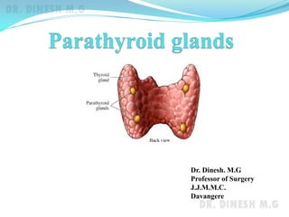
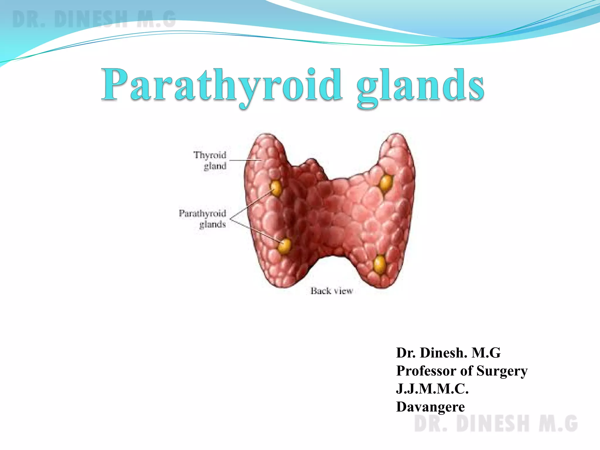
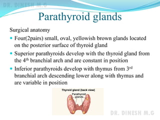
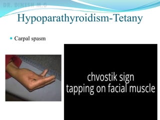
Dr. Dinesh. M.G
Professor of Surgery
J.J.M.M.C.
Davangere
Parathyroid glands
Surgical anatomy
 Four(2pairs) small, oval, yellowish brown glands located
on the posterior surface of thyroid gland
 Superior parathyroids develop with the thyroid gland from
the 4th branchial arch and are constant in position
 Inferior parathyroids develop with thymus from 3rd
branchial arch descending lower along with thymus and
are variable in position
Parathyroid glands
Physiology
Chief cells of parathyroid produce parathormone(PTH)
PTH raises plasma calcium levels by
 Increasing calcium absorption from intestine
 Releasing calcium from bones by osteoclastic stimulation
 Increasing the renal resorption of calcium
Calcitonin secreted by parafollicular cells of thyroid has
opposite action on calcium i.e. it lowers the serum calcium
levels.
Hypoparathyroidism
 Usually due to damage to parathyroid gland during
thyroidectomy
 Due to decreased PTH hypocalcemia develops leading to
tetany.
Clinical features
 Circumoral tingling and numbness
 Chvostek’s sign
 Trousseau’s sign
 Carpopedal smasm
 Laryngeal stridor
Hypoparathyroidism-Tetany
 Carpal spasm
Hypoparathyroidism-Tetany
Diagnosis
 Serum calcium levels below normal(9-11mg%)
Treatment
 10 ml of intravenous calcium gluconate slowly in acute
cases
 Oral calcium with Vit-D3 in permanent
hypoparathyroidism
Hyperparathyroidism
 Increased secretion of PTH leading to hypercalcemia and
its clinical manifestations
Types
 Primary hyperparathyroidism
 Adenoma (solitary)
 Hyperplasia
 carcinoma
 Secondary hyperparathyroidism
 Decreased calcium levels in CRF & Vit-D deficiency
 Tertiary hyperparathyroidism
 Prolonged stimulation by hypocalcemia
Hyperparathyroidism
Clinical features
 Asymptomatic- detected by biochemical screening
 Symptomatic cases
 Renal stones
 Diseases of bones
 Bone pains
 Pathological fractures
 Cysts and pseudotumours of bones
 Osteoporosis and subperiosteal erosions in skull and phalanges
 Psychic moans
 Abdominal groans
 Peptic ulcers
 Pancreatitis
Hyperparathyroidism
Diagnosis
 Biochemical investigations
 Raised calcium and PTH levels
 Decreased serum phosphorus levels
 Raised serum alkaline phosphatase levels
 Radiological investigations
 X-ray skull and phalanges
 Usg neck
 CT and MRI
 Thallium-Technetium subtraction isotope scan
 Selective angiography and venous sampling
Hyperparathyroidism
Treatment
 Surgical removal of overactive glands
 Adenoma-Excision
 Hyperplasia –Excision of all 4 parathyroids and
autotransplantation of one parathyroid in forearm muscles
 Carcinoma – radical excision along with thyroid
Pituitary gland

Parathyroid and Pituitary Glands

  • 1.
    Dr. Dinesh. M.G Professorof Surgery J.J.M.M.C. Davangere
  • 2.
    Parathyroid glands Surgical anatomy Four(2pairs) small, oval, yellowish brown glands located on the posterior surface of thyroid gland  Superior parathyroids develop with the thyroid gland from the 4th branchial arch and are constant in position  Inferior parathyroids develop with thymus from 3rd branchial arch descending lower along with thymus and are variable in position
  • 3.
    Parathyroid glands Physiology Chief cellsof parathyroid produce parathormone(PTH) PTH raises plasma calcium levels by  Increasing calcium absorption from intestine  Releasing calcium from bones by osteoclastic stimulation  Increasing the renal resorption of calcium Calcitonin secreted by parafollicular cells of thyroid has opposite action on calcium i.e. it lowers the serum calcium levels.
  • 4.
    Hypoparathyroidism  Usually dueto damage to parathyroid gland during thyroidectomy  Due to decreased PTH hypocalcemia develops leading to tetany. Clinical features  Circumoral tingling and numbness  Chvostek’s sign  Trousseau’s sign  Carpopedal smasm  Laryngeal stridor
  • 5.
  • 6.
    Hypoparathyroidism-Tetany Diagnosis  Serum calciumlevels below normal(9-11mg%) Treatment  10 ml of intravenous calcium gluconate slowly in acute cases  Oral calcium with Vit-D3 in permanent hypoparathyroidism
  • 7.
    Hyperparathyroidism  Increased secretionof PTH leading to hypercalcemia and its clinical manifestations Types  Primary hyperparathyroidism  Adenoma (solitary)  Hyperplasia  carcinoma  Secondary hyperparathyroidism  Decreased calcium levels in CRF & Vit-D deficiency  Tertiary hyperparathyroidism  Prolonged stimulation by hypocalcemia
  • 8.
    Hyperparathyroidism Clinical features  Asymptomatic-detected by biochemical screening  Symptomatic cases  Renal stones  Diseases of bones  Bone pains  Pathological fractures  Cysts and pseudotumours of bones  Osteoporosis and subperiosteal erosions in skull and phalanges  Psychic moans  Abdominal groans  Peptic ulcers  Pancreatitis
  • 9.
    Hyperparathyroidism Diagnosis  Biochemical investigations Raised calcium and PTH levels  Decreased serum phosphorus levels  Raised serum alkaline phosphatase levels  Radiological investigations  X-ray skull and phalanges  Usg neck  CT and MRI  Thallium-Technetium subtraction isotope scan  Selective angiography and venous sampling
  • 10.
    Hyperparathyroidism Treatment  Surgical removalof overactive glands  Adenoma-Excision  Hyperplasia –Excision of all 4 parathyroids and autotransplantation of one parathyroid in forearm muscles  Carcinoma – radical excision along with thyroid
  • 11.