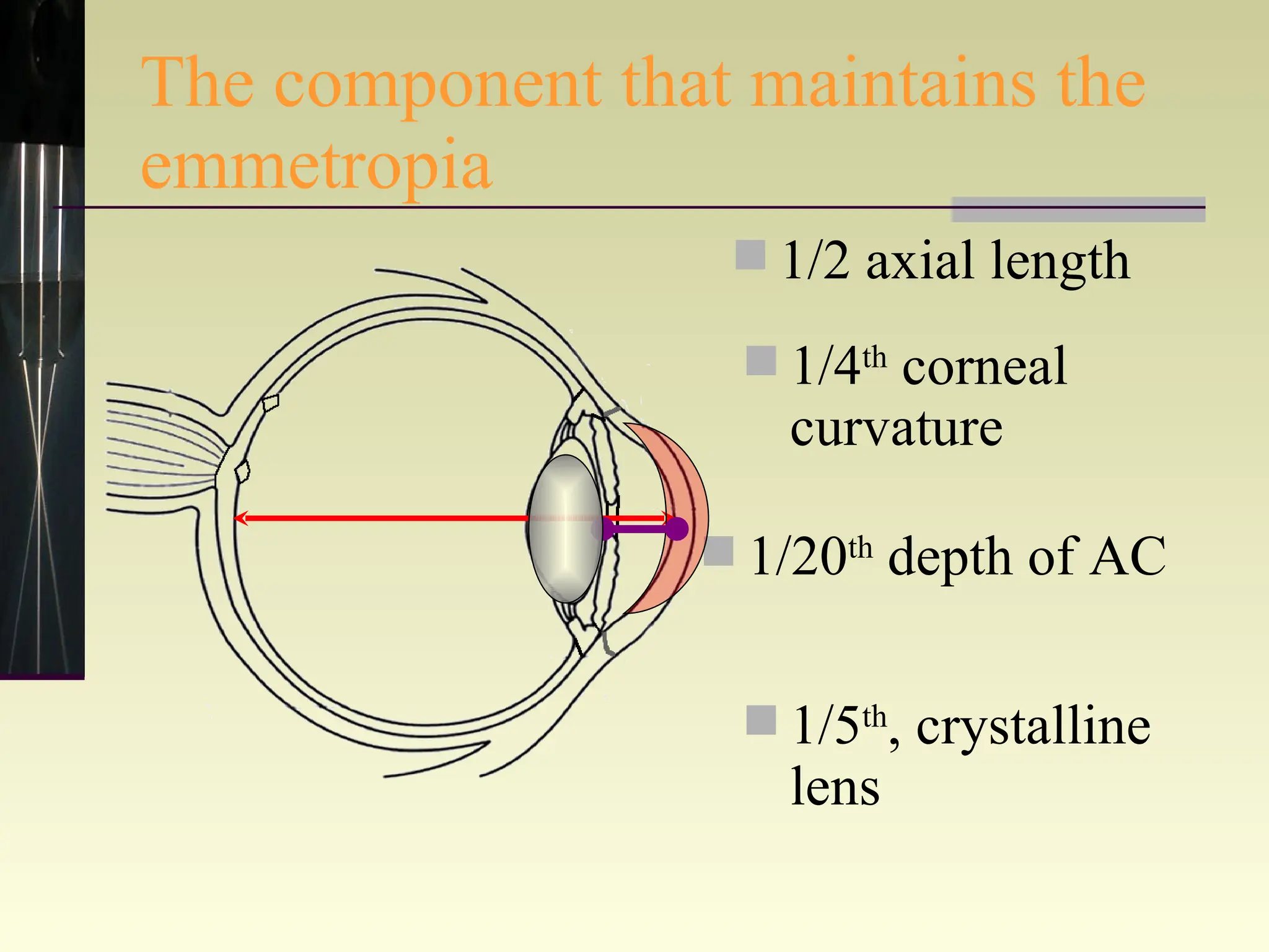
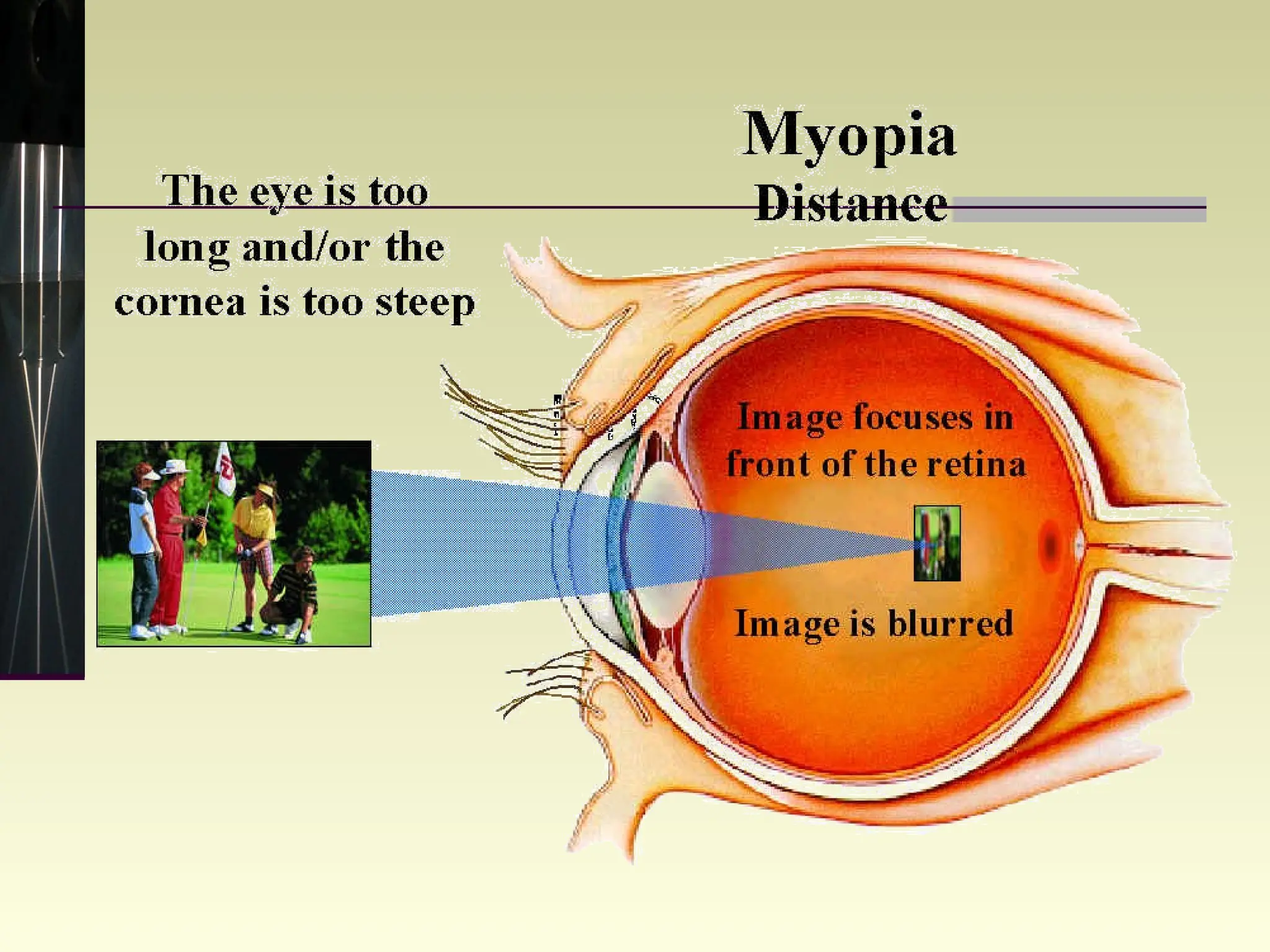
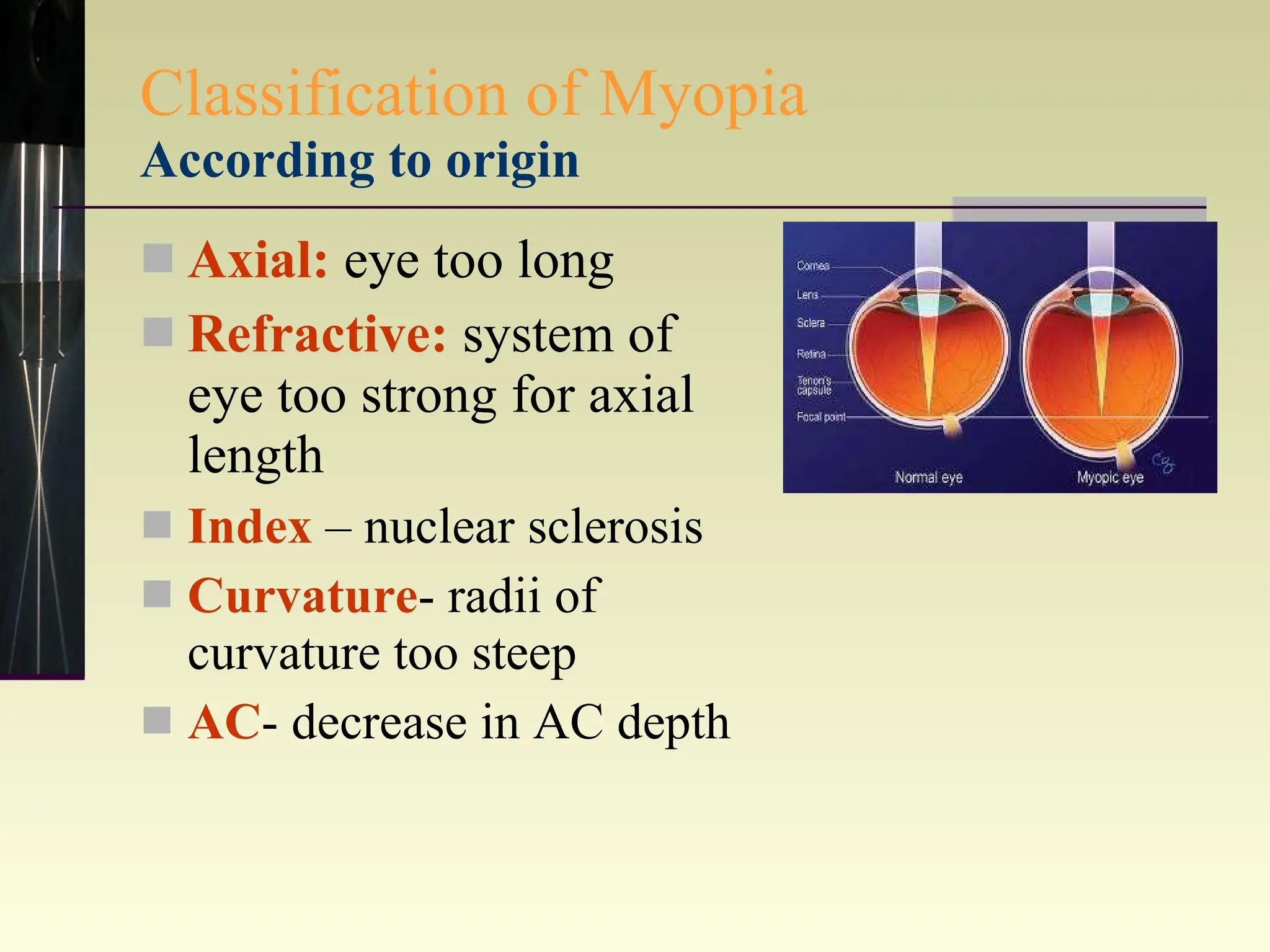
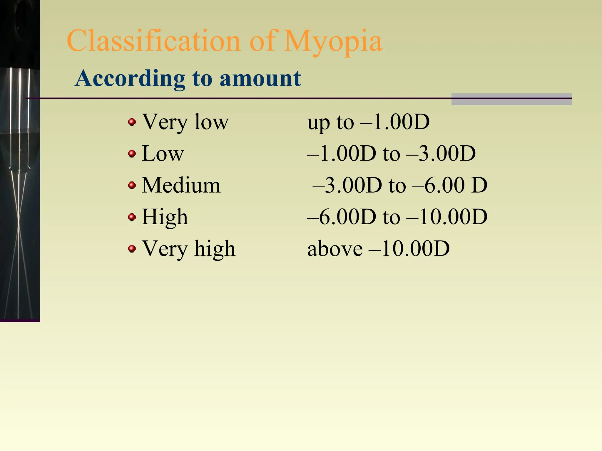
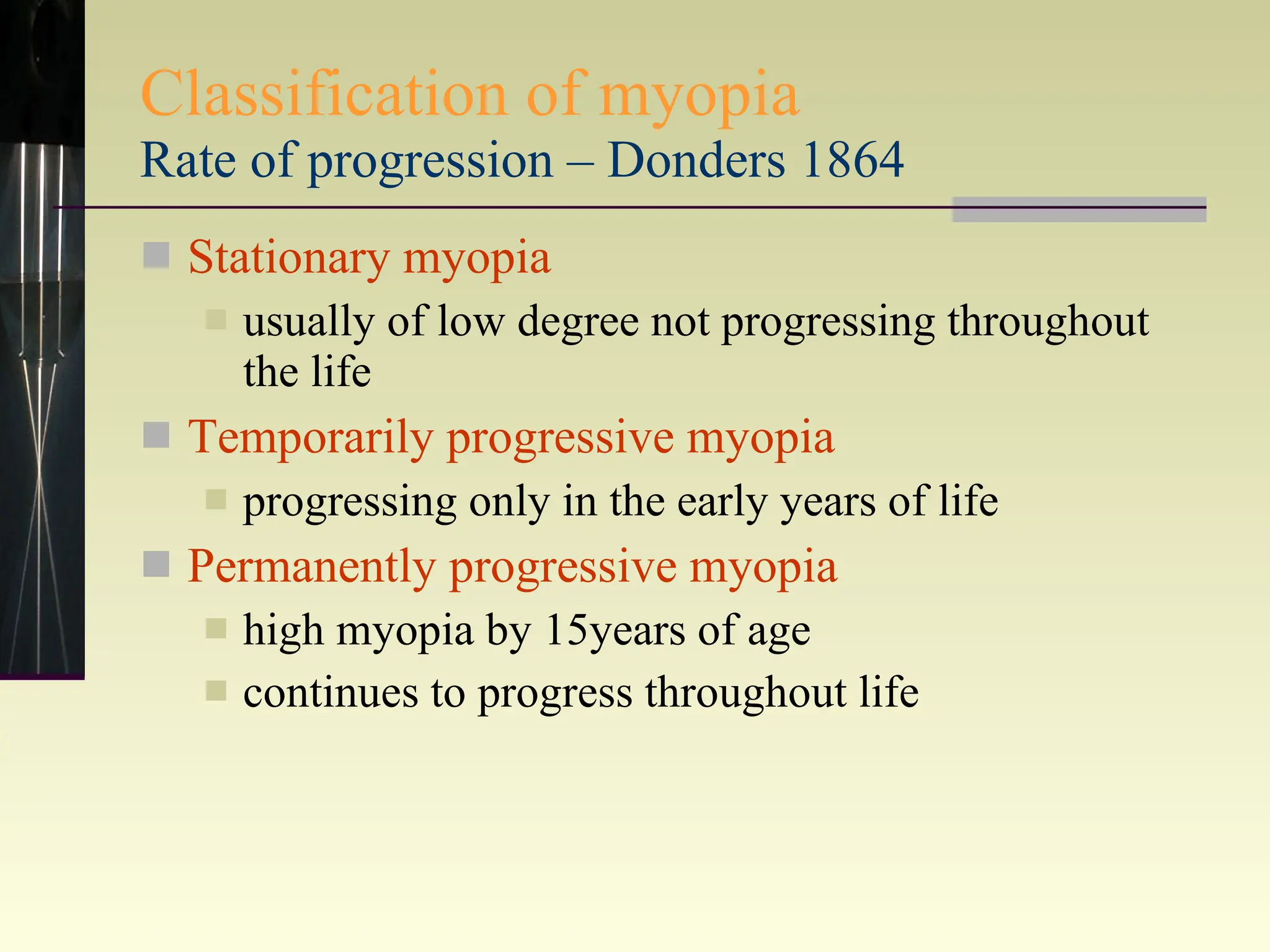
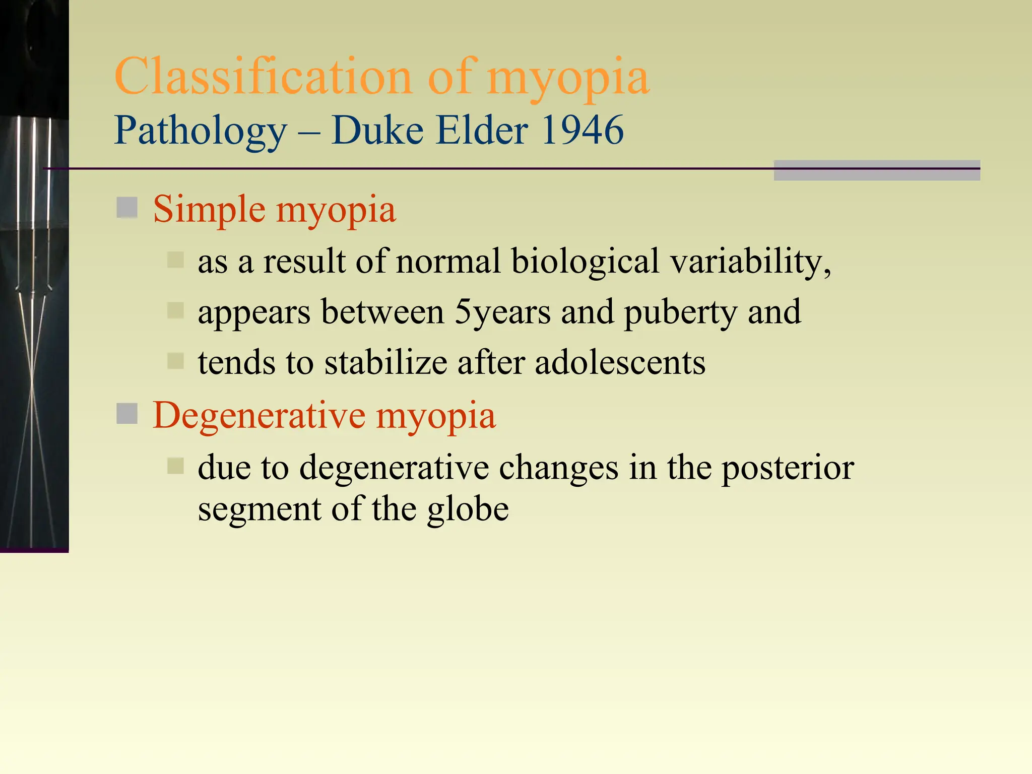
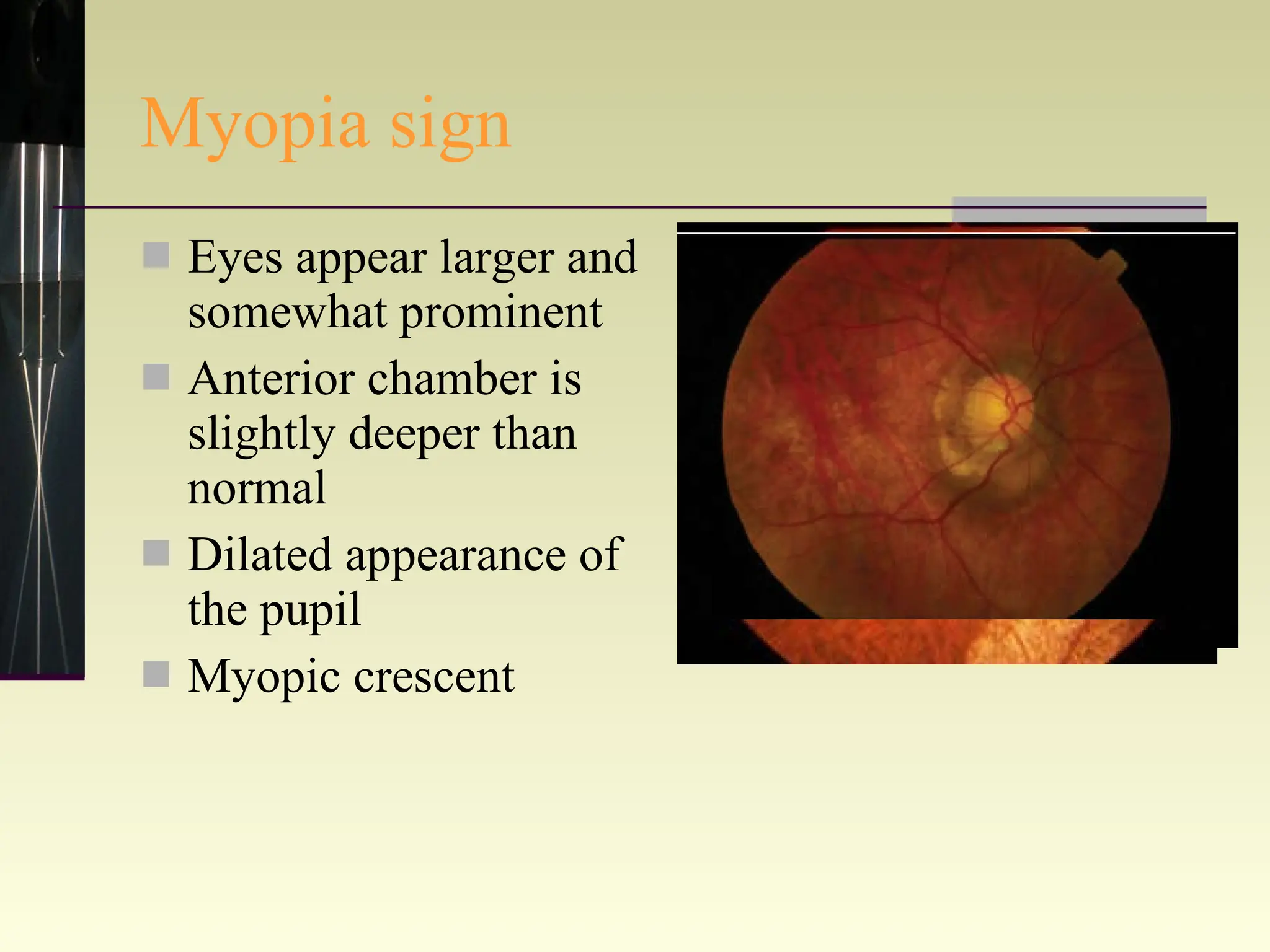
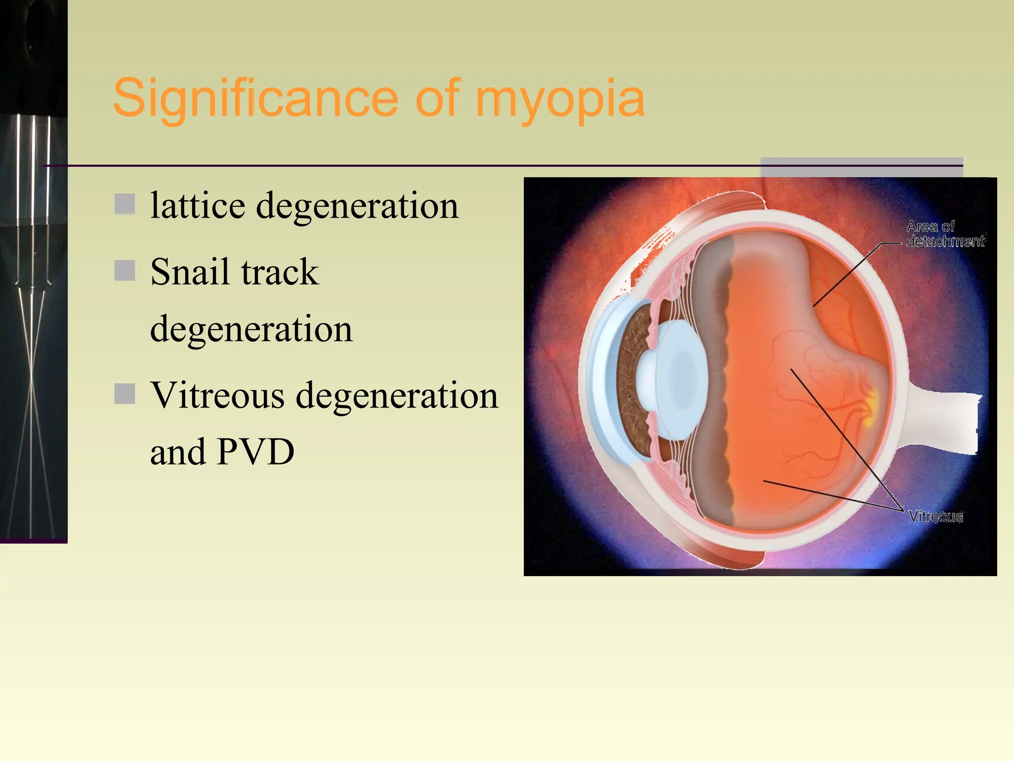
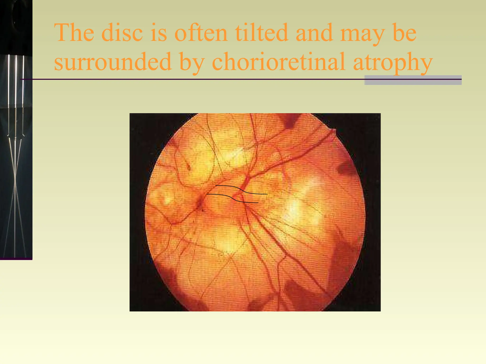
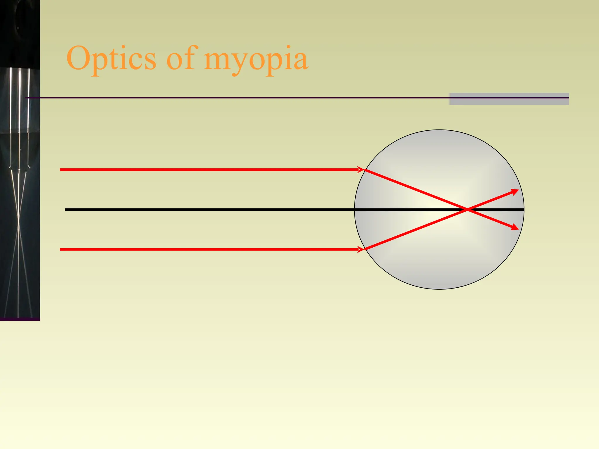
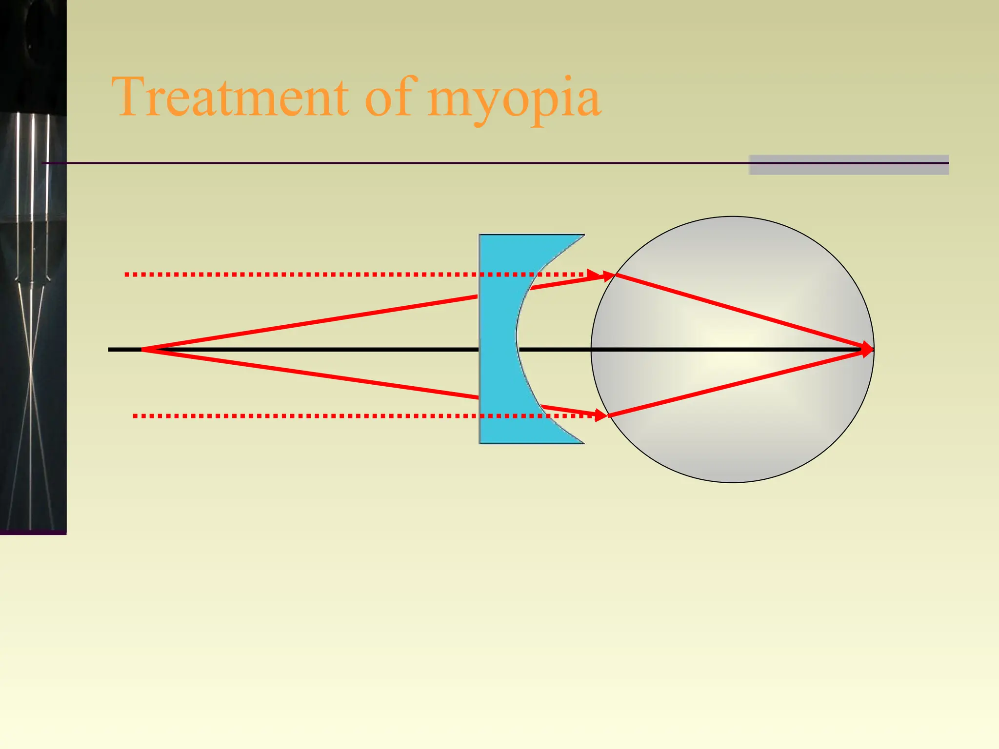
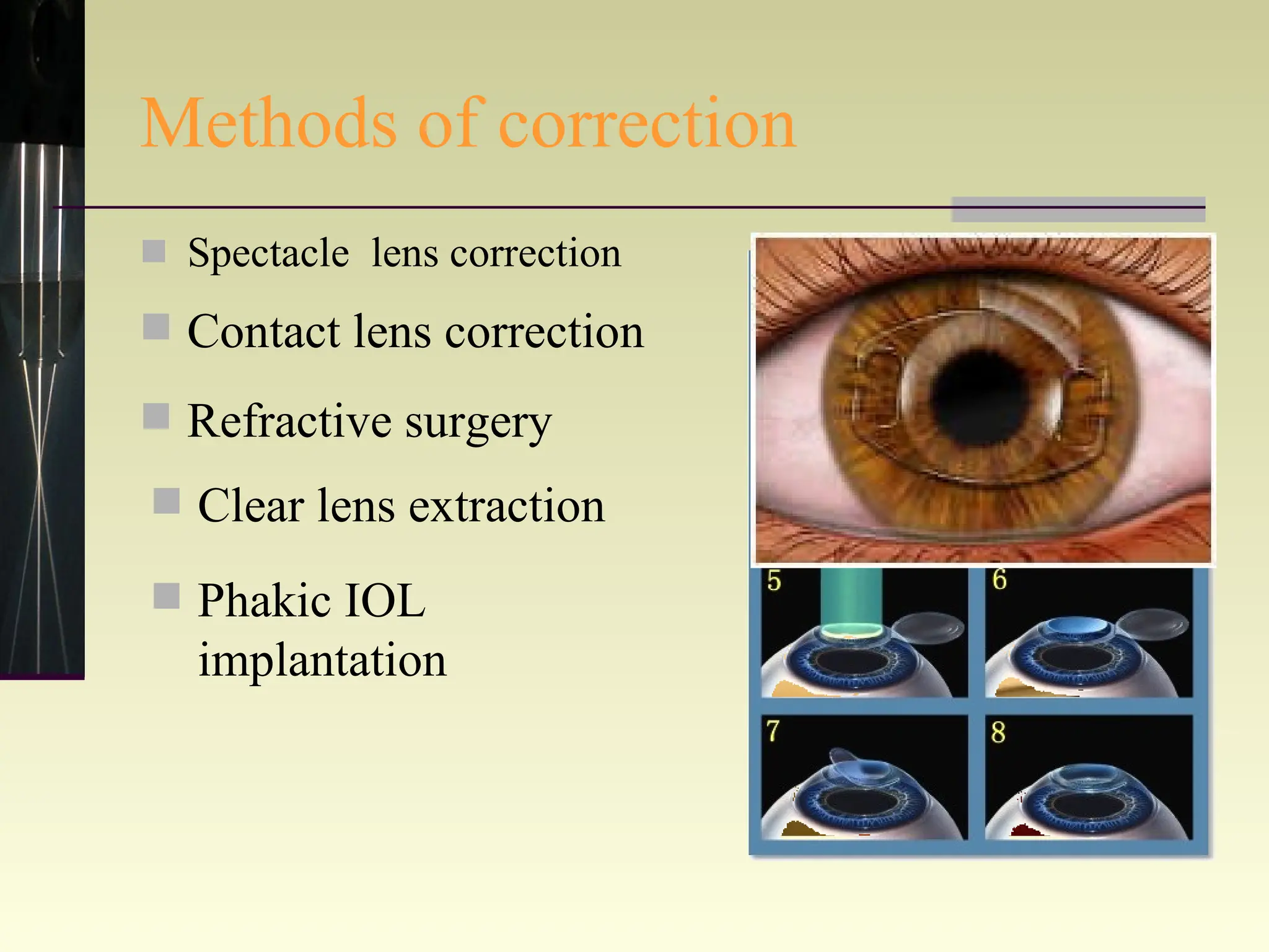
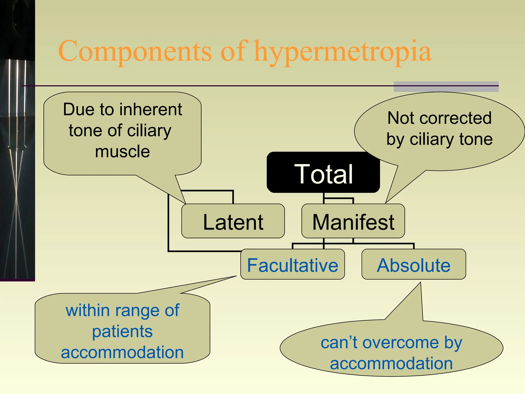
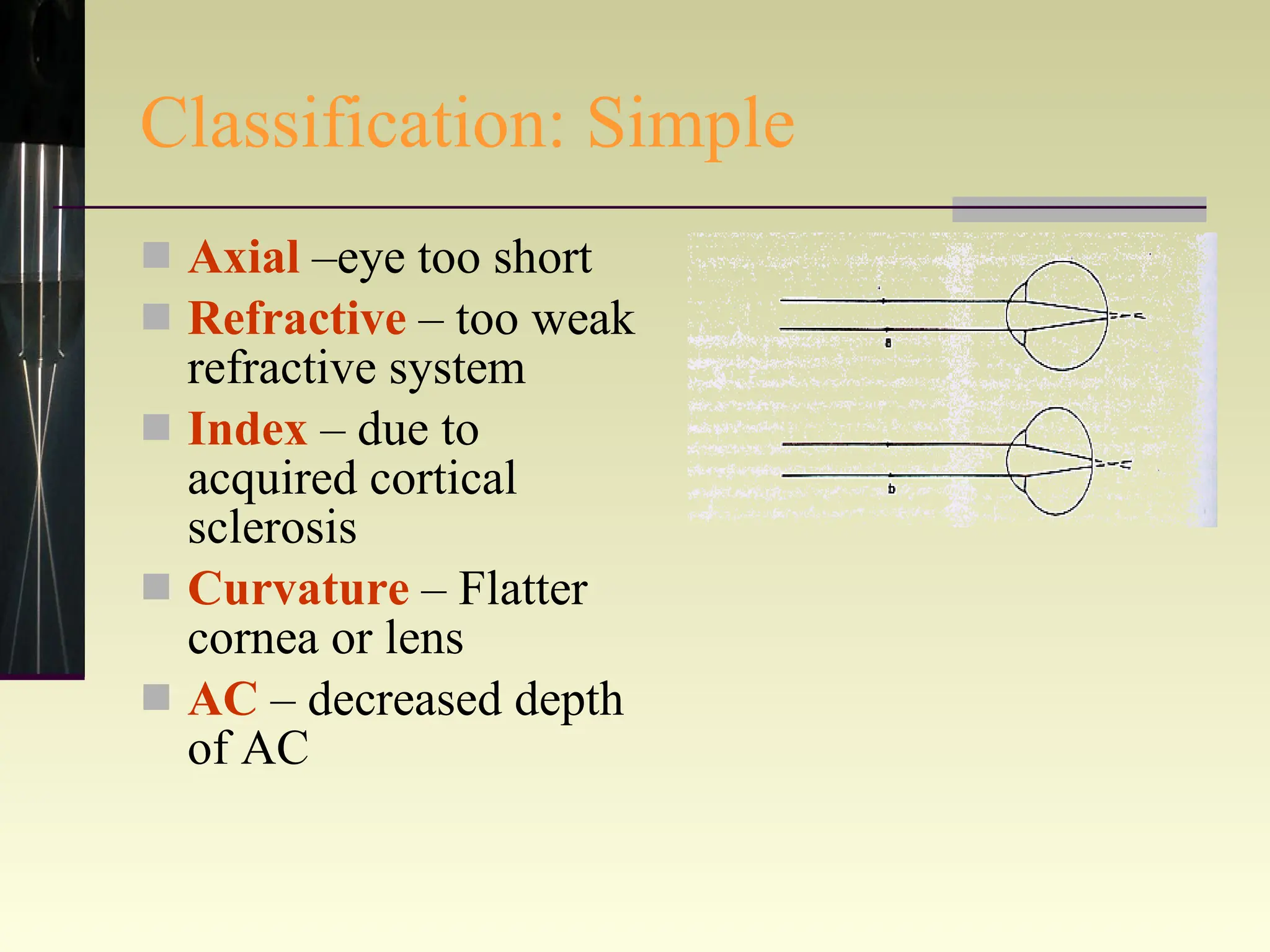
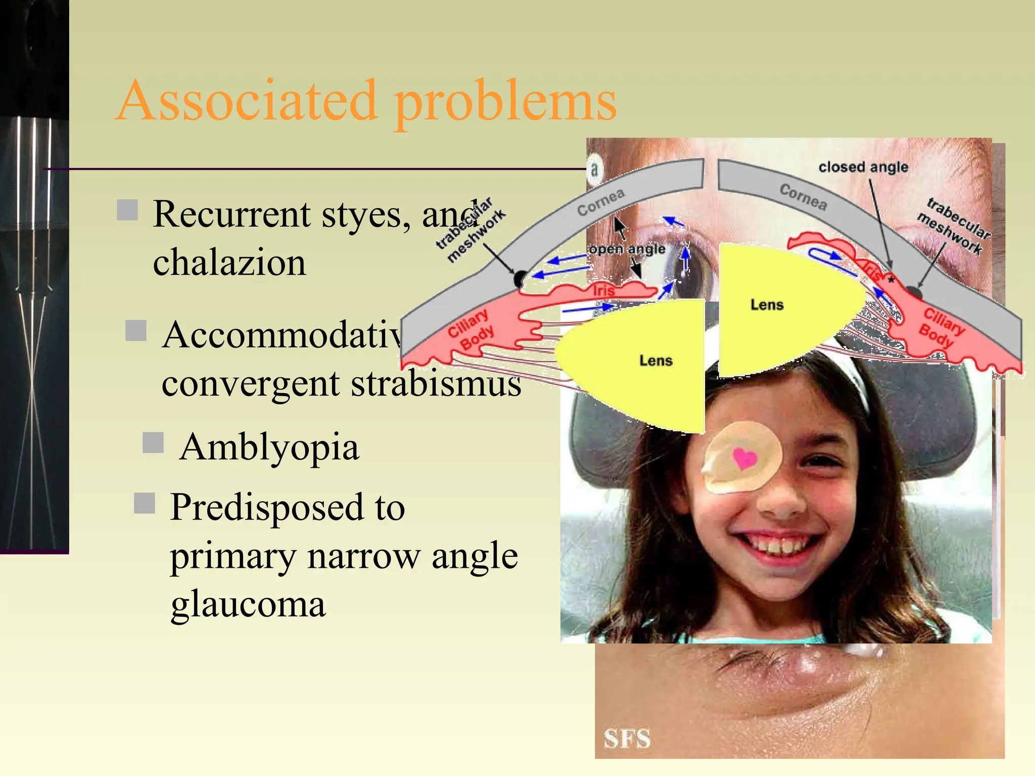
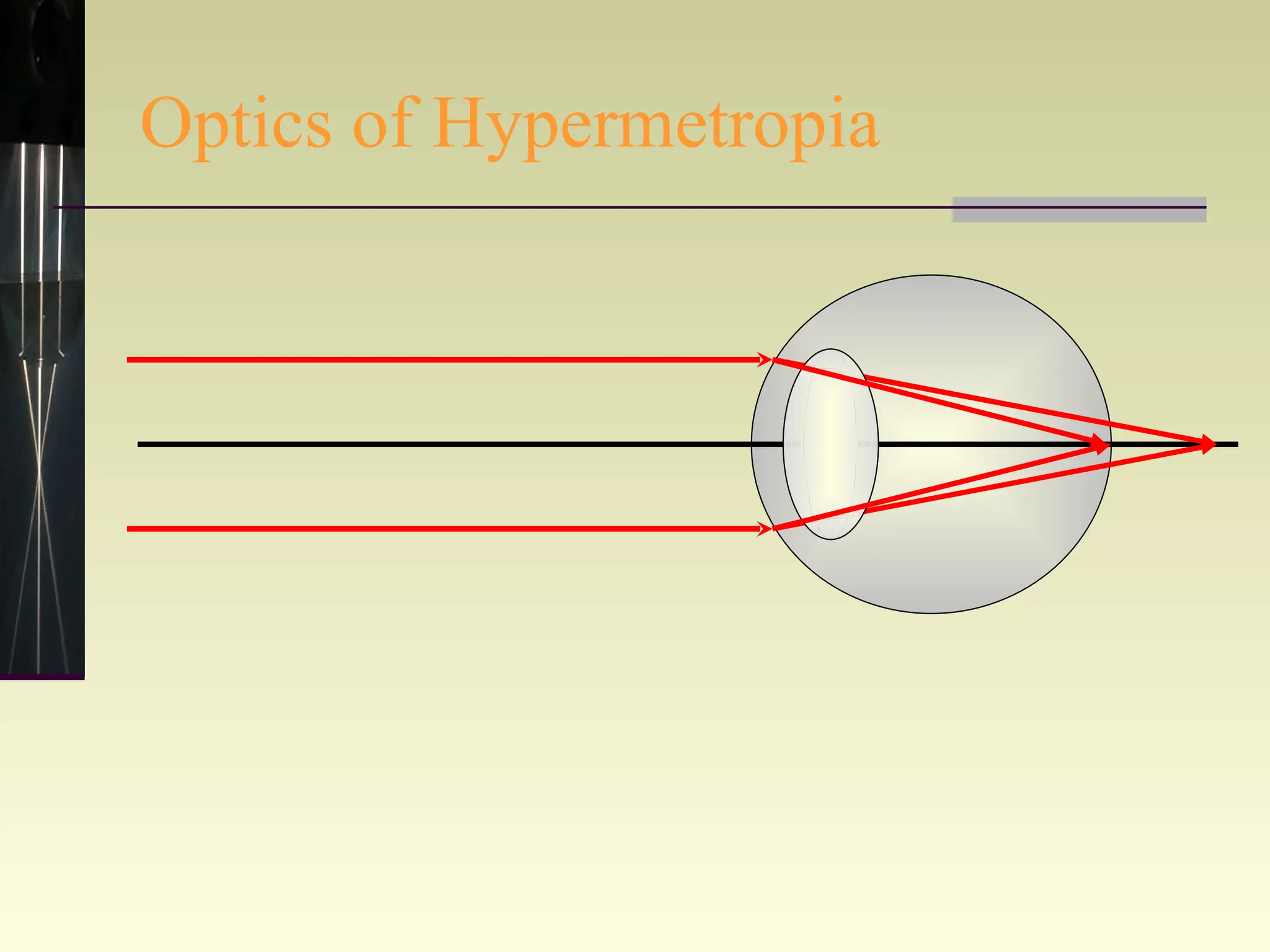
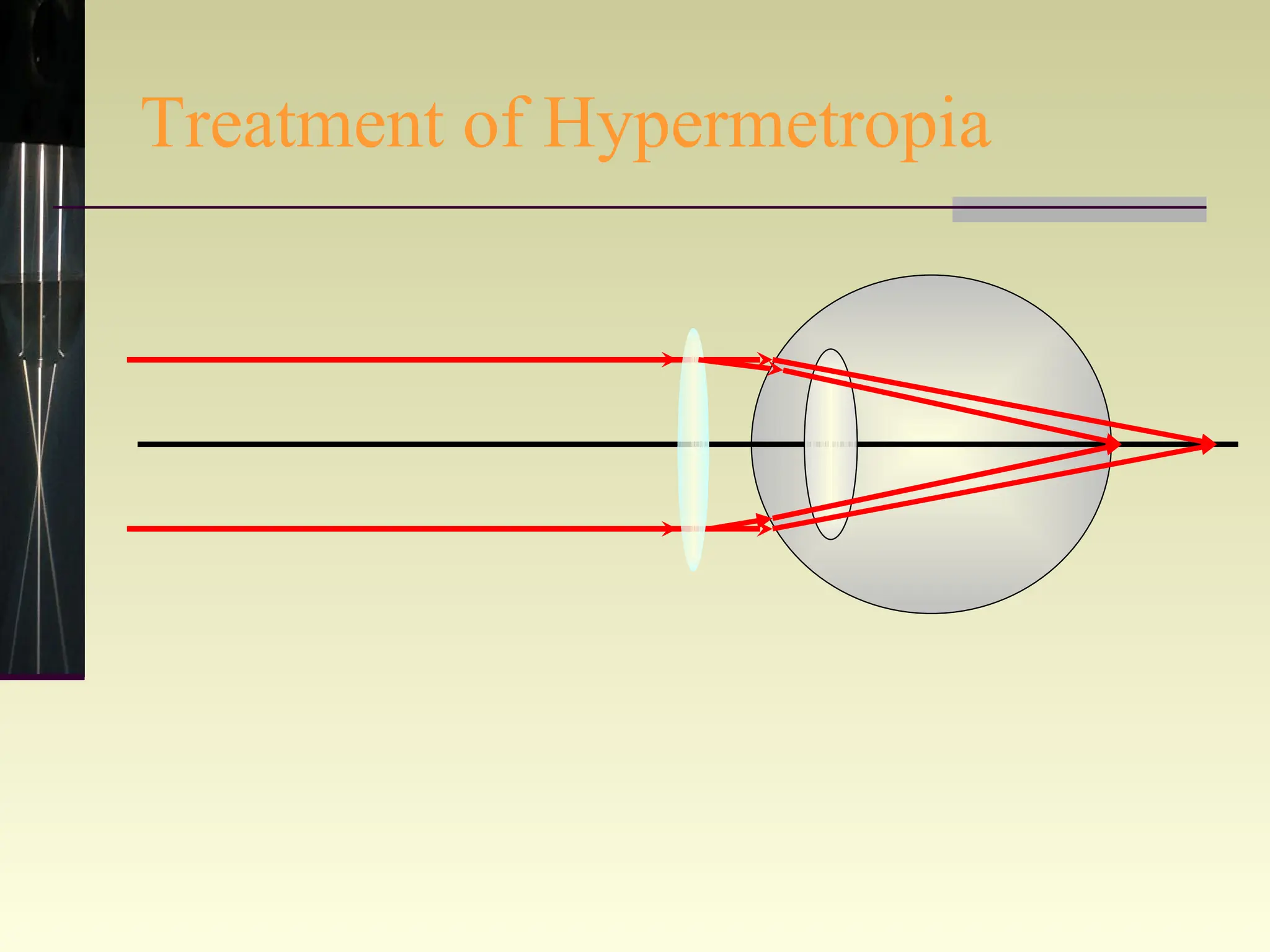
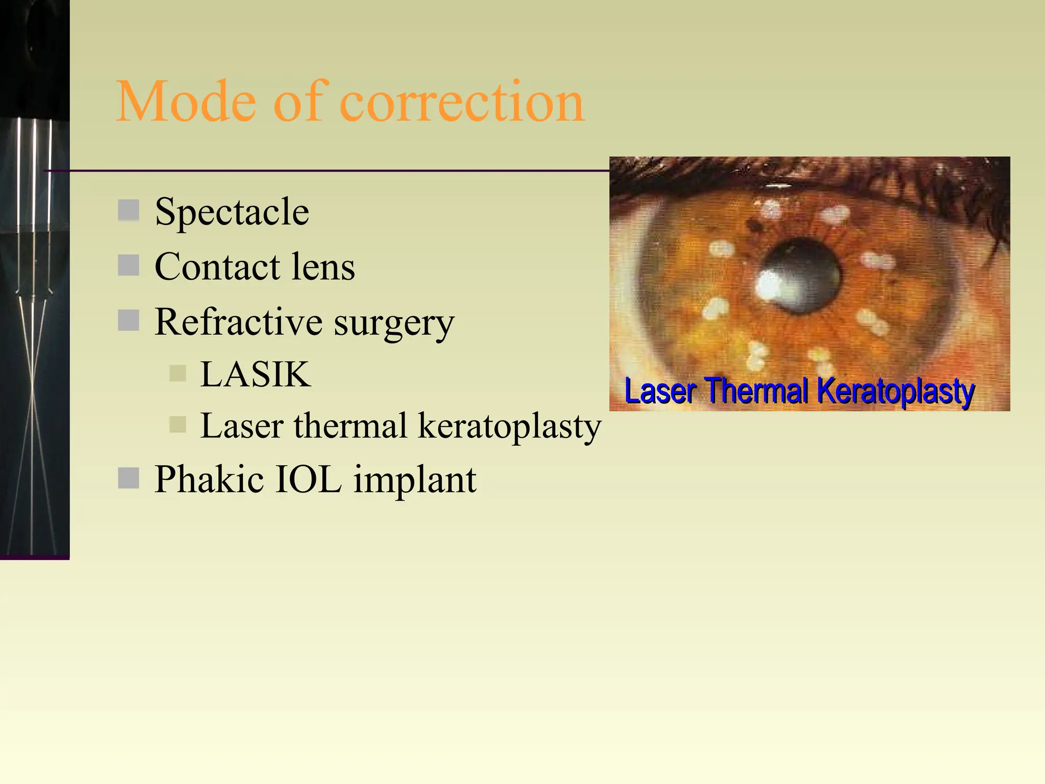
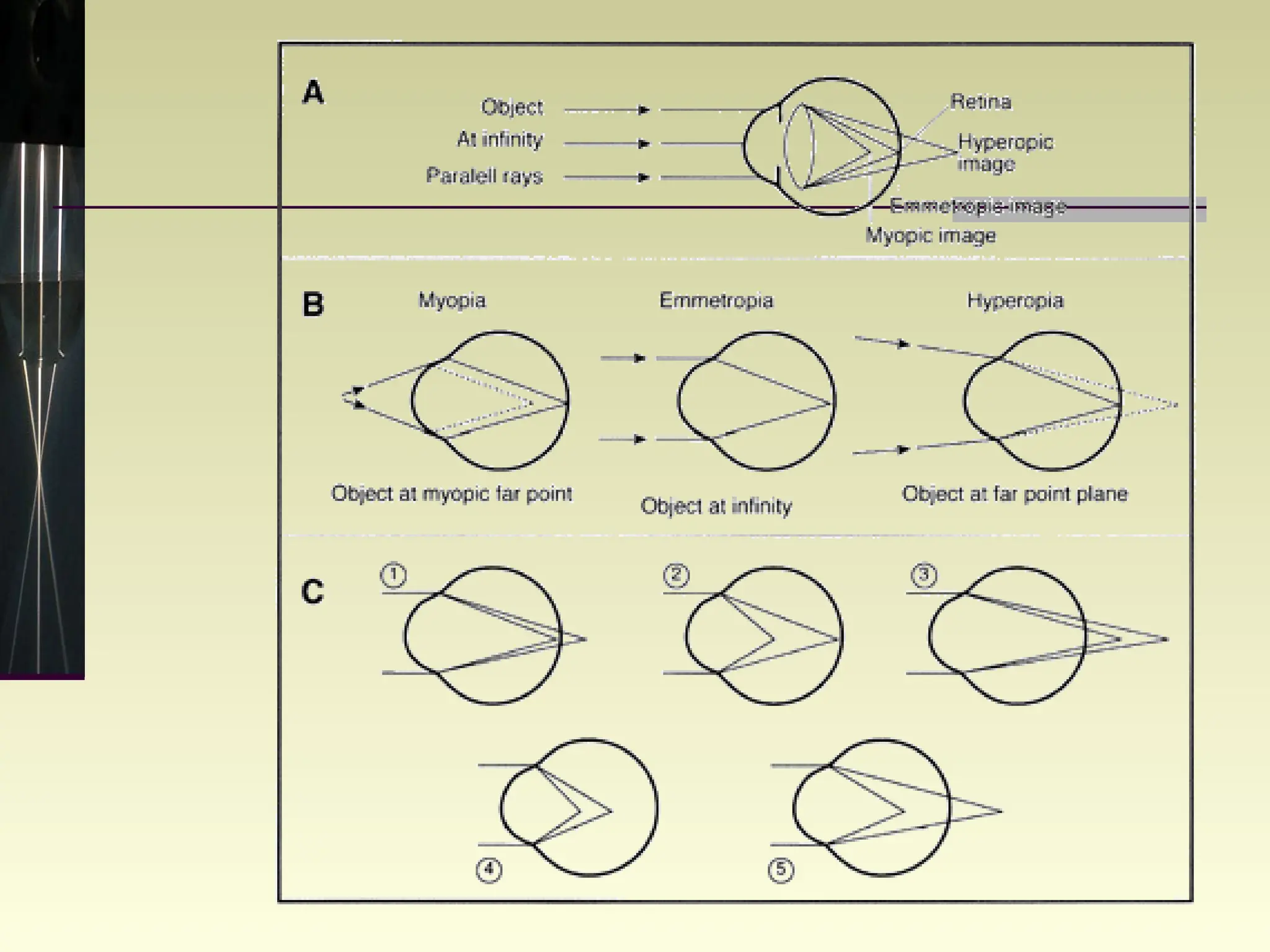
REFRACTIVE ERROR Gauri S. Shrestha, M.Optom, FIACLE
Refraction and its components during the growth of the eye Two distinct phases in the  growth of the eye   Infantile phase  (a rapid and marked)  Juvenile phase  (slower and much slighter)
Refraction and its components during the growth of the eye At birth anteroposterior length of the eye= 16 mm At 2 years of age, anteroposterior length = 22mm Eye assumes hyperopia during 1-3 years Astigmatism= against the rule B/W 3-14 years, elongation of eye= 0.1 mm/yr
Emmetropia The parallel rays of light coming from infinity are focused at the retina with accommodation being at rest
The component that maintains the emmetropia 1/2 axial length 1/5 th , crystalline lens 1/4 th  corneal curvature 1/20 th  depth of AC
Ametropia The parallel rays of light coming from infinity  (with accommodation at rest)  are focussed either in front or behind the retina in one or both the meridian Myopia:  image is focussed in front of retina  Hypermetropia:  image is focussed behind the retina  Astigmatism:  refraction varies in different meridian resulting in loss of point focus
MYOPIA
Classification of Myopia According to origin Axial:  eye too long Refractive:  system of eye too strong for axial length  Index  – nuclear sclerosis Curvature - radii of curvature too steep AC - decrease in AC depth
Classification of Myopia   According to amount Very low  up to –1.00D Low  –1.00D to –3.00D Medium   –3.00D to –6.00 D High  –6.00D to –10.00D Very high  above –10.00D
Classification of myopia Rate of progression – Donders 1864 Stationary myopia  usually of low degree not progressing throughout the life Temporarily progressive myopia progressing only in the early years of life Permanently progressive myopia   high myopia by 15years of age  continues to progress throughout life
Classification of myopia Pathology – Duke Elder 1946 Simple myopia  as a result of normal biological variability,  appears between 5years and puberty and  tends to stabilize after adolescents Degenerative myopia   due to degenerative changes in the posterior segment of the globe
Myopia symptom Poor distant vision Asthenopic Symptoms Watching T.V. from close distance Keeping books close to face Squinting of eyes Poor academic performance
Myopia sign Eyes appear larger and somewhat prominent Anterior chamber is slightly deeper than normal Dilated appearance of the pupil Myopic crescent
Significance of myopia lattice degeneration  Snail track degeneration Vitreous degeneration and PVD
The disc is often tilted and may be surrounded by chorioretinal atrophy
Peripheral chorioretinal atrophy (paving stone degeneration)
Optics of myopia
Treatment of myopia
Methods of correction Spectacle  lens correction Refractive surgery Contact lens correction Clear lens extraction Phakic IOL implantation
Hypermetropia People with hyperopia can see distant objects, but have difficulty seeing objects that are of close
Components of hypermetropia Due to inherent tone of ciliary  muscle Not corrected by ciliary tone within range of patients accommodation can’t overcome by accommodation Total Latent Manifest Facultative Absolute
Classification: Simple Axial  –eye too short Refractive  – too weak refractive system Index  – due to acquired cortical sclerosis Curvature  – Flatter cornea or lens AC  – decreased depth of AC
Classification: Degree Very low  +0.25D to +1.00D Low  +1.25D to +3.00D Medium  +3.00D to +5.00D High  > +5.00D
Classification: pathological Deformational   Short axial length eg; microphthalmus, optic edema Absence of element : aphakia Displacement of lens : posterior Paralysis of accommodation
Symptom Blur near vision Intermittent sudden blurring of vision Asthenopia during near work
Sign small to normal Eye ball size Cornea smaller than the normal Shallow AC and narrow angles Small optic disc Short AP length of the eyeball
Associated problems Recurrent styes, and chalazion Accommodative convergent strabismus Amblyopia Predisposed to primary narrow angle glaucoma
Optics of Hypermetropia
Treatment of Hypermetropia
Mode of correction Spectacle Contact lens Refractive surgery LASIK Laser thermal keratoplasty Phakic IOL implant Laser Thermal Keratoplasty
Astigmatism A variation of power in the different meridians of the eye Causes Corneal At birth – against the Rule, Adult – With the Rule , elder – Against the Rule Lens  – tilt of lens, difference in thickness of lens  Eccentric fovea Fundus irregularity
Classification: meridian With the rule astigmatism Greatest power lies in vertical meridian (60-120º) Against the rule astigmatism Greatest power lies in horizontal meridian (30-150º) Oblique astigmatism Greatest power lies in oblique meridian (30-60 or 120-150º)
Classification; component Total astigmatism = Corneal astigmatism + Internal astigmatism Corneal astigmatism = anterior cornea Internal astigmatism = Posterior corneal, Lens, Retinal   = -0.50DC X 090
Classification: regularity Regular astigmatism Irregular astigmatism 90º ≠ 90º Either the two principle axes  are/aren’t  right angle to each other  or The curvature of any one meridian  is/ isn’t  uniform
Causes of irregular astigmatism Cornea Injuries of cornea, congenital opacities, pterygium, wound healing, keratoconus Lens Inequality in density, deformity, incipient cataract, Coloboma, sublaxation, Tilting
Sturm's Conoid CIRCLE OF LEAST CONFUSION F 1 F 2 INTERVAL OF STURM Rays of light entering cannot converge to a point focus but forms a focal lines
Clinical classification: Sturm’s conoid Compound myopic astigmatism Simple myopic astigmatism Mixed astigmatism Simple hyperopic astigmatism Compound hyperopic astigmatism
Symptom and sign Asthenopia Distortion or blurring of vision Narrowed palpebral fissure and contracted eyebrows head tilt possible
Correction Glasses Contact lens Refractive surgery Arcuate keratotomy LASIK
Assessment History: Blurring of vision D or N Asthenopia : near or distance Distortion Head tilt Diplopia Family history: glasses, squint, amblyopia
Assessment Examination:  VA, Pin hole examination refraction,  keratometry,  axial length,  cycloplegic refraction,  anterior segment examination,  posterior segment examination
 
Thank you

Anomalies of refraction

  • 1.
    REFRACTIVE ERROR GauriS. Shrestha, M.Optom, FIACLE
  • 2.
    Refraction and itscomponents during the growth of the eye Two distinct phases in the growth of the eye Infantile phase (a rapid and marked) Juvenile phase (slower and much slighter)
  • 3.
    Refraction and itscomponents during the growth of the eye At birth anteroposterior length of the eye= 16 mm At 2 years of age, anteroposterior length = 22mm Eye assumes hyperopia during 1-3 years Astigmatism= against the rule B/W 3-14 years, elongation of eye= 0.1 mm/yr
  • 4.
    Emmetropia The parallelrays of light coming from infinity are focused at the retina with accommodation being at rest
  • 5.
    The component thatmaintains the emmetropia 1/2 axial length 1/5 th , crystalline lens 1/4 th corneal curvature 1/20 th depth of AC
  • 6.
    Ametropia The parallelrays of light coming from infinity (with accommodation at rest) are focussed either in front or behind the retina in one or both the meridian Myopia: image is focussed in front of retina Hypermetropia: image is focussed behind the retina Astigmatism: refraction varies in different meridian resulting in loss of point focus
  • 7.
  • 8.
    Classification of MyopiaAccording to origin Axial: eye too long Refractive: system of eye too strong for axial length Index – nuclear sclerosis Curvature - radii of curvature too steep AC - decrease in AC depth
  • 9.
    Classification of Myopia According to amount Very low up to –1.00D Low –1.00D to –3.00D Medium –3.00D to –6.00 D High –6.00D to –10.00D Very high above –10.00D
  • 10.
    Classification of myopiaRate of progression – Donders 1864 Stationary myopia usually of low degree not progressing throughout the life Temporarily progressive myopia progressing only in the early years of life Permanently progressive myopia high myopia by 15years of age continues to progress throughout life
  • 11.
    Classification of myopiaPathology – Duke Elder 1946 Simple myopia as a result of normal biological variability, appears between 5years and puberty and tends to stabilize after adolescents Degenerative myopia due to degenerative changes in the posterior segment of the globe
  • 12.
    Myopia symptom Poordistant vision Asthenopic Symptoms Watching T.V. from close distance Keeping books close to face Squinting of eyes Poor academic performance
  • 13.
    Myopia sign Eyesappear larger and somewhat prominent Anterior chamber is slightly deeper than normal Dilated appearance of the pupil Myopic crescent
  • 14.
    Significance of myopialattice degeneration Snail track degeneration Vitreous degeneration and PVD
  • 15.
    The disc isoften tilted and may be surrounded by chorioretinal atrophy
  • 16.
    Peripheral chorioretinal atrophy(paving stone degeneration)
  • 17.
  • 18.
  • 19.
    Methods of correctionSpectacle lens correction Refractive surgery Contact lens correction Clear lens extraction Phakic IOL implantation
  • 20.
    Hypermetropia People withhyperopia can see distant objects, but have difficulty seeing objects that are of close
  • 21.
    Components of hypermetropiaDue to inherent tone of ciliary muscle Not corrected by ciliary tone within range of patients accommodation can’t overcome by accommodation Total Latent Manifest Facultative Absolute
  • 22.
    Classification: Simple Axial –eye too short Refractive – too weak refractive system Index – due to acquired cortical sclerosis Curvature – Flatter cornea or lens AC – decreased depth of AC
  • 23.
    Classification: Degree Verylow +0.25D to +1.00D Low +1.25D to +3.00D Medium +3.00D to +5.00D High > +5.00D
  • 24.
    Classification: pathological Deformational Short axial length eg; microphthalmus, optic edema Absence of element : aphakia Displacement of lens : posterior Paralysis of accommodation
  • 25.
    Symptom Blur nearvision Intermittent sudden blurring of vision Asthenopia during near work
  • 26.
    Sign small tonormal Eye ball size Cornea smaller than the normal Shallow AC and narrow angles Small optic disc Short AP length of the eyeball
  • 27.
    Associated problems Recurrentstyes, and chalazion Accommodative convergent strabismus Amblyopia Predisposed to primary narrow angle glaucoma
  • 28.
  • 29.
  • 30.
    Mode of correctionSpectacle Contact lens Refractive surgery LASIK Laser thermal keratoplasty Phakic IOL implant Laser Thermal Keratoplasty
  • 31.
    Astigmatism A variationof power in the different meridians of the eye Causes Corneal At birth – against the Rule, Adult – With the Rule , elder – Against the Rule Lens – tilt of lens, difference in thickness of lens Eccentric fovea Fundus irregularity
  • 32.
    Classification: meridian Withthe rule astigmatism Greatest power lies in vertical meridian (60-120º) Against the rule astigmatism Greatest power lies in horizontal meridian (30-150º) Oblique astigmatism Greatest power lies in oblique meridian (30-60 or 120-150º)
  • 33.
    Classification; component Totalastigmatism = Corneal astigmatism + Internal astigmatism Corneal astigmatism = anterior cornea Internal astigmatism = Posterior corneal, Lens, Retinal = -0.50DC X 090
  • 34.
    Classification: regularity Regularastigmatism Irregular astigmatism 90º ≠ 90º Either the two principle axes are/aren’t right angle to each other or The curvature of any one meridian is/ isn’t uniform
  • 35.
    Causes of irregularastigmatism Cornea Injuries of cornea, congenital opacities, pterygium, wound healing, keratoconus Lens Inequality in density, deformity, incipient cataract, Coloboma, sublaxation, Tilting
  • 36.
    Sturm's Conoid CIRCLEOF LEAST CONFUSION F 1 F 2 INTERVAL OF STURM Rays of light entering cannot converge to a point focus but forms a focal lines
  • 37.
    Clinical classification: Sturm’sconoid Compound myopic astigmatism Simple myopic astigmatism Mixed astigmatism Simple hyperopic astigmatism Compound hyperopic astigmatism
  • 38.
    Symptom and signAsthenopia Distortion or blurring of vision Narrowed palpebral fissure and contracted eyebrows head tilt possible
  • 39.
    Correction Glasses Contactlens Refractive surgery Arcuate keratotomy LASIK
  • 40.
    Assessment History: Blurringof vision D or N Asthenopia : near or distance Distortion Head tilt Diplopia Family history: glasses, squint, amblyopia
  • 41.
    Assessment Examination: VA, Pin hole examination refraction, keratometry, axial length, cycloplegic refraction, anterior segment examination, posterior segment examination
  • 42.
  • 43.

Editor's Notes

  • #4 This represents a decrease in ocular refraction of approximately 3 D for the whole period between 3 and 14 years, when growth appears to be complete.