
More Related Content
Similar to DR SONAL Myopia and astigmatism.pptx
Similar to DR SONAL Myopia and astigmatism.pptx (20)
Recently uploaded
Recently uploaded (20)
DR SONAL Myopia and astigmatism.pptx
- 1. MYOPIA AND ASTIGMATISM Dr. Sonal Patil Assistant Professor GMC Jalgaon
- 2. EMMETROPIA:
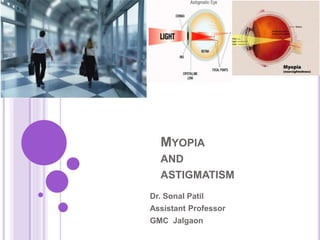
- 3. AMETROPIA Parallel rays of light coming from infinity (with accommodation at rest) are focussed either in front or behind the retina in one or both the meridian. 1. • Myopia: (Near sightedness): image is focused in front of retina 2. • Hyperopia(Hypermetropia)/( Far sightedness): image is focussed behind the retina. 3. • Astigmatism: refraction varies in different meridian resulting in loss of point focus, causes image blur on the retina
- 4. MYOPIA Myopia (Greek) = To close the eye (SHORT SIGHTEDNESS) Far point of the eye closer to retina than infinity
- 5. ETIOLOGICAL CLASSIFICATION 1. Axial myopia results from increase in anteroposterior length of the eyeball. It is the commonest form. 2. Curvatural myopia occurs due to increased curvature of the cornea, lens or both. 3. Positional myopia is produced by anterior placement of crystalline lens in the eye. 4. Index myopia results from increase in the refractive index of crystalline lens associated with nuclear sclerosis. 5. Myopia due to excessive accommodation occurs in patients with spasm of Accommodation.
- 6. CLINICAL VARIETIES OF MYOPIA 1. Congenital myopia 2. Simple or developmental myopia 3. Pathological or degenerative myopia 4. Acquired myopia which may be: (i) post-traumatic; (ii) post-keratitic; (iii) drug-induced, (iv)pseudomyopia; (v) space myopia; (vii) night myopia; and (viii) consecutive myopia.
- 7. CONGENITAL MYOPIA Congenital myopia is present since birth usually diagnosed by the age of 2-3 years mostly unilateral and manifests as anisometropia. Rarely bilateral. The child may develop convergent squint in order to preferentially see clear at its far point which is about 10- 12 cms. associated with other congenital anomalies such as cataract, microphthalmos, aniridia, megalocornea, and congenital separation of retina. Early correction of congenital myopia is desirable
- 8. SIMPLE MYOPIA Simple or developmental myopia is the commonest variety. It is considered as a physiological error not associated with any disease of the eye. Its prevalence increases from 2% at 5 years to 14% at 15 years of age. Since the sharpest rise occurs at school going age i.e between 8 year to 12 years so, it is also called school myopia. Etiology. It results from normal biological variation in the development of eye which may or may not be genetically determined.
- 9. Axial type of simple myopia may signify just a physiological variation in the length of the eyeball or it may be associated with precocious neurological growth during childhood. Curvatural type of simple myopia is considered to be due to underdevelopment of the eyeball. Role of diet in early childhood has also been reported without any conclusive results. Role of genetics. Genetics plays some role in the biological variation of the development of eye, as prevelance of myopia is more in children with both parents myopic (20%) than the children with one parent myopic (10%) and children with no parent myopic (5%). Theory of excessive near work in childhood was also put forward, but did not gain much importance.
- 10. SYMPTOMS Poor vision for distance (short-sightedness) is the main symptom of myopia. Asthenopic symptoms may occur in patients with small degree of myopia. Half shutting of the eyes may be complained by parents of the child. The child does so to achieve the greater clarity of stenopaeic vision.
- 11. SIGNS Prominent eyeballs. The myopic eyes typically are large and somewhat prominent. Anterior chamber is slightly deeper than normal. Pupils are somewhat large and a bit sluggishly reacting. Fundus is normal; rarely temporal myopic crescent may be seen. Magnitude of refractive errror. Simple myopia usually occur between 5 and 10 year of age and it keeps on increasing. In simple myopia, usually the error does not exceed 6 to 8. Diagnosis is confirmed by performing retinoscopy
- 12. . PATHOLOGICAL MYOPIA Pathological/degenerative/progressive myopia, is a rapidly progressive error which starts in childhood at 5-10 years of age and results in high myopia during early adult life which is usually associated with degenerative changes in the eye. Etiology. It results from a rapid axial growth of the eyeball which is outside the normal biological variations of development. Hypothesis has emerged to explain the etiology of pathological myopia. Role of heredity. It is now confirmed that genetic factors play a major role in the etiology, as the progressive myopia is (i) familial; (ii) more common in certain races like Chinese, Japanese, Arabs and Jews, (iii) uncommon among Negroes, Nubians and Sudanese. It is presumed that heredity-linked growth of retina is the determinant in the development of myopia. The sclera due to its distensibility follows the retinal growth but the choroid undergoes degeneration due to stretching, which in turn causes degeneration of retina.
- 13. 2. Role of general growth process, though minor, cannot be denied on the progress of myopia. Lengthening of the posterior segment of the globe commences only during the period of active growth and probably ends with the termination of the active growth. Therefore, the factors (such as nutritional deficiency, debilitating diseases, endocrinal disturbances and indifferent general health) which affect the general growth process will also influence the progress of myopia.
- 15. Symptoms: Defective vision Muscae volitantes Exophoria Flashes of light There may be apparent convergent squint Night blindness may be complained by very high myopes having marked chorioretinal degenerative changes.
- 16. Signs Prominent eyeballs,Large cornea Anterior chamber - deeper than normal Pupils- Large, sluggishly reacting Fundus- normal; rarely temporal myopic crescent may be seen Magnitude of refractive error Does not progress after adolescent (when a degree of 5 to 6D may be attained).
- 17. 1. Large and pale optic disc 2. Myopic crescent where choroid and retina is distracted away from disc margins, a white or grayish white crescentic area in the fundus of the eye located on the temporal side of the optic disc; caused by atrophy of the choroid, permitting the sclera to become visible.
- 18. Super traction crescent may be present on nasal side (retina pulled over disc margin
- 19. Degenerative changes in retina and choroid are common in progressive myopia .These are characterised by white atrophic patches at the macula with a little heaping up of pigment around them. Foster-Fuchs' spot(dark red circular patch due to sub-retinal neovas- cularization and choroidal haemorrhage) may be present at the macula. Cystoid degeneration may be seen at the periphery. In an advanced case there occurs total retinal atrophy, particularly in the central area.
- 20. Foster Fuchs spots: Fuch’s spots are a round or elliptical, circumscribed lesion/dark spots in the macular or perimacular area. It occurs as a result of breaks in Bruch’s membrane and the development of a neovascular membrane, giving rise to a hemorrhage that becomes pigmented.
- 21. Post. Staphyloma: Staphylomas are localized ectasia of the sclera, choroid, and RPE. It can be easily seen on B-scan or a CT Scan. Staphylomas can eventually lead to atrophy and loss of vision. ) Posterior staphyloma due to ectasia of sclera at posterior pole may be apparent as an excavation with the vessels bending backward over its margins
- 22. Degenerative changes in vitreous include: liquefaction, vitreous opacities, and posterior vitreous detachment (PVD) appearing as Weiss' reflex. . Visual fields show contraction and in some cases ring scotoma may be seen. ERG reveals subnormal electroretinogram due to chorioretinal atrophy
- 23. Complications (i) Retinal detachment; (ii) complicated cataract; (iii)vitreous haemorrhage; (iv) choroidal haemorrhage (v)Strabismus fixus convergence.
- 24. 2. Surgical treatment of myopia is becoming very popular now-a-days. 3. General measures empirically believed to effect the progress of myopia (unproven usefulness) include balanced diet rich in vitamins and proteins and early management of associated debilitating disease.
- 25. 4. Low vision aids (LVA) are indicated in patients of progressive myopia with advanced degenerative changes, where useful vision cannot be obtained with spectacles and contact lenses. 5. Prophylaxis (genetic counselling). As the pathological myopia has a strong genetic basis, the hereditary transfer of disease may be decreased by advising against marriage between two individuals with progressive myopia.
- 26. REFRACTIVE SURGERY OF MYOPIA 1. Radial keratotomy (RK) refers to making deep (90 percent of corneal thickness) radial incisions in the peripheral part of cornea leaving the central 4 mm optical zone . These incisions on healing; flatten the central cornea thereby reducing its refractive power. This procedure gives very good correction in low to moderate myopia (2 to 6 D). Disadvantages. Note: Because of its disadvantages RK is not recommended presently. (i) Cornea is weakened, so chances of globe rupture following trauma are more after RK than after PRK. This point is particularly important for patients who are at high risk of blunt trauma, e.g., sports persons, athletes and military personnel. (ii) Rarely, uneven healing may lead to irregular astigmatism. (iii) Patients may feel glare at night.
- 27. PHOTOREFRACTIVE KERATECTOMY 2. Photorefractive keratectomy (PRK). In this technique, to correct myopia a central optical zone of anterior corneal stroma is photoablated using excimer laser (193-nm UV flash) to cause flattening of the central cornea . Like RK, the PRK also gives very good correction for –2 to –6 D of myopia. Disadvantages. Note: Because of its disadvantages PRK is not recommended presently: (i) Postoperative recovery is slow. Healing of the epithelial defect may delay return of good vision and patient may experience pain or discomfort for several weeks. (ii) There may occur some residual corneal haze in the centre affecting vision. (iii) PRK is more expensive than RK.
- 28. LASER IN-SITU KERATOMILEUSIS Laser in-situ keratomileusis (LASIK). In this technique first a flap of 130-160 micron thickness of anterior corneal tissue is raised. After creating acorneal flap midstromal tissue is ablated directly with an excimer laser beam, ultimately flattening the cornea Currently this procedure is being considered the refractive surgery of choice for myopia of up to – 12 D.
- 29. Patient selection criteria are: Patients above 20 years of age, Stable refraction for at least 12 months. Motivated patient. Absence of corneal pathology. Presence of ectasia or any other corneal pathology and a corneal thickness less than 450 mm is an absolute contraindication for LASIK. Advances in LASIK. Recently many advances have been made in LASIK surgery. Some of the important advances are: Customized (C) LASIK. C-LASIK is based on the wave front technology. This technique, in addition to spherical and cylindrical correction, also corrects the aberrations present in the eye and gives vision beyond 6/6 i.e., 6/5 or 6/4 Epi-(E) LASIK. In this technique instead of corneal stromal flap only the epithelial sheet is separated mechanically with the use of a customized device (Epiedge Epikeratome). Being an advanced surface ablation procedure, it is devoid of complications related to corneal stromal flap.
- 30. Advantages of LASIK. (i) Minimal or no postoperative pain. (ii) Recovery of vision is very early as compared to PRK. (iii) No risk of perforation during surgery and later rupture of globe due to trauma unlike RK. (iv) No residual haze unlike PRK where subepithelial scarring may occur. (v) LASIK is effective in correcting myopia of – 12 D.
- 31. DISADVANTAGES 1. LASIK is much more expensive. 2. It requires greater surgical skill than RK and PRK. 3. There is potential risk of flap related complications which include (i) intraoperative flap amputation, (ii) wrinkling of the flap on repositioning, (iii) postoperative flap dislocation/subluxation, (iv) epithelization of flap-bed interface, and (v) irregular astigmatism.
- 32. 4. Extraction of clear crystalline lens (Fucala's operation) has been advocated for myopia of –16 to –18 D, especially in unilateral cases. Recently, clear lens extraction with intraocular lens implantion of appropriate power is being recommended as the refractive surgery for myopia of more than 12 D. 5. Phakic intraocular lens or intraocular contact lens (ICL) implantation is also being considered for correction of myopia of >12D. In this technique, a special type of intraocular lens is implanted in the anterior chamber or posterior chamber anterior to the natural crystalline lens.
- 33. 6. Intercorneal ring (ICR) implantation into the peripheral cornea at approximately 2/3 stromal depth is being considered. It results in a vaulting effect that flattens the central cornea, decreasing myopia. The ICR procedure has the advantage of being reversible. 7. Orthokeratology a non-surgical reversible method of molding the cornea with overnight wear unique rigid gas permeable contact lenses, is also being considered for correction of myopia upto –5D. It can be used even in the patients below 18 year of age.
- 34. ASTIGMATISM Astigmatism is a type of refractive error where in the refraction varies in the different meridia. Consequently,the rays of light entering in the eye cannot convergeto a point focus but form focal lines. Broadly, thereare two types of astigmatism: Regular Irregular.
- 35. REGULAR ASTIGMATISM Astigmatism is regular when the refractive power changes uniformly from one meridian to another (i.e., there are two principal meridia). Etiology 1. Corneal astigmatism is the result of abnormalities of curvature of cornea. It constitutes the most common cause of astigmatism. 2. Lenticular astigmatism is rare. It may be: i. Curvatural due to abnormalities of curvature of lens as seen in lenticonus. ii. Positional due to tilting or oblique placement of lens as seen in subluxation. iii. Index astigmatism may occur rarely due to variable refractve index of lens in different meridia. 3. Retinal astigmatism due to oblique placement of macula may also be seen occasionally
- 36. TYPES OF REGULAR ASTIGMATISM Depending upon the axis and the angle between thetwo principal meridia, regular astigmatism can be classified into the following types : 1. With-the-rule astigmatism. In this type the two principal meridia are placed at right angles to one another but the vertical meridian is more curved than the horizontal. Thus, correction of this astigmatism will require the concave cylinders at 180° ± 20° or convex cylindrical lens at 90° ± 20°. This is called 'with-the-rule' astigmatism, because similar astigmatic condition exists normally (the vertical meridian is normally rendered 0.25 D more convex than the horizontal meridian by the pressure of eyelids).
- 37. 2. Against-the-rule astigmatism refers to an astigmatic condition in which the horizontal meridian is more curved than the vertical meridian. Therefore, correction of this astigmatism will require the presciption of convex cylindrical lens at 180° ± 20° or concave cylindrical lens at 90° ± 20° axis
- 38. Oblique astigmatism: Two principal meridia are not the horizontal and vertical though these are at right angles to one another.
- 39. Bi-oblique astigmatism: Two principal meridia are not the horizontal and vertical and they are not at right angle to each other eg: one may be at 30 degree and other at 100 degree.
- 40. As already mentioned, in regular astigmatism the parallel rays of light are not focused on a point but form two focal lines. The configuration of rays refracted through the astigmatic surface (toric surface) is called Sturm’s conoid and the distance between the two focal lines is known as focal interval of Sturm
- 41. STURM'S CONOID The configuration of rays refracted through a toric surface is called the Sturm’s conoid. The shape of bundle of the light rays at different levels in Sturm's conoid is as follows: At point A, the vertical rays (V) are converging more than the horizontal rays (H); so the section here is a horizontal oval or an oblate ellipse. At point B, (first focus) the vertical rays have come to a focus while the horizontal rays are still converging and so they form a horizontal line. At point C, the vertical rays are diverging and their divergence is less than the convergence of the horizontal rays; so a horizontal oval is formed here. At point D, the divergence of vertical rays is exactly equal to the convergence of the horizontal rays from the axis. So here the section is a circle, which is called the circle of least diffusion. At point E, the divergence of vertical rays is more than the convergence of horizontal rays; so the section here is a vertical oval. At point F, (second focus), the horizontal rays have come to a focus while the vertical rays are divergent and so a vertical line is formed here. Beyond F, (as at point G) both horizontal and vertical rays are diverging and so the section will always be a vertical oval or prolate ellipse. The distance between the two foc (B and F) is called the focal interval of Sturm.
- 42. OPTICS OF REGULAR ASTIGMATISM Refractive types of regular astigmatism Depending upon the position of the two focal lines in relation to retina, the regular astigmatism is further classified into three types: 1. Simple astigmatism, wherein the rays are focused on the retina in one meridian and either in front simple myopic astigmatism Or behind simple hypermetropic astigmatism the retina in the other meridian.
- 43. 2. Compound astigmatism. In this type the rays of light in both the meridia are focused either in front or behind the retina and the condition is labelled as compound myopic or compound hypermetropic astigmatism, respectively . 3. Mixed astigmatism refers to a condition wherein the light rays in one meridian are focused in front and in other meridian behind the retina . Thus in one meridian eye is myopic and in another hypermetropic. Such patients have comparatively less symptoms as 'circle of least diffusion' is formed on the retina
- 44. Symptoms Symptoms of regular astigmatism include: (i) defective vision; (ii) blurring of objects; (iii) depending upon the type and degree of astigmatism, objects may appear proportionately elongated; and (iv) asthenopic symptoms, which are marked especially in small amount of astigmatism, consist of a dull ache in the eyes, headache, early tiredness of eyes and sometimes nausea and even drowsiness.
- 45. Signs 1. Different power in two meridia is revealed on retinoscopy or autorefractometry. 2. Oval or tilted optic disc may be seen on ophthalmoscopy in patients with high degree of astigmatism. 3. Head tilt. The astigmatic patients may (very exceptionally) develop a torticollis in an attempt to bring their axes nearer to the horizontal or vertical meridians. 4. Half closure of the lid. Like myopes, the astigmatic patients may half shut the eyes to achieve the greater clarity of stenopaeic vision.
- 46. Investigations 1. Retinoscopy reveals different power in two different axis 2. Keratometry. Keratometry and computerized corneal topotograpy reveal different corneal curvature in two different meridia in corneal astigmatism 3. Astigmatic fan test and (4) Jackson's cross cylinder test. These tests are useful in confirming the power and axis of cylindrical lenses
- 47. Optical treatment: 1. Optical treatment of regular astigmatism comprises the prescribing appropriate cylindrical lens, discovered after accurate refraction. i. Spectacles with full correction of cylindrical power and appropriate axis should be used for distance and near vision. ii. Contact lenses. Rigid contact lenses may correct upto 2-3 of regular astigmatism, while soft contact lenses can correct only little astigmatism. For higher degrees of astigmatism toric contact lenses are needed. In order to maintain the correct axis of toric lenses, ballasting or truncation is required
- 48. Surgical treatment: Refractive surgery for astigmatism Refractive surgical techniques employed for myopia can be adapted to correct astigmatism alone or simultaneously with myopia as follows: 1. Astigmatic keratotomy (AK) refers to making transverse cuts in the mid periphery of the steep corneal meridian. AK can be performed alone (for astigmatism only) or along with RK (for associated myopia). 2. Photo-astigmatic refractive keratotomy (PARK) is performed using excimer laser. 3. LASIK procedure can also be adapted to correct astigmatism upto 5D.
- 49. BASED ON FOCUS OF THE PRINCIPAL MERIDIANS
- 50. IRREGULAR ASTIGMATISM: Irregular change of refractive power in different meridia. There are multiple meridia which admin no geomerical analysis.
- 51. Etiological types 1. Curvatural irregular astigmatism is found in patients with extensive corneal scars or keratoconus. 2. Index irregular astigmatism due to variable refractive index in different parts of the crystalline lens may occur rarely during maturation of cataract.
- 52. causes of Irregular astigmatism: Corneal: Scars ,injury, Keratoconus , pterygium, marginal degenration , Lenticular: incipient Cataract, coloboma, subluxation, tilting, inequality in density. Retinal-[scarring of macula,tumours of retina,choroid]
- 53. SYMPTOMS: Defective vision, Distortion of objects and Polyopia.
- 54. Investigations 1. Placido's disc test reveales distorted circles 2. Photokerotoscopy and computerized corneal topography give photographic record of irregular corneal curvature
- 55. 1. Optical treatment of irregular astigmatism consists of contact lens which replaces the anterior surface of the cornea for refraction. 2. Phototherapeutic keratectomy (PTK) performed with excimer laser may be helpful in patients with superficial corneal scar responsible for irregular astigmatism. 3. Surgical treatment is indicated in extensive corneal scarring (when vision does not improve with contact lenses) and consists of penetrating keratoplasty
- 56. THANKS