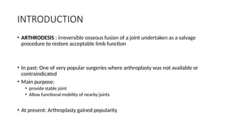
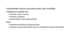
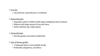
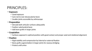
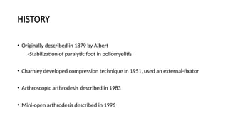
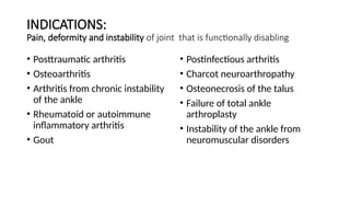
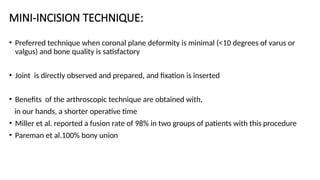
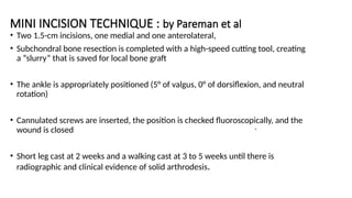
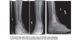
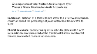
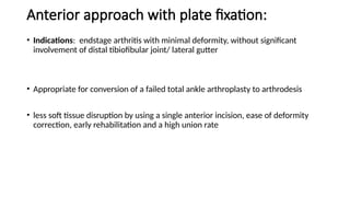
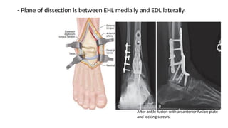
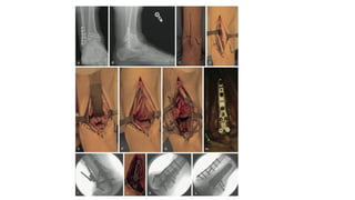
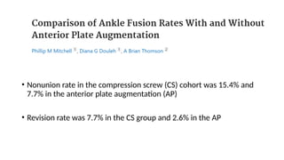
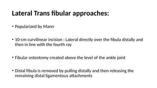
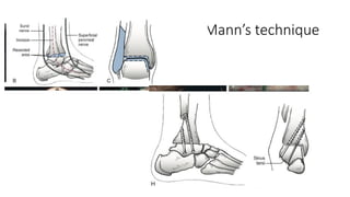
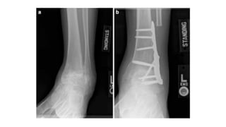
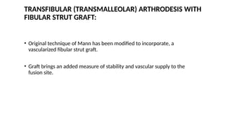
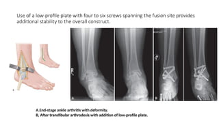
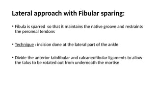
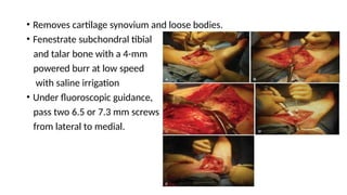
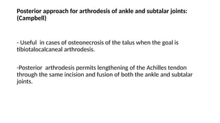
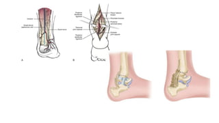
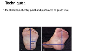
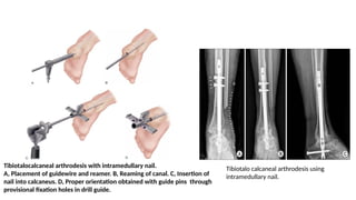
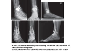
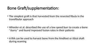
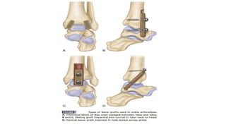
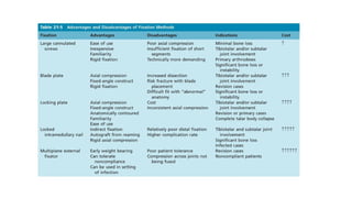
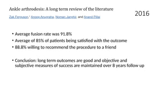
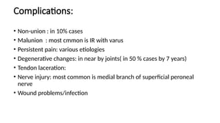
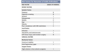
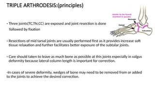
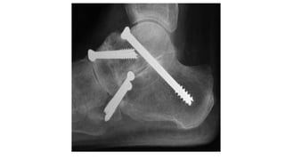
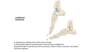
ANKLE ARTHRODESIS
MODERATOR: Asst Prof Dr Bhim Sigdel
PRESENTER: Dr Sandeep Oli (3rd year resident)
PoAHS
2081/03/02
Contents:
• Introduction
• Techniques
• Special considerations
• Complications
INTRODUCTION
• ARTHRODESIS : irreversible osseous fusion of a joint undertaken as a salvage
procedure to restore acceptable limb function
• In past: One of very popular surgeries where arthroplasty was not available or
contraindicated
• Main purpose:
• provide stable joint
• Allow functional mobility of nearby joints
• At present: Arthroplasty gained popularity
• Substantially reduces and relieves pain, joint instability
• Satisfactory solution for:
• Infection, tumor, trauma
• Paralytic conditions
• Osteoarthritis, rheumatoid arthritis
• But:
• Disturbed mechanics of adjacent joints
• Increase energy requirements (eg. For ambulation in lower extremities)
• Can be:
• Intraarticular, extraarticular or combined
• Extraarticular:
• Especially useful in children (with large cartilaginous joint surfaces)
• Patients with large amount of necrotic bone
• Active infection (eg. Tuberculosis)
• Intraarticular:
• Permits greater correction of deformity
• Use of bone grafts:
• if adequate bone is not available locally
• Preferably autogenous, cancellous
PRINCIPLES:
• Exposure
• Good exposure
• Care not to over devascularise bone
• Smaller joints accessible by arthroscope
• Preparation
• Denude both articular surfaces adequately
• Feather subchondral bone
• Add bone grafts in larger joints
• Coaptation
• Surfaces apposed in optimal position with good contact and proper axial and rotational alignment
• Fixation
• Rigid stability and compression by internal or external fixation
• Bone grafts augmentation in larger joints for osseous bridging
• Protect until union
HISTORY
• Originally described in 1879 by Albert
-Stabilization of paralytic foot in poliomyelitis
• Charnley developed compression technique in 1951, used an external-fixator
• Arthroscopic arthrodesis described in 1983
• Mini-open arthrodesis described in 1996
INDICATIONS:
Pain, deformity and instability of joint that is functionally disabling
• Posttraumatic arthritis
• Osteoarthritis
• Arthritis from chronic instability
of the ankle
• Rheumatoid or autoimmune
inflammatory arthritis
• Gout
• Postinfectious arthritis
• Charcot neuroarthropathy
• Osteonecrosis of the talus
• Failure of total ankle
arthroplasty
• Instability of the ankle from
neuromuscular disorders
CONTRAINDICATION
• Vascular impairment of limb
• Skin Infection
• Severe osteoporosis
• Pre existing ipsilateral hindfoot arthrosis or contralateral ankle arthrosis
EVALUATION
History and Physical Examination:
• location of pain
• Examination of ipsilateral Hip and knee joint
• Patients current impairments
• Functional desires of patient
• Proper counselling
• Assessment of medical comorbidities (peripheral neuropathy, DM, peripheral
vascular disease, Active tobacco addiction)
• Bone quality: sclerosis, osteopenia ,bone loss
• Skin : any previous scar
• Timing of surgery: in case of old fractures allow it to completely
vascularize the bone fragments
• Subtalar arthritis: sinus tarsi tenderness in forced passive plantar
flexion
• Vascular status of limb, smoking status directly affects the recovery
and prognosis.
RADIOGRAPHIC EVALUATION:
• Weight bearing AP and Lateral radiographs.
• Assessment of deformity and planning of correction.
• Evaluation of limb length discrepancy.
• Hindfoot alignment view can be used for assessing deformity distal to ankle joint.
• Quality of bone stock and presence of cysts should be checked.
• CT scan can be further helpful in assessment of any defects in the region of
planned fusion.
Optimum position of foot and ankle fusions:
• Dorsiflexon/ plantar flexion: neutral
• Varus/ valgus: 5 degree valgus
• Rotation: 5-10 degree ext rotation
Basic surgical principles: ( mann’s)
• Respect the soft tissues
• Retract carefully
• Meticulously avoid local cutanaeous nerve injury and entrapment during all
stages of procedure
• Remove all cartilage and penetrate into subchondral bone
• Create congruent cancellous surfaces
• Use bone graft and substitutes to fill the defects
• Align hindfoot and leg and forefoot to hind foot: plantigrade
• Stabilize with rigid fixation
• Immobilize and non weight bearing till bony bridging
ARTHRODESIS OF ANKLE:
•Arthroscopic: in well aligned ankle (varus/valgus < 5
degree)
•Mini incision: deformity (<10 degree)
•Open
A, Two anterior portals for ankle arthroscopy are marked in relation to anterior tibial
and other extensor tendons where they cross anterior aspect of ankle. B, Anteromedial
portal site. C, Anterolateral portal site. D, Posterior portals.
ARTHROSCOPIC ARTHRODESIS:
• Avoids large incisions in patients with a poor soft-tissue envelope and minimal
deformity.
• Portal: Standard anterolateral and anteromedial
• Noninvasive distraction
• Curets and shavers are used to remove articular cartilage.
• An arthroscopic burr is then used to abrade the subchondral bone .
• Percutaneous 6.5-mm or 7.0-mm screws can be placed across the joint with the
help of fluoroscopy.
ARTHROSCOPIC ARTHRODESIS:
Advantages:
• Maintenance of malleolar congruency
• Less risk of malunion
• More bone surface and anatomic support for fusion
• Less chance of disruption of the blood supply resulting early healing of the fusion
• Less postoperative pain because of less soft-tissue stripping
• Radiographic evidence of ankle fusion was achieved in 95% of
patients without any serious complications
MINI-INCISION TECHNIQUE:
• Preferred technique when coronal plane deformity is minimal (<10 degrees of varus or
valgus) and bone quality is satisfactory
• Joint is directly observed and prepared, and fixation is inserted
• Benefits of the arthroscopic technique are obtained with,
in our hands, a shorter operative time
• Miller et al. reported a fusion rate of 98% in two groups of patients with this procedure
• Pareman et al.100% bony union
MINI INCISION TECHNIQUE : by Pareman et al
• Two 1.5-cm incisions, one medial and one anterolateral,
• Subchondral bone resection is completed with a high-speed cutting tool, creating
a “slurry” that is saved for local bone graft
• The ankle is appropriately positioned (5° of valgus, 0° of dorsiflexion, and neutral
rotation)
• Cannulated screws are inserted, the position is checked fluoroscopically, and the
wound is closed
• Short leg cast at 2 weeks and a walking cast at 3 to 5 weeks until there is
radiographic and clinical evidence of solid arthrodesis.
.
A and B, End-stage ankle arthritis. C and D, After mini-incision ankle arthrodesis;
use of “home run” screw from posterolateral tibial into talar neck/head distally
ADVANTAGES..
• Offers decreased soft-tissue insult
• Decreased bone stripping
• Easy application
• Rapid healing time for the treatment of severe degenerative changes of the ankle
with minimal deformity.
Conclusion: addition of a third 7.0-mm screw to a 2-screw ankle fusion
construct raised the percentage of joint surface lost from 5.91% to
9.5%
Clinical Relevance: consider using extra-articular plates with 1 or 2
intra-articular screws instead of the traditional 3-screw construct if
there is an elevated concern for nonunion
Anterior approach with plate fixation:
• Indications: endstage arthritis with minimal deformity, without significant
involvement of distal tibiofibular joint/ lateral gutter
• Appropriate for conversion of a failed total ankle arthroplasty to arthrodesis
• less soft tissue disruption by using a single anterior incision, ease of deformity
correction, early rehabilitation and a high union rate
- Plane of dissection is between EHL medially and EDL laterally.
After ankle fusion with an anterior fusion plate
and locking screws.
• Nonunion rate in the compression screw (CS) cohort was 15.4% and
7.7% in the anterior plate augmentation (AP)
• Revision rate was 7.7% in the CS group and 2.6% in the AP
Lateral Trans fibular approaches:
• Popularized by Mann
• 10-cm curvilinear incision : Lateral directly over the fibula distally and
then in line with the fourth ray
• Fibular osteotomy created above the level of the ankle joint
• Distal fibula is removed by pulling distally and then releasing the
remaining distal ligamentous attachments
Mann’s technique
TRANSFIBULAR (TRANSMALLEOLAR) ARTHRODESIS WITH
FIBULAR STRUT GRAFT:
• Original technique of Mann has been modified to incorporate, a
vascularized fibular strut graft.
• Graft brings an added measure of stability and vascular supply to the
fusion site.
Use of a low-profile plate with four to six screws spanning the fusion site provides
additional stability to the overall construct.
A.End-stage ankle arthritis with deformity.
B, After transfibular arthrodesis with addition of low-profile plate.
Lateral approach with Fibular sparing:
• Fibula is sparred so that it maintains the native groove and restraints
the peroneal tendons
• Technique : incision done at the lateral part of the ankle
• Divide the anterior talofibular and calcaneofibular ligaments to allow
the talus to be rotated out from underneath the mortise
• Removes cartilage synovium and loose bodies.
• Fenestrate subchondral tibial
and talar bone with a 4-mm
powered burr at low speed
with saline irrigation
• Under fluoroscopic guidance,
pass two 6.5 or 7.3 mm screws
from lateral to medial.
Posterior approach for arthrodesis of ankle and subtalar joints:
(Campbell)
- Useful in cases of osteonecrosis of the talus when the goal is
tibiotalocalcaneal arthrodesis.
-Posterior arthrodesis permits lengthening of the Achilles tendon
through the same incision and fusion of both the ankle and subtalar
joints.
TIBIOTALOCALCANEAL ARTHRODESIS:
• A lateral approach with or without the onlay fibular graft can be used
• Posterior approach may be appropriate in patients with compromised skin and
soft tissues in the area of a lateral approach.
• Biomechanical studies shows locking plate fixation to have higher rigidity than
intramedullary nails and provide higher initial stiffness
Technique :
• Identification of entry point and placement of guide wire
Tibiotalocalcaneal arthrodesis with intramedullary nail.
A, Placement of guidewire and reamer. B, Reaming of canal. C, Insertion of
nail into calcaneus. D, Proper orientation obtained with guide pins through
provisional fixation holes in drill guide.
Tibiotalo calcaneal arthrodesis using
intramedullary nail.
Tibiocalcaneal arthrodesis with a thin-wire and Ilizarov
external fixation:
• Described by Eylon et al.
• Thin-wire fixation to the leg is done, beginning with ring
fixators at the proximal tibia and supramalleolar region.
• A talar half ring is anchored with two wires positioned
50-60⁰ from each other through the talar neck and body.
• A calcaneal-forefoot extended half-ring is added and
anchored with the wires through the calcaneus and
through the metatarsals
• Final compression is applied.
Charnley Method (1951)
• Compression arthrodesis
• First to describe use of external fixator
• Open debridement of ankle joint cartilage
via anterior approach + Ex-fix
• One pin through distal tibia
• One pin through neck of talus
• Use of connecting bars keeps the arthrodesis compressed.
• Provides arthrodesis in only one plane ( doesn’t provide rotational
stability),hence little consideration of biomechanical principles.
SPECIAL CONSIDERATIONS
OSTEONECROSIS OF THE TALUS:
• Blair’s procedure: Tibiotalar arthrodesis with a sliding bone graft
• Fuses the distal tibia to the talar neck in situations in which the body of the talus has
been lost or is osteonecrotic.
• uses an anterior tibial sliding graft, allows nearly normal appearance of the foot with
little shortening of the extremity, and permits some flexion-extension motion of the foot
on the leg.
Morris et al. modified the technique and used transcalcaneotibial pin for 6 weeks to
improve stability.
Blair fusion. A, Approach to the ankle. B, Excision of body of talus.
C, Sliding bone graft. D, Graft in final position.
FAILED TOTAL ANKLE ARTHROPLASTY:
• Total ankle arthroplasty is on rise.
• Managing failed total ankle arthroplasty is challenging.
• Tibiotalar and tibiotalocalcaneal arthrodesis using massive cancellous
allografts.
• Tibiotalar arthrodesis for patients with good bone stock and an intact
subtalar joint
• Tibiotalocalcaneal arthrodesis for those with poor bone quality, subtalar
• If the subtalar joint is to be preserved.
arthrodesis through an anterior approach with strut iliac crest grafting and
double plating .
(benefit of the use of the previous anterior incision, which makes removal of the
implant easier)
• If the subtalar joint is to be fused, the same anterior approach can be used with
grafting as described, or a lateral transfibular approach gives improved access to
the subtalar joint.
A and B, Total ankle arthroplasty with loosening, periarticular cyst, and medial and
lateral anterior impingement.
C and D, After arthrodesis with femoral head allograft and double-plate fixation
CHARCOAT NEUROPATHY:
• Timing of the surgery is important,
• surgery is best done before a difficult deformity leads to skin breakdown and
infection.
• Multidisciplinary care plan, including revascularization procedures, infection
treatment, and an off-loading regimen when needed
• Pantalar arthrodesis may be indicated as a salvage procedure in patients with
neuro arthropathies to avoid amputation
Bone Graft/supplementation:
• The simplest graft is that harvested from the resected fibula in the
transfibular approach
• Wheeler et al. described the use of a low speed burr to create a bone
‘’slurry’’ and found improved fusion rates in their patients
• A RIA can be used to harvest bone from the hindfoot or tibial shaft
during reaming
2016
• Average fusion rate was 91.8%
• Average of 85% of patients being satisfied with the outcome
• 88.8% willing to recommend the procedure to a friend
• Conclusion: long term outcomes are good and objective and
subjective measures of success are maintained over 8 years follow up
Complications:
• Non-union : in 10% cases
• Malunion : most cmmon is IR with varus
• Persistent pain: various etiologies
• Degenerative changes: in near by joints( in 50 % cases by 7 years)
• Tendon laceration:
• Nerve injury: most common is medial branch of superficial peroneal
nerve
• Wound problems/infection
TRIPLE ARTHRODESIS:(principles)
- Three joints(TC,TN,CC) are exposed and joint resection is done
followed by fixation
- Resections of mid tarsal joints are usually performed first as it provides increase soft
tissue relaxation and further facilitates better exposure of the subtalar joints.
- Care should taken to leave as much bone as possible at this joints especially in valgus
deformity because lateral column length is important for correction.
-In cases of severe deformity, wedges of bone may need to be removed from or added
to the joints to achieve the desired correction.
TRIPPLE ARTHRODESIS:
Indications:
• Failure after 6 months of appropriate Nonoperative management.
• Arthritis involving the subtalar and either or both the TN and CC joints
• Hindfoot instability involving the subtalar and transverse tarsal joints secondary to
neuromuscular disorders , nerve injury, posterior tibial tendon deficiency, or rheumatoid
arthritis.
• Fixed symptomatic varus or valgus malignment of the hindfoot
• Symptomatic unresectable or previously resected calcaneonavicular coalition
• Severe rigid symptomatic pes planovalgus deformity not responsive to previous operative
treatments.
Triple arthrodesis is performed:
(1) to obtain stable and static realignment of the foot,
(2) to remove deforming forces,
(3) to arrest progression of deformity,
(4) to eliminate pain,
(5) to eliminate the use of a short leg brace or to provide sufficient correction to allow
fitting of a long leg brace to control the knee joint,
(6) to obtain a more normal-appearing foot.
Triple arthrodesis: (Technique)
Few modifications:
(Lambrinudi arthrodesis:)
• For correction of isolated fixed equinus deformity in patients older than 10 years
• Wedge of bone is removed from the plantar distal part of the talus
• The talus remains in complete equinus at the ankle joint while the remainder of the
foot is repositioned to the desired degree of plantarflexion
• Tendon resection or transfer may be necessary to prevent varus or valgus deformity
if active muscle power remains
A, Colored area indicates part of talus to be resected.
B, Sharp distal margin of remaining part of talus has been wedged into
prepared trough in navicular,and, and raw osseous surfaces of talus, calcaneus, and cuboid
have been apposed.
LAMBRINUDI
PROCEDURE
Pantalar arthrodesis:
• Indications: more extensive ankle and hind foot degeneration or
deformity, inclusion
• Approach is similar to TTC fusion
2017
Conclusion: Higher overall complication rate after AA, but a higher
reoperation rate for revision after TAA
Alternative to ankle arthrodesis:
• Non operative treatment
• Debridement
• Osteochondral allograft ankle joint resurfacing
• Distraction arthroplasty
• Total ankle arthroplasty
TAKE HOME MESSAGE:
• Principle of arthrodesis is aimed at correction of painful, deformed, or
unstable joint to produce a painless plantigrade foot
• Mini incision technique is preferred technique when coronal plane deformity is
minimal (<10 degrees of varus or valgus) and bone quality is satisfactory
References:
• Campbell’s Operative Orthopaedics – 13th
edition
• Mann’s surgery of foot and ankle – 9th
edition
THANK YOU..

ANKLE AND FOOT ARTHODESIS final.pptx final

  • 1.
    ANKLE ARTHRODESIS MODERATOR: AsstProf Dr Bhim Sigdel PRESENTER: Dr Sandeep Oli (3rd year resident) PoAHS 2081/03/02
  • 2.
    Contents: • Introduction • Techniques •Special considerations • Complications
  • 3.
    INTRODUCTION • ARTHRODESIS :irreversible osseous fusion of a joint undertaken as a salvage procedure to restore acceptable limb function • In past: One of very popular surgeries where arthroplasty was not available or contraindicated • Main purpose: • provide stable joint • Allow functional mobility of nearby joints • At present: Arthroplasty gained popularity
  • 4.
    • Substantially reducesand relieves pain, joint instability • Satisfactory solution for: • Infection, tumor, trauma • Paralytic conditions • Osteoarthritis, rheumatoid arthritis • But: • Disturbed mechanics of adjacent joints • Increase energy requirements (eg. For ambulation in lower extremities)
  • 5.
    • Can be: •Intraarticular, extraarticular or combined • Extraarticular: • Especially useful in children (with large cartilaginous joint surfaces) • Patients with large amount of necrotic bone • Active infection (eg. Tuberculosis) • Intraarticular: • Permits greater correction of deformity • Use of bone grafts: • if adequate bone is not available locally • Preferably autogenous, cancellous
  • 6.
    PRINCIPLES: • Exposure • Goodexposure • Care not to over devascularise bone • Smaller joints accessible by arthroscope • Preparation • Denude both articular surfaces adequately • Feather subchondral bone • Add bone grafts in larger joints • Coaptation • Surfaces apposed in optimal position with good contact and proper axial and rotational alignment • Fixation • Rigid stability and compression by internal or external fixation • Bone grafts augmentation in larger joints for osseous bridging • Protect until union
  • 7.
    HISTORY • Originally describedin 1879 by Albert -Stabilization of paralytic foot in poliomyelitis • Charnley developed compression technique in 1951, used an external-fixator • Arthroscopic arthrodesis described in 1983 • Mini-open arthrodesis described in 1996
  • 8.
    INDICATIONS: Pain, deformity andinstability of joint that is functionally disabling • Posttraumatic arthritis • Osteoarthritis • Arthritis from chronic instability of the ankle • Rheumatoid or autoimmune inflammatory arthritis • Gout • Postinfectious arthritis • Charcot neuroarthropathy • Osteonecrosis of the talus • Failure of total ankle arthroplasty • Instability of the ankle from neuromuscular disorders
  • 9.
    CONTRAINDICATION • Vascular impairmentof limb • Skin Infection • Severe osteoporosis • Pre existing ipsilateral hindfoot arthrosis or contralateral ankle arthrosis
  • 10.
    EVALUATION History and PhysicalExamination: • location of pain • Examination of ipsilateral Hip and knee joint • Patients current impairments • Functional desires of patient • Proper counselling • Assessment of medical comorbidities (peripheral neuropathy, DM, peripheral vascular disease, Active tobacco addiction)
  • 11.
    • Bone quality:sclerosis, osteopenia ,bone loss • Skin : any previous scar • Timing of surgery: in case of old fractures allow it to completely vascularize the bone fragments • Subtalar arthritis: sinus tarsi tenderness in forced passive plantar flexion • Vascular status of limb, smoking status directly affects the recovery and prognosis.
  • 12.
    RADIOGRAPHIC EVALUATION: • Weightbearing AP and Lateral radiographs. • Assessment of deformity and planning of correction. • Evaluation of limb length discrepancy. • Hindfoot alignment view can be used for assessing deformity distal to ankle joint. • Quality of bone stock and presence of cysts should be checked. • CT scan can be further helpful in assessment of any defects in the region of planned fusion.
  • 13.
    Optimum position offoot and ankle fusions: • Dorsiflexon/ plantar flexion: neutral • Varus/ valgus: 5 degree valgus • Rotation: 5-10 degree ext rotation
  • 14.
    Basic surgical principles:( mann’s) • Respect the soft tissues • Retract carefully • Meticulously avoid local cutanaeous nerve injury and entrapment during all stages of procedure • Remove all cartilage and penetrate into subchondral bone • Create congruent cancellous surfaces • Use bone graft and substitutes to fill the defects • Align hindfoot and leg and forefoot to hind foot: plantigrade • Stabilize with rigid fixation • Immobilize and non weight bearing till bony bridging
  • 15.
    ARTHRODESIS OF ANKLE: •Arthroscopic:in well aligned ankle (varus/valgus < 5 degree) •Mini incision: deformity (<10 degree) •Open
  • 16.
    A, Two anteriorportals for ankle arthroscopy are marked in relation to anterior tibial and other extensor tendons where they cross anterior aspect of ankle. B, Anteromedial portal site. C, Anterolateral portal site. D, Posterior portals.
  • 17.
    ARTHROSCOPIC ARTHRODESIS: • Avoidslarge incisions in patients with a poor soft-tissue envelope and minimal deformity. • Portal: Standard anterolateral and anteromedial • Noninvasive distraction • Curets and shavers are used to remove articular cartilage. • An arthroscopic burr is then used to abrade the subchondral bone . • Percutaneous 6.5-mm or 7.0-mm screws can be placed across the joint with the help of fluoroscopy.
  • 18.
    ARTHROSCOPIC ARTHRODESIS: Advantages: • Maintenanceof malleolar congruency • Less risk of malunion • More bone surface and anatomic support for fusion • Less chance of disruption of the blood supply resulting early healing of the fusion • Less postoperative pain because of less soft-tissue stripping
  • 19.
    • Radiographic evidenceof ankle fusion was achieved in 95% of patients without any serious complications
  • 20.
    MINI-INCISION TECHNIQUE: • Preferredtechnique when coronal plane deformity is minimal (<10 degrees of varus or valgus) and bone quality is satisfactory • Joint is directly observed and prepared, and fixation is inserted • Benefits of the arthroscopic technique are obtained with, in our hands, a shorter operative time • Miller et al. reported a fusion rate of 98% in two groups of patients with this procedure • Pareman et al.100% bony union
  • 21.
    MINI INCISION TECHNIQUE: by Pareman et al • Two 1.5-cm incisions, one medial and one anterolateral, • Subchondral bone resection is completed with a high-speed cutting tool, creating a “slurry” that is saved for local bone graft • The ankle is appropriately positioned (5° of valgus, 0° of dorsiflexion, and neutral rotation) • Cannulated screws are inserted, the position is checked fluoroscopically, and the wound is closed • Short leg cast at 2 weeks and a walking cast at 3 to 5 weeks until there is radiographic and clinical evidence of solid arthrodesis. .
  • 23.
    A and B,End-stage ankle arthritis. C and D, After mini-incision ankle arthrodesis; use of “home run” screw from posterolateral tibial into talar neck/head distally
  • 24.
    ADVANTAGES.. • Offers decreasedsoft-tissue insult • Decreased bone stripping • Easy application • Rapid healing time for the treatment of severe degenerative changes of the ankle with minimal deformity.
  • 25.
    Conclusion: addition ofa third 7.0-mm screw to a 2-screw ankle fusion construct raised the percentage of joint surface lost from 5.91% to 9.5% Clinical Relevance: consider using extra-articular plates with 1 or 2 intra-articular screws instead of the traditional 3-screw construct if there is an elevated concern for nonunion
  • 26.
    Anterior approach withplate fixation: • Indications: endstage arthritis with minimal deformity, without significant involvement of distal tibiofibular joint/ lateral gutter • Appropriate for conversion of a failed total ankle arthroplasty to arthrodesis • less soft tissue disruption by using a single anterior incision, ease of deformity correction, early rehabilitation and a high union rate
  • 27.
    - Plane ofdissection is between EHL medially and EDL laterally. After ankle fusion with an anterior fusion plate and locking screws.
  • 29.
    • Nonunion ratein the compression screw (CS) cohort was 15.4% and 7.7% in the anterior plate augmentation (AP) • Revision rate was 7.7% in the CS group and 2.6% in the AP
  • 30.
    Lateral Trans fibularapproaches: • Popularized by Mann • 10-cm curvilinear incision : Lateral directly over the fibula distally and then in line with the fourth ray • Fibular osteotomy created above the level of the ankle joint • Distal fibula is removed by pulling distally and then releasing the remaining distal ligamentous attachments
  • 31.
  • 33.
    TRANSFIBULAR (TRANSMALLEOLAR) ARTHRODESISWITH FIBULAR STRUT GRAFT: • Original technique of Mann has been modified to incorporate, a vascularized fibular strut graft. • Graft brings an added measure of stability and vascular supply to the fusion site.
  • 34.
    Use of alow-profile plate with four to six screws spanning the fusion site provides additional stability to the overall construct. A.End-stage ankle arthritis with deformity. B, After transfibular arthrodesis with addition of low-profile plate.
  • 35.
    Lateral approach withFibular sparing: • Fibula is sparred so that it maintains the native groove and restraints the peroneal tendons • Technique : incision done at the lateral part of the ankle • Divide the anterior talofibular and calcaneofibular ligaments to allow the talus to be rotated out from underneath the mortise
  • 36.
    • Removes cartilagesynovium and loose bodies. • Fenestrate subchondral tibial and talar bone with a 4-mm powered burr at low speed with saline irrigation • Under fluoroscopic guidance, pass two 6.5 or 7.3 mm screws from lateral to medial.
  • 37.
    Posterior approach forarthrodesis of ankle and subtalar joints: (Campbell) - Useful in cases of osteonecrosis of the talus when the goal is tibiotalocalcaneal arthrodesis. -Posterior arthrodesis permits lengthening of the Achilles tendon through the same incision and fusion of both the ankle and subtalar joints.
  • 39.
    TIBIOTALOCALCANEAL ARTHRODESIS: • Alateral approach with or without the onlay fibular graft can be used • Posterior approach may be appropriate in patients with compromised skin and soft tissues in the area of a lateral approach. • Biomechanical studies shows locking plate fixation to have higher rigidity than intramedullary nails and provide higher initial stiffness
  • 40.
    Technique : • Identificationof entry point and placement of guide wire
  • 41.
    Tibiotalocalcaneal arthrodesis withintramedullary nail. A, Placement of guidewire and reamer. B, Reaming of canal. C, Insertion of nail into calcaneus. D, Proper orientation obtained with guide pins through provisional fixation holes in drill guide. Tibiotalo calcaneal arthrodesis using intramedullary nail.
  • 42.
    Tibiocalcaneal arthrodesis witha thin-wire and Ilizarov external fixation: • Described by Eylon et al. • Thin-wire fixation to the leg is done, beginning with ring fixators at the proximal tibia and supramalleolar region. • A talar half ring is anchored with two wires positioned 50-60⁰ from each other through the talar neck and body. • A calcaneal-forefoot extended half-ring is added and anchored with the wires through the calcaneus and through the metatarsals • Final compression is applied.
  • 43.
    Charnley Method (1951) •Compression arthrodesis • First to describe use of external fixator • Open debridement of ankle joint cartilage via anterior approach + Ex-fix • One pin through distal tibia • One pin through neck of talus • Use of connecting bars keeps the arthrodesis compressed. • Provides arthrodesis in only one plane ( doesn’t provide rotational stability),hence little consideration of biomechanical principles.
  • 45.
    SPECIAL CONSIDERATIONS OSTEONECROSIS OFTHE TALUS: • Blair’s procedure: Tibiotalar arthrodesis with a sliding bone graft • Fuses the distal tibia to the talar neck in situations in which the body of the talus has been lost or is osteonecrotic. • uses an anterior tibial sliding graft, allows nearly normal appearance of the foot with little shortening of the extremity, and permits some flexion-extension motion of the foot on the leg. Morris et al. modified the technique and used transcalcaneotibial pin for 6 weeks to improve stability.
  • 46.
    Blair fusion. A,Approach to the ankle. B, Excision of body of talus. C, Sliding bone graft. D, Graft in final position.
  • 47.
    FAILED TOTAL ANKLEARTHROPLASTY: • Total ankle arthroplasty is on rise. • Managing failed total ankle arthroplasty is challenging. • Tibiotalar and tibiotalocalcaneal arthrodesis using massive cancellous allografts. • Tibiotalar arthrodesis for patients with good bone stock and an intact subtalar joint • Tibiotalocalcaneal arthrodesis for those with poor bone quality, subtalar
  • 48.
    • If thesubtalar joint is to be preserved. arthrodesis through an anterior approach with strut iliac crest grafting and double plating . (benefit of the use of the previous anterior incision, which makes removal of the implant easier) • If the subtalar joint is to be fused, the same anterior approach can be used with grafting as described, or a lateral transfibular approach gives improved access to the subtalar joint.
  • 49.
    A and B,Total ankle arthroplasty with loosening, periarticular cyst, and medial and lateral anterior impingement. C and D, After arthrodesis with femoral head allograft and double-plate fixation
  • 50.
    CHARCOAT NEUROPATHY: • Timingof the surgery is important, • surgery is best done before a difficult deformity leads to skin breakdown and infection. • Multidisciplinary care plan, including revascularization procedures, infection treatment, and an off-loading regimen when needed • Pantalar arthrodesis may be indicated as a salvage procedure in patients with neuro arthropathies to avoid amputation
  • 52.
    Bone Graft/supplementation: • Thesimplest graft is that harvested from the resected fibula in the transfibular approach • Wheeler et al. described the use of a low speed burr to create a bone ‘’slurry’’ and found improved fusion rates in their patients • A RIA can be used to harvest bone from the hindfoot or tibial shaft during reaming
  • 55.
    2016 • Average fusionrate was 91.8% • Average of 85% of patients being satisfied with the outcome • 88.8% willing to recommend the procedure to a friend • Conclusion: long term outcomes are good and objective and subjective measures of success are maintained over 8 years follow up
  • 56.
    Complications: • Non-union :in 10% cases • Malunion : most cmmon is IR with varus • Persistent pain: various etiologies • Degenerative changes: in near by joints( in 50 % cases by 7 years) • Tendon laceration: • Nerve injury: most common is medial branch of superficial peroneal nerve • Wound problems/infection
  • 58.
    TRIPLE ARTHRODESIS:(principles) - Threejoints(TC,TN,CC) are exposed and joint resection is done followed by fixation - Resections of mid tarsal joints are usually performed first as it provides increase soft tissue relaxation and further facilitates better exposure of the subtalar joints. - Care should taken to leave as much bone as possible at this joints especially in valgus deformity because lateral column length is important for correction. -In cases of severe deformity, wedges of bone may need to be removed from or added to the joints to achieve the desired correction.
  • 59.
    TRIPPLE ARTHRODESIS: Indications: • Failureafter 6 months of appropriate Nonoperative management. • Arthritis involving the subtalar and either or both the TN and CC joints • Hindfoot instability involving the subtalar and transverse tarsal joints secondary to neuromuscular disorders , nerve injury, posterior tibial tendon deficiency, or rheumatoid arthritis. • Fixed symptomatic varus or valgus malignment of the hindfoot • Symptomatic unresectable or previously resected calcaneonavicular coalition • Severe rigid symptomatic pes planovalgus deformity not responsive to previous operative treatments.
  • 61.
    Triple arthrodesis isperformed: (1) to obtain stable and static realignment of the foot, (2) to remove deforming forces, (3) to arrest progression of deformity, (4) to eliminate pain, (5) to eliminate the use of a short leg brace or to provide sufficient correction to allow fitting of a long leg brace to control the knee joint, (6) to obtain a more normal-appearing foot.
  • 62.
  • 63.
    Few modifications: (Lambrinudi arthrodesis:) •For correction of isolated fixed equinus deformity in patients older than 10 years • Wedge of bone is removed from the plantar distal part of the talus • The talus remains in complete equinus at the ankle joint while the remainder of the foot is repositioned to the desired degree of plantarflexion • Tendon resection or transfer may be necessary to prevent varus or valgus deformity if active muscle power remains
  • 64.
    A, Colored areaindicates part of talus to be resected. B, Sharp distal margin of remaining part of talus has been wedged into prepared trough in navicular,and, and raw osseous surfaces of talus, calcaneus, and cuboid have been apposed. LAMBRINUDI PROCEDURE
  • 65.
    Pantalar arthrodesis: • Indications:more extensive ankle and hind foot degeneration or deformity, inclusion • Approach is similar to TTC fusion
  • 66.
    2017 Conclusion: Higher overallcomplication rate after AA, but a higher reoperation rate for revision after TAA
  • 67.
    Alternative to anklearthrodesis: • Non operative treatment • Debridement • Osteochondral allograft ankle joint resurfacing • Distraction arthroplasty • Total ankle arthroplasty
  • 68.
    TAKE HOME MESSAGE: •Principle of arthrodesis is aimed at correction of painful, deformed, or unstable joint to produce a painless plantigrade foot • Mini incision technique is preferred technique when coronal plane deformity is minimal (<10 degrees of varus or valgus) and bone quality is satisfactory
  • 69.
    References: • Campbell’s OperativeOrthopaedics – 13th edition • Mann’s surgery of foot and ankle – 9th edition
  • 70.

Editor's Notes

  • #6 No localized problems, dermatitis, open wound, sepsis Handle tendons, surrounding structure with great care Remove sclerotic bones . Malalignment leads to gait abnormalities, reduced willingness to use the limb and abnormal forces placed across adjacent joints, which may predispose them to osteoarthritis or instability structural allografts, tricortical iliac crest graft, or vascularized fibular grafts may be required
  • #7 Biomechanical aspects of the Ankle joint: -First, it is primarily a hinge joint and, although there is a continuously changing axis of rotation throughout the range of motion of the Tibiotalar joint, fixation in a neutral position does not produce severe biomechanical consequences in the limb. -Second, the talus sits within a well-defined, stable architecture of the ankle joint, supported by the medial malleolus, the congruent tibial plafond, and the lateral malleolus, all of which provide bone surfaces for healing of the arthrodesis. -Third, normal gait requires only 10 to 12 degrees of ankle extension and 20 degrees of ankle flexion so loss of some motion is not critical. Biomechanics after Ankle fusion: -In a fused ankle joint, there is increased stress in the subtalar joint, the chopart joint line and the knee joint. The adjacent joints develop a compensatory hypermobility, in particular the transverse tarsal articulation. -If the ankle is mal-positioned in excessive internal rotation, There is increased stress in the subtalar joint, the midfoot, the knee and the hip. There may be overuse problems of the hip and the knee because of compensatory external rotation of the hip. -In excessively externally rotated position the foot rolls over the medial side. Increased stress acts there with a frequent development of hallux valgus, and problems on the medial side of the knee. -Fusing the ankle in varus position increases the stress on the lateral side of the foot. this locks the transverse tarsal articulation making the transition from the hindfoot to the midfoot rigid, thereby overloading the small joints of the midfoot. -A plantarflexed ankle fusion leads to a functional lengthening of the limb. There is increased stress on the midfoot. -Increased dorsiflexed position concentrates the ground impact on a small area of the heel, which is easily mechanically overloaded and painful.
  • #8 - Absence of normal alignment and arthritis in the subtalar complex
  • #9 -peripheral neuropathy : as in diabetes because of increased likelihood of non-union. ……likely to require surgical treatment in the foreseeable future TYPES OF ARTHRODESIS: Intra-articular: -permits greater correction of deformity or malalignment Extra-articular: -severe osteopenia ,pre-existing septic joint -for children and with large cartilaginous surface. -Treating pt with necrotic bone or active infection. Mini-open/Arthroscopic arthrodesis: -minimal deformity
  • #10 -status of social support system and care after surgery.
  • #12 Amount of joint space loss in AP view Lat view : antero-posterior subluxation of ankle should be noted as well as any tilt of tibial plafond Coronal plane deformity should be assessed. Evaluate the hind foot joints, talonavicular joint because it is responsible for most of the ankle movement after ankle arthrodesis. Hindfoot alignment view showing measurement of heel alignment angle and heel alignment ratio. Heel alignment angle is angle between tibial axis and calcaneal axis. Heel alignment ratio is calculated by dividing width of calcaneus medial to tibial axis by greatest width of calcaneus. - Xray beam angled 20 degree to the floor.
  • #13 -Buck et al suggested the optimum position for fusion to be neutral or slight dorsiflexion of 5°, mild hindfoot valgus of 5° to 8°, external rotation of 5 to 10° to match the other foot and slight translation of talus posteriorly on tibia. -Neutral or slight dorsiflexion is important in India for squatting for toilet -Extension results in excessive pressure and intractable pain under the heel -Varus and ant. Translation of tibia lead to a vaulting type of gait and knee pain.
  • #15 1)Open : improved visualization, improve access for bony resection, large correction, accurate screw placement. :large incisions with significant soft tissue stripping 2)Arthroscopic : indicated in <10⁰ of varus and valgus and good bone quality :faster time to union, less blood loss, less morbidity ,faster mobilization. :but does not allow for large deformity correction. Fixation devices: Home run screw: 1⁰ stabilizer against dorsi/plantar flexion forces Parallel vs. crossed screws : 2 crossed create more rigid construct Two vs. three screws : cadaveric studies have shown that three screw configurations provide increased compression and resist torque better - Fusion occurs between 8-12 weeks post-operatively.
  • #16 Anteromedial - medial to tibialis anterior and lateral to medial malleolus/saphaneous vein Anterolateral -  just lateral to peroneus tertius and superficial peroneal nerve and medial to lateral malleolus Anterocentral - medial to EDL and lateral to EHL Posterolateral – located 2 cm proximal to tip of Lateral malleolus -medial to peroneal tendons and lateral to Achilles tendon 5) Posteromedial - just medial to Achilles tendon
  • #18 - Can be done as OPD procedure with the use of a popliteal block for postoperative analgesia -A final advantage is preservation of the malleoli if the decision is made to take down the fusion for total ankle arthroplasty. -Fair evidence exists for arthroscopic arthrodesis of ankles without deformity of more than 15 degrees. -Incomplete evidence for its use in ankles with greater deformity. -Although results are good from this procedure, the complication rate can be as high as 55%, but most of these complications are minor.
  • #19 -  resulted in good/excellent functional outcomes at a mean of 86 months post-operatively in nearly three-quarters of our patient cohort
  • #21 -Three screws are ideal, but sometimes only two are possible. The most desirable position is the so-called “home run”. Screws positions are : 1st screw : from the posterolateral tibia into the talar neck/body 2nd screw : proximomedial screw directed into the posterior body of the talus 3rd screw : proximal anterolateral to distal medial screw or a distal lateral screw from the lateral process of the talus directed proximal, posterior, and medial. -Miniarthrotomy incisions, one just medial to tibial tendon and one lateral to peroneus tertius tendon
  • #23 -Three screws are ideal, but sometimes only two are possible. The most desirable position is the so-called “home run”. Screws positions are : 1st screw : from the posterolateral tibia into the talar neck/body 2nd screw : proximomedial screw directed into the posterior body of the talus 3rd screw : proximal anterolateral to distal medial screw or a distal lateral screw from the lateral process of the talus directed proximal, posterior, and medial.
  • #25 2019
  • #26 Plaass etal. Described an anterior double-plating technique for severe osteoarthritis ,non-union of ankle arthrodesis, and failed TAR Stiffer two-plate system may improve clinical fusion rates, esp. in patients with suboptimal bone quality
  • #27 -The position of the patient, medial and lateral skin incisions, soft-tissue dissection, removal of the body of the talus, and fixation of the head and neck of the talus to the anterior tibia are the same as previously described.
  • #29 2017
  • #32 Once the fibula has been removed, the tibiotalar joint is prepared for arthrodesis. A laminar spreader placed into the joint often helps provide access. We use a sharp curette to remove remaining cartilage, a 4-mm powered burr run at low speed to avoid thermal necrosis to fenestrate the joint surfaces, and then a 2.5-mm drill bit to penetrate any remaining subchondral bone. Bone grafting is then performed as needed, typically using the fibula for graft. A reduction of the joint is achieved by placing a stack of towels behind the distal tibia and pushing the talus posteriorly under the tibia into appropriate alignment. As mentioned previously, optimal position of the ankle is neutral dorsiflexion/plantarflexion, 0–5 degrees of hindfoot valgus, and usually 5–10 degrees of external rotation (to match the contralateral limb). We provisionally hold this reduction with Kirschner wires and then confirm the alignment both with careful observation and fluoroscopic assessment. Definitive fixation is then placed, typically with either cannulated screws or with a lateral plate. A careful layered closure is then performed, followed by placement of a well-padded plaster splint.
  • #33 -Colman and Pomeroy reported a 96% fusion rate in 48 patients, with an average time to fusion of 82 days.
  • #34 -Extended approach to the lateral ankle taking care to protect the superficial peroneal nerve. -Periosteum over the anterior half of the fibula elevated. -The ankle joint capsule is entered by extending the distal approach several centimeters in a j-shaped fashion toward the cuboid. - Elevate the periosteum and capsule over the anterior aspect of the tibial plafond. -Remove any anterior marginal osteophytes from the tibia and talus. -Transect the fibula proximal to the ankle plafond and remove approx. 1 cm with a second parallel cut. -Make a cut in the sagittal plane to remove the medial two thirds of the fibula, preserving the lateral one third with its periosteal attachment. -Use a laminar spreader to allow removal of residual joint contents -If correction of a valgus deformity is necessary, make a separate medial longitudinal approach to remove the medial malleolus -Insert multiple partially threaded 7.5-mm or 8.0-mm cancellous screws from posterolateral in the tibia into the talar head and neck and from posteromedial into the talar body. An additional screw from the sinus tarsi into the tibia is helpful
  • #36 -modification of Mann lateral Transfibular approach -Acc. to Smith etal. Intact fibula provides additional surface area for fusion ,blocks valgus drift in cases of delayed union and may serve as guide to proper rotation and positioning -Preservation of the fibula also enables conversion to total ankle arthroplasty
  • #37 -pass one screw with washer in an anterior position from the base of the talar neck to the tibia -start the second screw at the lateral process of the talus and direct it into the distal tibia posteriorly.
  • #38 - Flaps of bone from posterior tibia and superior calcaneus turned using osteotome and bone graft is also added -The procedure also may be kept extraarticular. -This technique is rarely used without fixation but may be appropriate when current instrumentation and equipment are not available
  • #40 With an osteotome , turn large flaps of bone distally from the posterior aspect of the tibia and proximally from the superior aspect of the calcaneus , overlapping them successively Additional bone grafts can be used -In severe subluxations of the subtalar joint, the calcaneus is so far lateral that the nail will purchase only the lateral third of the talus. In this situation, a compression device probably is a better choice of fixation.
  • #41 -In certain circumstances, arthrodesis of both the ankle and subtalar joints is necessary or advantageous. Numerous designs and constructs of compression screws, intramedullary nails ,blade plates , and locking plates can be used.
  • #42 - The position of the patient, medial and lateral skin incisions, soft-tissue dissection, removal of the body of the talus, and fixation of the head and neck of the talus to the anterior tibia are the same as previously described -A posterior approach can be used if wide exposure is necessary for removal of total ankle components or in patients with osteonecrosis of the talus or significant deformities that require correction. -After the arthrodesis site is prepared, determine the position by holding the patella straight up and placing the foot in neutral dorsiflexion-plantar flexion, 8 to 10 degrees of valgus at the heel, and slight posterior displacement of the calcaneus on the tibia. Hold the foot on the tibia in the proper position. -Stephenson et al. described a simple, reproducible method of determining the correct entry site. -In the sagittal plane, a line is drawn from the second toe to the center of the heel. -In the coronal plane, a line is drawn at the junction of the anterior and middle thirds of the heel pad. -The intersection of these lines indicates the correct entry portal for the nail
  • #43 -Guide wire passed through will pass through the centre of tibia and just anterior to the posterior facet of the calcaneum -Ream the canal with successively number upto 13mm -Following insert the nail over the guide wire (nail lengths diameters and lengths available are = 10-11.5 mm and 16,20 and 25 cms respectively) -Placement of screws: always place locking screws from calcaneum to tibia to achieve compression at each joint level -Drill guide and sleeve assembly should be posterior to calcaneal tuberosity
  • #44 -Talar screw should be inserted from calcaneum posteroinferior lateral to anteromedial in the talar dome perpendicular to subtalar joint, this screw may engage the anterior tibial plafond. -Cuboid screw inserted from posteromedial the of calcaneus to anterolateral in the cuboid. -Insert a third transverse distal locking screw -To lock the nail proximally, insert a proximal screw from medial to lateral using the drill guide or a freehand technique -Before final seating of the nail, place bone graft from the morselized malleoli in the arthrodesis and in the sinus tarsi area of the calcaneus. -The tip of the nail should rest anywhere from slightly inside the cortex of the calcaneus to approximately 1 cm outside the plantar surface of the calcaneus
  • #45 Distraction Arthroplasty: -based on the concept that mechanical unloading of the joint and the intermittent flow of intraarticular synovial fluid encourage cartilage healing. Ideal candidate: young pt., symptoms not relieved with conservative measures and who is unwilling to have an arthrodesis of joint. Key elements of the procedure: : Inman Axis has to be used for hinge placement : forefoot wire to be avoided :no more than 5-6 mm of distraction to be done in OT :ROM exercises should be started in early post op. Preop evaluation: joint space,periarticular deformity and determine supramalleolar osteotomy Joint preparation: Frame application: 2 ring fixation is needed for tibia and foot, extra proximal tibial ring needed in case of supramalleolar ostetomy if done. 5 mm distraction is to be applied Post op care: 2 weeks sutures removal and maintain 5 mm distraction :12 weeks frame removed ambulation started
  • #46 Compression relies on intact Achilles tendon
  • #47 Calandruccio external fixator: -triangular compression device -open debridement,ex-fix placed -2 pins through neck and body of talus and 2 pins through tibia -provides additional compression and more rigid fixation -fusion site buttressed with bimalleolar onlay grafts
  • #48 -Klein et al. described a slot-graft inlay technique for arthrodesis in a group of high-risk patients and reported union in 13 of 17 feet, as well as low pain scores, high satisfaction scores, and a low complication rate.
  • #49 -Hold the foot in 0 degrees of dorsiflexion, 5 degrees of valgus, and 10 degrees of external rotation, and fix the proximal part of the graft to the tibia with a screw -Insert a Steinmann pin vertically through the calcaneus and 3 to 10 cm into the distal tibia for added stability.
  • #50 joint arthritis, or severe bone loss. For patients with infection, extensive debridement and implant removal, insertion of an antibiotic-impregnated spacer, stabilization with a brace or external fixator, and intravenous antibiotic therapy were done before arthrodesis.
  • #51 A key decision point in these cases is the presence or absence of subtalar arthritis. Make an anterior longitudinal incision beginning 8 cm proximal to the ankle and ending at the medial cuneiform
  • #52 -If sepsis is detected, place an antibiotic-impregnated cement spacer within the defect and initiate a staged protocol. -If no sepsis is detected and the residual talar bone is sufficient to achieve stable internal fixation and the subtalar joint is intact, proceed with tibiotalar fusion, choosing a suitable bone graft to fill the bone defect such that limb length can be preserved. -If the remaining talar bone is inadequate to obtain stability of tibiotalar fusion, or if the subtalar joint is degenerative or eroded by the total ankle arthroplasty, proceed with ankle-hindfoot arthrodesis and include the subtalar joint in the arthrodesis construct.
  • #53 Timing of the surgery is important, because many of these patients have severe, unbraceable deformities, and surgery is best done before a difficult deformity leads to skin breakdown and infection Pantalar arthrodesis (fusion of the tibiotalar, subtalar, talonavicular, and calcaneocuboid joints) Patients with a dense neuropathy and a history of Charcot arthropathy or other diabetic target organ disease (retinopathy, nephropathy) who sustain an ankle fracture may best be treated with arthrodesis at the time of fracture. -Although reported to provide pain relief, correct ankle and hindfoot malalignment, and improve function in some patients, Pantalar arthrodesis, as either a single or staged procedure, is a difficult operation and major complications are frequent
  • #54 - FIG.A and B, Severe varus deformity of hindfoot and ankle with Charcot arthropathy in a 55-year-old diabetic patient. C-E, After hindfoot Pantalar arthrodesis fixed with lateral ankle fusion plate; tibial bone graft was obtained with reamer-irrigatoraspirator (RIA) technique
  • #55 -Certainly for defects or gaps in the fusion site, bone graft of some type is advantageous. -In other cases, where healthy cancellous surfaces are opposed, often no supplementation is necessary. -If the fibula is not used as an onlay graft a small ace tabular reamer can be applied to the fibula before resection to produce a morselized graft ---With the mini-incision technique, we generally supplement with bone graft harvested from the proximal tibia.
  • #56 Chuinard-Peterson procedure A tricortical block of iliac crest, split carefully between the two tables, can be wedged into a 2.5-cm wide trough in the tibia and talus with the cancellous side facing the tibia bed. A sliding graft approximately 2 cm wide, 1 cm deep, and 8 to 10 cm long, can be taken from the anterior, lateral, or medial tibia and impacted into a tunnel created in the talar neck or talar bed. A central bone graft has been recommended for tubercular or rheumatoid ankles; the hole bored across the ankle also can be filed with cancellous bone graft from the iliac crest. The medial and lateral malleoli can be used as local bone graft or placed as onlay graft. Free vascularized autogenous bone graft can be used for reconstruction of ankles with segmental bone loss caused by osteomyelitis, tumor, or trauma. Additional procedures: Adams – Horowitz-Goldwaith : A transfibular arthrodesis osteotomizes the fibula 8-12 cm proximal to its distal tip. The bone is used as strut across ankle joint. Cordebar-Glissan: A transmalleolar arthrodesis through a medial malleolus , it incorporates small section of bone ,which is fixed to tibia. Mead: This procedure uses the medial malleolus as an onlay bone graft.
  • #58 J ortho - gold standard for treatment of end stage ankle osteoarthritis, rheumatoid deformity and post-traumatic ankle arthritis is arthrodesis of the tibiotalar joint
  • #59 -Overall assessment of vit D level,albumin,HbA1c level -Satisfactory immobilization of a delayed union in a protected weight-bearing boot or cast is necessary -Better results have been reported with revision arthrodesis Non-union : neuropathic atrophy or presence of preop infection Rx: pulse electromagnetic field used for bone growth stimulation Malunion : can have deleterious effects on foot and adjacent joints Rx: pads, inserts and shoe modifications for minor deformity and for severe malposition's with osteotomy. Infection Persistent pain : subtalar inflammation or arthrosis is often the cause - screws penetrating the subtalar joint can also be painful 5) Degenerative changes : of subtalar and lisfrancs joint Tendon laceration : of TP and FHL Nerve injury Wound problems Lateral instability AVN of talus
  • #61 -Positional corrections can usually be achieved by rotating the foot along the natural contours of the joint surfaces prior to fusion.
  • #62 -Arthritis in adjacent joints (ankle and tarsometatarsal joints) has been reported after triple arthrodesis in up to 58% of patients in a 21-year follow-up -The literature points out that because of the added stress across the ankle joint as a result of a triple arthrodesis, approximately 30% of patients demonstrate ankle degeneration at 5 years -When a triple arthrodesis is carried out, increased stress is placed proximally on the ankle joint and distally on the midfoot. Therefore it is imperative that a more limited arthrodesis always be considered when feasible -Untreated posterior tibial tendon dysfunction leads to a severe peritalar subluxation and valgus/abduction collapse of the foot.
  • #64  reserved for severe deformity in children 12 years old and older; occasionally, it may be required in children 8 to 12 years old with progressive, uncontrollable deformity. It is therefore essential that the hindfoot be placed in about 5 degrees of valgus, the transverse tarsal joint in 0 to 5 degrees of abduction, and the forefoot in less than 10 degrees of varus.
  • #65 -straight lateral incision 1 cm inferior to the tip of the LM extending it distally over the anterolateral border of the calcaneus and cuboid -Protect the peroneal tendons and the common branch of the sural nerve at the posterior or proximal end of the incision. -Abduct and adduct the midfoot to locate the calcaneocuboid joint and invert and evert the subtalar joint for location -Lift the proximal tendons of the origin of EDB and EHB from the sinus tarsi and dissect along the lateral or inferior border of the muscle belly. -Extend the dissection, lifting the extensor digitorum brevis from proximal to distal until the calcaneocuboid joint is well exposed. -Identify the subtalar joint and remove the deep components of the inferior extensor retinaculum from the floor of the sinus tarsi. Release the talocalcaneal interosseous ligament. -When the posterior facet has been prepared for arthrodesis, evaluate the position of the foot with the posterior facet apposed. If the subluxation is severe (more than one third of the lateral surface of the calcaneus is exposed and abutting against the fibula), the subtalar portion of the triple arthrodesis alone would not correct this deformity. If subluxation of the joint is not severe (less than one third of the lateral surface of the calcaneus is exposed), adequate dissection, distraction, and manual translation can correct the deformity. Place the calcaneus in the proper position of 8 to 10 degrees of valgus and the tibiotalar joint at 90 degrees. -To expose the talonavicular joint, make a straight or gently curved anteromedial incision extending from the distal anterior aspect of the medial malleolus distally to the anterior tibial tendon. -Abduct and adduct the foot until the talonavicular joint can be clearly identified. -Raise a full thickness flap dorsally with deep peroneal nerve and DPA and plantarly. -Reduce all the joints in an anatomic plantigrade position with hindfoot in 8 to 10 degrees of valgus -Fix the talonavicular joint first from inferomedial to superolateral. -Fix the calcaneocuboid joint with separate 2 cm incision between the base of 4th and 5th MT and drill across the calcaneocuboid joint towards the posterior aspect of the tuberosity of calcaneus -Do not sacrifice proper alignment in any plane because the surfaces are not completely apposed. -Place the hindfoot and midfoot in proper position and fill any gaps with bone graft.
  • #67 A – mosquito clamp is used to deliver peroneus longus and brevis tendons out of a 2-cm longitudinal incision approx. 10 cm proximal to the ankle B- 8 cm medial longitudinal incision is centred over talonavicular joint C – All eburnated or subchondral bone is removed from talonavicular joint to expose bleeding, cancellous bony surfaces D- lamina spreader is placed in talonavicular joint for exposure of calcaneocuboid joint by sharply release of the capsule of the calcaneocuboid joint and the bifurcate ligament -Reduce the subtalar joint and fi it with a single 6.5-mm or larger cannulated screw extending from the posterior calcaneus into the talar body. E- posteriorly directed 5 mm screw is inserted percutaneously across calcaneocuboid joint beginning at the dorsal anterior cuboid. F – completed fixation includes two cannulated screws in talonavicular joint and one larger 6.5mm screw through posterior calcaneus into talar body.