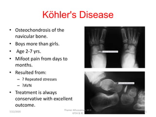
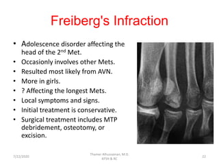
Introduction To
Pediatric Foot
Thamer Alhussainan, M.D.
7/22/2020
Thamer Alhussainan, M.D.
KFSH & RC
1
Pediatric Foot
• Introduction.
• Normal variations.
• Osteochondroses.
• Congenital and developmental foot deformities.
7/22/2020
Thamer Alhussainan, M.D.
KFSH & RC
2
Introduction
• The foot is a complex and unique structure providing:
– Sufficient support for the body weight
– Flexibility to accommodate the uneven surfaces.
• The movement of the foot is complex and coupled,
starting from the ankle joint and ending in the
forefoot.
7/22/2020
Thamer Alhussainan, M.D.
KFSH & RC
3
Introduction
• This complex foot movement is the resultant of the
unique configuration of the subtalar joint complex.
• The talus, calcaneum, navicular, and cuboid bones are
the hindfoot structures.
• The cuniforms and its articulation with the metatarsals
are considered the midfoot.
• The metatarsals and phalanges are considered the
forefoot segment
7/22/2020
Thamer Alhussainan, M.D.
KFSH & RC
4
Introduction
• Terminology to describe the foot alignment:
– Ankle:
• Equinus.
• Calcaneus.
– Hindfoot:
• Varus.
• Valgus.
– Midfoot:
• Cavus
• Planus.
– Foorefoot:
• Adduction.
• Abduction.
• Supination.
• Pronation
7/22/2020
Thamer Alhussainan, M.D.
KFSH & RC
5
Introduction
• Ossific development of the foot begins in utero.
• At birth, talus, calcaneus, cuboid, metatarsals, and phalanges are
ossified.
• The lateral cuneiform ossifies 4/12-20/12.
• The medial cuneiform ossifies at 24/12.
• The intermediate cuneiform ossifies at 36/24.
• The navicular ossifies last 3-5 yrs.
7/22/2020
Thamer Alhussainan, M.D.
KFSH & RC
6
Introduction
7/22/2020
Thamer Alhussainan, M.D.
KFSH & RC
7
Introduction
7/22/2020
Thamer Alhussainan, M.D.
KFSH & RC
8
Introduction
• The foot has its own growth pattern.
• The foot grows rapidly between birth and 5 yrs and
then slows down
• The girls complete foot growth at 12 yrs, and the
boys at 14 yrs.
7/22/2020
Thamer Alhussainan, M.D.
KFSH & RC
9
Normal variations
7/22/2020
Thamer Alhussainan, M.D.
KFSH & RC
10
Normal variations
• Clinically, the foot position might be affected by the
intrauterine crowding.
• The foot is correctable.
• The most common positional variations are
metatarsus adductus and calcaneovlagus feet.
7/22/2020
Thamer Alhussainan, M.D.
KFSH & RC
11
Normal variations
• Accessory ossification centers are very common and
might be confused with fracture lines…
– Clinical correlation.
– X-ray of the other side.
7/22/2020
Thamer Alhussainan, M.D.
KFSH & RC
12
Os Trigonum
• It is the lateral border for the FHL grove in the back
of the talus
7/22/2020
Thamer Alhussainan, M.D.
KFSH & RC
13
Os Trigonum
• Between the ages 8-11 yrs, the secondary
ossification center appears, and it fuses within 1
year.
• Injury to Os Trigonum occurs with forceful plantar
flexion.
• Clinical and radiological evaluation.
7/22/2020
Thamer Alhussainan, M.D.
KFSH & RC
14
Os Trigonum
7/22/2020
Thamer Alhussainan, M.D.
KFSH & RC
15
Os Trigonum
• Conservative treatment should be tried first.
• Surgical excision for resistant cases.
7/22/2020
Thamer Alhussainan, M.D.
KFSH & RC
16
Accessory Navicular
• It is the most accessory bone associated with
symptoms.
• Prevalence 16-26%.
• Type 1: small ossicle within tibialis posterior tendon
(true sessamoid).
• Type 2: small ossicle medial and plantar to navucular
and connected with cartilage.
• Type 3: fused type 2.
7/22/2020
Thamer Alhussainan, M.D.
KFSH & RC
17
Accessory Navicular
• Symptoms are caused by
repeated micro fractures and
chronic inflammation.
• Originally was correlated to
flatfoot development, but
recently the association is
rejected.
7/22/2020
Thamer Alhussainan, M.D.
KFSH & RC
18
Accessory Navicular
• MRI and bone scans can differentiate the symptomatic
ones.
• Treatment is mainly conservative:
– Shoe wear modification.
– Insoles.
– Casting.
– Local injections.
• Surgical treatment in resistant cases:
– Drilling.
– Excision with tibialis posterior preservation.
– Kinder procedure (excision with tibialis posterior lateral
placement).
7/22/2020
Thamer Alhussainan, M.D.
KFSH & RC
19
Osteochondroses
7/22/2020
Thamer Alhussainan, M.D.
KFSH & RC
20
Köhler's Disease
• Osteochondrosis of the
navicular bone.
• Boys more than girls.
• Age 2-7 yrs.
• Mifoot pain from days to
months.
• Resulted from:
– ? Repeated stresses
– ?AVN
• Treatment is always
conservative with excellent
outcome.
7/22/2020
Thamer Alhussainan, M.D.
KFSH & RC
21
Freiberg's Infraction
• Adolescence disorder affecting the
head of the 2nd Met.
• Occasionly involves other Mets.
• Resulted most likely from AVN.
• More in girls.
• ? Affecting the longest Mets.
• Local symptoms and signs.
• Initial treatment is conservative.
• Surgical treatment includes MTP
debridement, osteotomy, or
excision.
7/22/2020
Thamer Alhussainan, M.D.
KFSH & RC
22
Congenital and Developmental
Foot Deformities
7/22/2020
Thamer Alhussainan, M.D.
KFSH & RC
23
Metatarsus Adductus
• It is considered as a postural foot deformity
secondary to intrauterine crowding.
• In its simple form, the forefoot is adducted in
relation to a neutral and flexible hindfoot.
• It is associated to torticollis and DDH.
• The incidence varies between 0.1%-1%.
7/22/2020
Thamer Alhussainan, M.D.
KFSH & RC
24
Metatarsus Adductus
• The deformity can be classified into:
– Mild: the deformity can be actively corrected.
– Moderate : the deformity can be passively corrected.
– Severe: the deformity is not completely correctable.
7/22/2020
Thamer Alhussainan, M.D.
KFSH & RC
25
Metatarsus Adductus
• Treatment (after exclusion of DDH):
– Mild : observation.
– Moderate: ? Observation ? Stretching ? Casting.
– Severe: serial casting .
7/22/2020
Thamer Alhussainan, M.D.
KFSH & RC
26
Metatarsus Adductus
• Surgical treatment:
• Reserved for resistant cases after the age of 4 yrs.
7/22/2020
Thamer Alhussainan, M.D.
KFSH & RC
27
Metatarsus Adductus Complex
(Skewfoot)
• It a metatarsus adductus
associated with rigid heel
valgus.
• S-shaped foot, serpentine
foot, or Z-foot.
• Very rare foot deformity.
• Treatment is controversial.
7/22/2020
Thamer Alhussainan, M.D.
KFSH & RC
28
Talipes Calcaneovalgus
• It is the commonest foot
postural deformity.
• Excessive dorsiflexion and
limited plantar flexion.
• Associated with DDH (other foot
metatarsus adductus).
• Posteromedial bowing of the
tibia has to be ruled out.
• Natural history is excellent.
7/22/2020
Thamer Alhussainan, M.D.
KFSH & RC
29
Flexible Flatfoot
(Pes Planovalgus)
• Probably, it is the most common “deformity”
evaluated by pediatric orthopedists.
• The parents and pediatricians perceive this foot as an
“abnormal” and “deformed” foot.
• Flexible flatfoot is a normal foot variation.
7/22/2020
Thamer Alhussainan, M.D.
KFSH & RC
30
Flexible Flatfoot
(Pes Planovalgus)
“ flatfeet are usual in infants,
common in children, and within
the normal range in adults”
Lynn Staheli
Staheli LT, Chew DE, Corbett M: The longitudinal
arch. A survey of eight hundred and eighty-two feet
in normal children and adults. J Bone Joint Surg Am
1987; 69:426
7/22/2020
Thamer Alhussainan, M.D.
KFSH & RC
31
Flexible Flatfoot
(Pes Planovalgus)
• Clinical evaluation of flexible flatfoot is
not accurately defined.
• Assessment on standing position:
– Hindfoot valgus.
– Depressed or absent foot arch (head of
the talus is touching the ground).
– Abducted (and secondarily supinated)
forefoot.
• Deformities disappear in sitting position
or standing on tip toe.
• ? Achilles tendon shortening
7/22/2020
Thamer Alhussainan, M.D.
KFSH & RC
32
Flexible Flatfoot
(Pes Planovalgus)
• Radiographic measures for
flatfoot are not clearly defined.
• Lateral Talo-first Met angle
(Meyer’s angle) to measure the
midfoot sagging.
• AP Talo-Calcaneal angle (Kite’s
angle) to measure hindfoot
valgus
7/22/2020
Thamer Alhussainan, M.D.
KFSH & RC
33
Flexible Flatfoot
(Pes Planovalgus)
Should the flatfeet be treated?
7/22/2020
Thamer Alhussainan, M.D.
KFSH & RC
34
Flexible Flatfoot
(Pes Planovalgus)
NO
7/22/2020
Thamer Alhussainan, M.D.
KFSH & RC
35
Flexible Flatfoot
(Pes Planovalgus)
• Flexible flatfeet function as the normal feet with
arch, if there is no shortening in Achilles tendon.
• Rigid deformities should be excluded.
Harris R, Beath T: Army Foot Survey: An Investigation of Foot
Ailments in Canadian Soldiers, Ottawa: National Research
Council of Canada; 1947:1.
7/22/2020
Thamer Alhussainan, M.D.
KFSH & RC
36
Flexible Flatfoot
(Pes Planovalgus)
• Orthotics and shoe insert is only for pain relief not
for foot arch formation.
• Stretching exercises for the patients with Achilles
tendon shortening.
• Surgical treatment should be considered only for
symptomatic patients resistant to conservative
measures.
• Surgical outcomes for flatfeet is not favorable in
49%-77%.
7/22/2020
Thamer Alhussainan, M.D.
KFSH & RC
37
Flexible Flatfoot
(Pes Planovalgus)
• Surgical options:
– Achilles tendon lengthening with
• Arthrodesis (subtalar or triple).
• Lateral column lengthening (Evans procedure).
• Plication of talonavicular joint.
• Calcaneum-cuboid-cuniform (triple C ) osteotomy.
7/22/2020
Thamer Alhussainan, M.D.
KFSH & RC
38
Flexible Flatfoot
(Pes Planovalgus)
7/22/2020
Thamer Alhussainan, M.D.
KFSH & RC
39
Flexible Flatfoot
(Pes Planovalgus)
7/22/2020
Thamer Alhussainan, M.D.
KFSH & RC
40
Flexible Flatfoot
(Pes Planovalgus)
Further Readings
Scott Mubarak,
M.D.
Vincent
Mosca, M.D7/22/2020
Thamer Alhussainan, M.D.
KFSH & RC
41
Tarsal Coalition
• It is an abnormal connection between 2 or more
bones of the foot that produces limitation in the
ROM.
• The most common is talo-calcaneal coalition.
• Incidence is 0.03%-1.0%.
• “Peroneal Spastic foot”.
7/22/2020
Thamer Alhussainan, M.D.
KFSH & RC
42
Tarsal Coalition
• Familial
• Usually presents as a painful stiff
flatfoot at the age 12-13 yrs.
• Hindfoot valgus is not fully
correctable.
• Plain radiographs (AP, Lateral, oblique,
and Harris calcaneal view).
• Anterior talar beak
• CT scan is the definitive diagnostic
study.
7/22/2020
Thamer Alhussainan, M.D.
KFSH & RC
43
Tarsal Coalition
• Conservative treatment may be successful in almost
70% cases.
– Orthotics and casting.
• Surgical treatment for resistant cases:
– Excision and interposition:
• Calcaneonavicular coalition
– Fusions:
• Talocalcaneal coalition
7/22/2020
Thamer Alhussainan, M.D.
KFSH & RC
44
Vertical Talus
(congenital convex pes valgus)
• The foot resembles “ Rocker
Bottom”.
• ? Due to muscular imbalance.
• Mostly associated with
neuromuscular diseases (50%)
but it can present as isolated
deformity.
• Spina bifida, arthrogryposis,
trisomy 13-15-18.
• Unilateral or bilateral (50%).
7/22/2020
Thamer Alhussainan, M.D.
KFSH & RC
45
Vertical Talus
(congenital convex pes valgus)
• The hindfoot is fixed in equinus.
• The forefoot is fixed in dorsiflexion,
valgus position.
• The apex of the “Rocker” is the
talar head.
• The navicular is dislocated and sets
on top of talar neck.
• Deformity is not passively
correctable.
7/22/2020
Thamer Alhussainan, M.D.
KFSH & RC
46
Vertical Talus
(congenital convex pes valgus)
7/22/2020
Thamer Alhussainan, M.D.
KFSH & RC
47
Vertical Talus
(congenital convex pes valgus)
7/22/2020
Thamer Alhussainan, M.D.
KFSH & RC
48
Vertical Talus
(congenital convex pes valgus)
• Treatment of the vertical talus is usually surgical.
• The extent of the procedure is related to the severity
of the deformity.
– Single or double stage release.
– Dorsal release.
– STR with excision of navicular.
– STR with subtalar fusion.
– Triple arthrodesis or talectomy (salvage).
• Serial casting (reverse Ponseti casting) with minimal
soft tissue release shows promising results..
Dobbs M, et al. J Bone Joint Surg Am. 2006 Jun;88(6):1192-200.
7/22/2020
Thamer Alhussainan, M.D.
KFSH & RC
49
Clubfoot
7/22/2020
Thamer Alhussainan, M.D.
KFSH & RC
50
Clubfoot
• Congenital talipes equinovarus defomity.
• It is one of the commonest pediatric orthopedic
problems (1 :1000 births).
• Male: Female 3:1.
• Up-to 50 % bilateral.
• It can be isolated or associated with other congenital
anomalies.
7/22/2020
Thamer Alhussainan, M.D.
KFSH & RC
51
Clubfoot
• Family history is strongly associated.
• ? Genetics.
• Association with hip dysplasia is debatable .
7/22/2020
Thamer Alhussainan, M.D.
KFSH & RC
52
Clubfoot
Patho-anatomy
• The deformity in clubfoot
is formed by 4 elements :
– Cavus of the midfoot.
– Adduction of the forefoot.
– Varus of the hindfoot.
– Equinus of the ankle.
7/22/2020
Thamer Alhussainan, M.D.
KFSH & RC
53
Clubfoot
Patho-anatomy
• Talo-navicular joint is
subluxated medially .
• The posteromedial soft
tissues are contracted.
• Wide range of
neuromuscular changes
were found.
• Absent dorsalis pedis
vessels is common.
7/22/2020
Thamer Alhussainan, M.D.
KFSH & RC
54
Clubfoot
Clinical Assessment
• Pre natal US can detect clubfoot.
• Child with clubfoot should be examined carefully.
• General examinations should rule out associated
spine anomalies and neurological impairment.
• Other joint deformities can point out presence of
arthrogryposis.
7/22/2020
Thamer Alhussainan, M.D.
KFSH & RC
55
Clubfoot
Clinical Assessment
• Local examination of the clubfoot should focus on the
severity and rigidity of the deformity.
• Classify the clubfoot :
– Typical
– Atypical :
• Rigid deformity
• Associated with neuromuscular and genetic problems.
• Small and chubby foot.
• Short first ray.
• Deep and long medial crease.
7/22/2020
Thamer Alhussainan, M.D.
KFSH & RC
56
Clubfoot
Clinical Assessment
7/22/2020
Thamer Alhussainan, M.D.
KFSH & RC
57
Clubfoot
Clinical Assessment
7/22/2020
Thamer Alhussainan, M.D.
KFSH & RC
58
Clubfoot
Clinical Assessment
7/22/2020
Thamer Alhussainan, M.D.
KFSH & RC
59
Clubfoot
Management
Ignacio Ponseti
1914-2009
7/22/2020
Thamer Alhussainan, M.D.
KFSH & RC
60
Clubfoot
Management
http://www.global-
help.org/publications/books/help_cfponseti.pdf
7/22/2020
Thamer Alhussainan, M.D.
KFSH & RC
61
Clubfoot
Management
• The clubfoot is treated with serial
casting that will take the foot in a
step wise correction.
• The first element of clubfoot
deformity to be corrected is cavus.
• Correction of adduction will follow.
• The last element is equinus, and
usually requires Achilles tendon
tenotomy.
7/22/2020
Thamer Alhussainan, M.D.
KFSH & RC
62
Clubfoot
Management
• The total number of casting varies
between 4-10 according to the
severity of the deformity.
• The best final position that should be
targeted is 60-70 deg of abduction,
and 15 deg of dorsiflexion.
• After the tenotomy, the cast is applied
for 3 weeks to maintain the final
position.
7/22/2020
Thamer Alhussainan, M.D.
KFSH & RC
63
Clubfoot
Management
• The child is then fitted in “boots and
bar” splint that will hold the feet in
70 deg of abduction and 15 deg of
dorsiflexion.
• The splint should be worn for at
least 3 months continuously, and
then at bed times till 4 years.
7/22/2020
Thamer Alhussainan, M.D.
KFSH & RC
64
Clubfoot
Management
• Clubfoot treated by Ponseti method is expected to
have a good to excellent long term outcome in 78%
of patients (compared to 85% in people with no
clubfoot) .
• Additional procedures are required in 93% (usually
Achilles tendon tenotomy alone or with tibialis
anterior tendon transfer).
Cooper DM, Dietz FR. Treatment of idiopathic clubfoot: a thirty year
follow-up note. J Bone Joint Surg 1995;77A:1477e89.
7/22/2020
Thamer Alhussainan, M.D.
KFSH & RC
65
Clubfoot
Management
• Atypical clubfoot should be
recognized and treated with
the modifies Ponseti
technique.
• Response to conservative
treatment is less favorable
and relapse rate is high.
7/22/2020
Thamer Alhussainan, M.D.
KFSH & RC
66
Clubfoot
Management
• Surgical release of the clubfoot is reserved for the
resistant cases to conservative treatment.
• For easier surgical dissection, the procedure is
delayed till the foot reaches a reasonable size.
• It requires a delicate soft tissue dissection to avoid
injury to neurovascular structures.
7/22/2020
Thamer Alhussainan, M.D.
KFSH & RC
67
Clubfoot
Management
• Two types of skin incisions are used
for the complete clubfoot soft tissue
release:
– The posteromedial skin incision
(Turco incision).
– The posterior circumferential skin
incision (Cincinnati incision)
7/22/2020
Thamer Alhussainan, M.D.
KFSH & RC
68
Clubfoot
Management
• Surgical steps:
– Isolate and protect the
posterior NV bundle.
– Release 4 tendons, 4
joints capsule, and 4
ligaments.
– Plantar fasciotomy.
– Relocate and stabilize
the TN joint.
– Repair the tendons in
the corrected position.
7/22/2020
Thamer Alhussainan, M.D.
KFSH & RC
69
Clubfoot
Management
7/22/2020
Thamer Alhussainan, M.D.
KFSH & RC
70
Clubfoot
Management
• Up-to 4yrs, soft tissue releases alone can correct the
clubfoot deformity .
• In older ages bony procedures should be considered
due to the development of permanent bony changes.
– Cuboid wedge osteotomy.
– Midfoot osteotomies.
– Triple arthrodesis.
– Talectomy.
7/22/2020
Thamer Alhussainan, M.D.
KFSH & RC
71
Clubfoot
Management
• Complications:
• Under-correction.
• Relapse (recurrence).
• Overcorrection.
• Dorsal bunion.
• Stiffness.
• Wound problems.
7/22/2020
Thamer Alhussainan, M.D.
KFSH & RC
72
Clubfoot
Management
7/22/2020
Thamer Alhussainan, M.D.
KFSH & RC
73
Cavus foot
7/22/2020
Thamer Alhussainan, M.D.
KFSH & RC
74
Cavus foot
• A foot deformity characterized
by an elevated longitudinal
arch.
• Caused by fixed plantar flexion
of the forefoot (mainly the first
ray).
• Mostly, it is a developmental
foot problem secondary to
neurological conditions.
7/22/2020
Thamer Alhussainan, M.D.
KFSH & RC
75
Cavus foot
• First step in management is to diagnose or role out
an associated neurological diseases:
– Charcot-Marie-Tooth
– Freidreich's ataxia
– Cerebral palsy
– Polio
– spinal cord lesions
7/22/2020
Thamer Alhussainan, M.D.
KFSH & RC
76
Cavus foot
• Muscular imbalance between tibialis anterior and
peroneus longus causes the first ray to drop.
• The heel compensates in to varus position.
• The toe extensors will overact as ankle dorsiflexors to
compensate for Tib.Ant weakness causing clawing of
the toes
• Deformities starts as flexible and correctable, then
end as a rigid non correctable.
• Coleman block test is performed to assess the
hindfoot flexibility.
7/22/2020
Thamer Alhussainan, M.D.
KFSH & RC
77
Cavus foot
Management
• Conservative:
– Semi-rigid insole orthotic.
– Effective in mild cases only.
• Surgical :
– Bony:
• First metatarsal osteotomy.
• Midfoot osteotomy.
• Calcaneal valgus osteotomy.
• Claw toes reconstruction.
• Triple arthrodesis
– Soft tissue:
• Plantar fasciotomy.
• Tendon transfer.
7/22/2020
Thamer Alhussainan, M.D.
KFSH & RC
78
Thank You
7/22/2020
Thamer Alhussainan, M.D.
KFSH & RC
79

Introduction to pediatric foot

  • 1.
    Introduction To Pediatric Foot ThamerAlhussainan, M.D. 7/22/2020 Thamer Alhussainan, M.D. KFSH & RC 1
  • 2.
    Pediatric Foot • Introduction. •Normal variations. • Osteochondroses. • Congenital and developmental foot deformities. 7/22/2020 Thamer Alhussainan, M.D. KFSH & RC 2
  • 3.
    Introduction • The footis a complex and unique structure providing: – Sufficient support for the body weight – Flexibility to accommodate the uneven surfaces. • The movement of the foot is complex and coupled, starting from the ankle joint and ending in the forefoot. 7/22/2020 Thamer Alhussainan, M.D. KFSH & RC 3
  • 4.
    Introduction • This complexfoot movement is the resultant of the unique configuration of the subtalar joint complex. • The talus, calcaneum, navicular, and cuboid bones are the hindfoot structures. • The cuniforms and its articulation with the metatarsals are considered the midfoot. • The metatarsals and phalanges are considered the forefoot segment 7/22/2020 Thamer Alhussainan, M.D. KFSH & RC 4
  • 5.
    Introduction • Terminology todescribe the foot alignment: – Ankle: • Equinus. • Calcaneus. – Hindfoot: • Varus. • Valgus. – Midfoot: • Cavus • Planus. – Foorefoot: • Adduction. • Abduction. • Supination. • Pronation 7/22/2020 Thamer Alhussainan, M.D. KFSH & RC 5
  • 6.
    Introduction • Ossific developmentof the foot begins in utero. • At birth, talus, calcaneus, cuboid, metatarsals, and phalanges are ossified. • The lateral cuneiform ossifies 4/12-20/12. • The medial cuneiform ossifies at 24/12. • The intermediate cuneiform ossifies at 36/24. • The navicular ossifies last 3-5 yrs. 7/22/2020 Thamer Alhussainan, M.D. KFSH & RC 6
  • 7.
  • 8.
  • 9.
    Introduction • The foothas its own growth pattern. • The foot grows rapidly between birth and 5 yrs and then slows down • The girls complete foot growth at 12 yrs, and the boys at 14 yrs. 7/22/2020 Thamer Alhussainan, M.D. KFSH & RC 9
  • 10.
  • 11.
    Normal variations • Clinically,the foot position might be affected by the intrauterine crowding. • The foot is correctable. • The most common positional variations are metatarsus adductus and calcaneovlagus feet. 7/22/2020 Thamer Alhussainan, M.D. KFSH & RC 11
  • 12.
    Normal variations • Accessoryossification centers are very common and might be confused with fracture lines… – Clinical correlation. – X-ray of the other side. 7/22/2020 Thamer Alhussainan, M.D. KFSH & RC 12
  • 13.
    Os Trigonum • Itis the lateral border for the FHL grove in the back of the talus 7/22/2020 Thamer Alhussainan, M.D. KFSH & RC 13
  • 14.
    Os Trigonum • Betweenthe ages 8-11 yrs, the secondary ossification center appears, and it fuses within 1 year. • Injury to Os Trigonum occurs with forceful plantar flexion. • Clinical and radiological evaluation. 7/22/2020 Thamer Alhussainan, M.D. KFSH & RC 14
  • 15.
  • 16.
    Os Trigonum • Conservativetreatment should be tried first. • Surgical excision for resistant cases. 7/22/2020 Thamer Alhussainan, M.D. KFSH & RC 16
  • 17.
    Accessory Navicular • Itis the most accessory bone associated with symptoms. • Prevalence 16-26%. • Type 1: small ossicle within tibialis posterior tendon (true sessamoid). • Type 2: small ossicle medial and plantar to navucular and connected with cartilage. • Type 3: fused type 2. 7/22/2020 Thamer Alhussainan, M.D. KFSH & RC 17
  • 18.
    Accessory Navicular • Symptomsare caused by repeated micro fractures and chronic inflammation. • Originally was correlated to flatfoot development, but recently the association is rejected. 7/22/2020 Thamer Alhussainan, M.D. KFSH & RC 18
  • 19.
    Accessory Navicular • MRIand bone scans can differentiate the symptomatic ones. • Treatment is mainly conservative: – Shoe wear modification. – Insoles. – Casting. – Local injections. • Surgical treatment in resistant cases: – Drilling. – Excision with tibialis posterior preservation. – Kinder procedure (excision with tibialis posterior lateral placement). 7/22/2020 Thamer Alhussainan, M.D. KFSH & RC 19
  • 20.
  • 21.
    Köhler's Disease • Osteochondrosisof the navicular bone. • Boys more than girls. • Age 2-7 yrs. • Mifoot pain from days to months. • Resulted from: – ? Repeated stresses – ?AVN • Treatment is always conservative with excellent outcome. 7/22/2020 Thamer Alhussainan, M.D. KFSH & RC 21
  • 22.
    Freiberg's Infraction • Adolescencedisorder affecting the head of the 2nd Met. • Occasionly involves other Mets. • Resulted most likely from AVN. • More in girls. • ? Affecting the longest Mets. • Local symptoms and signs. • Initial treatment is conservative. • Surgical treatment includes MTP debridement, osteotomy, or excision. 7/22/2020 Thamer Alhussainan, M.D. KFSH & RC 22
  • 23.
    Congenital and Developmental FootDeformities 7/22/2020 Thamer Alhussainan, M.D. KFSH & RC 23
  • 24.
    Metatarsus Adductus • Itis considered as a postural foot deformity secondary to intrauterine crowding. • In its simple form, the forefoot is adducted in relation to a neutral and flexible hindfoot. • It is associated to torticollis and DDH. • The incidence varies between 0.1%-1%. 7/22/2020 Thamer Alhussainan, M.D. KFSH & RC 24
  • 25.
    Metatarsus Adductus • Thedeformity can be classified into: – Mild: the deformity can be actively corrected. – Moderate : the deformity can be passively corrected. – Severe: the deformity is not completely correctable. 7/22/2020 Thamer Alhussainan, M.D. KFSH & RC 25
  • 26.
    Metatarsus Adductus • Treatment(after exclusion of DDH): – Mild : observation. – Moderate: ? Observation ? Stretching ? Casting. – Severe: serial casting . 7/22/2020 Thamer Alhussainan, M.D. KFSH & RC 26
  • 27.
    Metatarsus Adductus • Surgicaltreatment: • Reserved for resistant cases after the age of 4 yrs. 7/22/2020 Thamer Alhussainan, M.D. KFSH & RC 27
  • 28.
    Metatarsus Adductus Complex (Skewfoot) •It a metatarsus adductus associated with rigid heel valgus. • S-shaped foot, serpentine foot, or Z-foot. • Very rare foot deformity. • Treatment is controversial. 7/22/2020 Thamer Alhussainan, M.D. KFSH & RC 28
  • 29.
    Talipes Calcaneovalgus • Itis the commonest foot postural deformity. • Excessive dorsiflexion and limited plantar flexion. • Associated with DDH (other foot metatarsus adductus). • Posteromedial bowing of the tibia has to be ruled out. • Natural history is excellent. 7/22/2020 Thamer Alhussainan, M.D. KFSH & RC 29
  • 30.
    Flexible Flatfoot (Pes Planovalgus) •Probably, it is the most common “deformity” evaluated by pediatric orthopedists. • The parents and pediatricians perceive this foot as an “abnormal” and “deformed” foot. • Flexible flatfoot is a normal foot variation. 7/22/2020 Thamer Alhussainan, M.D. KFSH & RC 30
  • 31.
    Flexible Flatfoot (Pes Planovalgus) “flatfeet are usual in infants, common in children, and within the normal range in adults” Lynn Staheli Staheli LT, Chew DE, Corbett M: The longitudinal arch. A survey of eight hundred and eighty-two feet in normal children and adults. J Bone Joint Surg Am 1987; 69:426 7/22/2020 Thamer Alhussainan, M.D. KFSH & RC 31
  • 32.
    Flexible Flatfoot (Pes Planovalgus) •Clinical evaluation of flexible flatfoot is not accurately defined. • Assessment on standing position: – Hindfoot valgus. – Depressed or absent foot arch (head of the talus is touching the ground). – Abducted (and secondarily supinated) forefoot. • Deformities disappear in sitting position or standing on tip toe. • ? Achilles tendon shortening 7/22/2020 Thamer Alhussainan, M.D. KFSH & RC 32
  • 33.
    Flexible Flatfoot (Pes Planovalgus) •Radiographic measures for flatfoot are not clearly defined. • Lateral Talo-first Met angle (Meyer’s angle) to measure the midfoot sagging. • AP Talo-Calcaneal angle (Kite’s angle) to measure hindfoot valgus 7/22/2020 Thamer Alhussainan, M.D. KFSH & RC 33
  • 34.
    Flexible Flatfoot (Pes Planovalgus) Shouldthe flatfeet be treated? 7/22/2020 Thamer Alhussainan, M.D. KFSH & RC 34
  • 35.
  • 36.
    Flexible Flatfoot (Pes Planovalgus) •Flexible flatfeet function as the normal feet with arch, if there is no shortening in Achilles tendon. • Rigid deformities should be excluded. Harris R, Beath T: Army Foot Survey: An Investigation of Foot Ailments in Canadian Soldiers, Ottawa: National Research Council of Canada; 1947:1. 7/22/2020 Thamer Alhussainan, M.D. KFSH & RC 36
  • 37.
    Flexible Flatfoot (Pes Planovalgus) •Orthotics and shoe insert is only for pain relief not for foot arch formation. • Stretching exercises for the patients with Achilles tendon shortening. • Surgical treatment should be considered only for symptomatic patients resistant to conservative measures. • Surgical outcomes for flatfeet is not favorable in 49%-77%. 7/22/2020 Thamer Alhussainan, M.D. KFSH & RC 37
  • 38.
    Flexible Flatfoot (Pes Planovalgus) •Surgical options: – Achilles tendon lengthening with • Arthrodesis (subtalar or triple). • Lateral column lengthening (Evans procedure). • Plication of talonavicular joint. • Calcaneum-cuboid-cuniform (triple C ) osteotomy. 7/22/2020 Thamer Alhussainan, M.D. KFSH & RC 38
  • 39.
  • 40.
  • 41.
    Flexible Flatfoot (Pes Planovalgus) FurtherReadings Scott Mubarak, M.D. Vincent Mosca, M.D7/22/2020 Thamer Alhussainan, M.D. KFSH & RC 41
  • 42.
    Tarsal Coalition • Itis an abnormal connection between 2 or more bones of the foot that produces limitation in the ROM. • The most common is talo-calcaneal coalition. • Incidence is 0.03%-1.0%. • “Peroneal Spastic foot”. 7/22/2020 Thamer Alhussainan, M.D. KFSH & RC 42
  • 43.
    Tarsal Coalition • Familial •Usually presents as a painful stiff flatfoot at the age 12-13 yrs. • Hindfoot valgus is not fully correctable. • Plain radiographs (AP, Lateral, oblique, and Harris calcaneal view). • Anterior talar beak • CT scan is the definitive diagnostic study. 7/22/2020 Thamer Alhussainan, M.D. KFSH & RC 43
  • 44.
    Tarsal Coalition • Conservativetreatment may be successful in almost 70% cases. – Orthotics and casting. • Surgical treatment for resistant cases: – Excision and interposition: • Calcaneonavicular coalition – Fusions: • Talocalcaneal coalition 7/22/2020 Thamer Alhussainan, M.D. KFSH & RC 44
  • 45.
    Vertical Talus (congenital convexpes valgus) • The foot resembles “ Rocker Bottom”. • ? Due to muscular imbalance. • Mostly associated with neuromuscular diseases (50%) but it can present as isolated deformity. • Spina bifida, arthrogryposis, trisomy 13-15-18. • Unilateral or bilateral (50%). 7/22/2020 Thamer Alhussainan, M.D. KFSH & RC 45
  • 46.
    Vertical Talus (congenital convexpes valgus) • The hindfoot is fixed in equinus. • The forefoot is fixed in dorsiflexion, valgus position. • The apex of the “Rocker” is the talar head. • The navicular is dislocated and sets on top of talar neck. • Deformity is not passively correctable. 7/22/2020 Thamer Alhussainan, M.D. KFSH & RC 46
  • 47.
    Vertical Talus (congenital convexpes valgus) 7/22/2020 Thamer Alhussainan, M.D. KFSH & RC 47
  • 48.
    Vertical Talus (congenital convexpes valgus) 7/22/2020 Thamer Alhussainan, M.D. KFSH & RC 48
  • 49.
    Vertical Talus (congenital convexpes valgus) • Treatment of the vertical talus is usually surgical. • The extent of the procedure is related to the severity of the deformity. – Single or double stage release. – Dorsal release. – STR with excision of navicular. – STR with subtalar fusion. – Triple arthrodesis or talectomy (salvage). • Serial casting (reverse Ponseti casting) with minimal soft tissue release shows promising results.. Dobbs M, et al. J Bone Joint Surg Am. 2006 Jun;88(6):1192-200. 7/22/2020 Thamer Alhussainan, M.D. KFSH & RC 49
  • 50.
  • 51.
    Clubfoot • Congenital talipesequinovarus defomity. • It is one of the commonest pediatric orthopedic problems (1 :1000 births). • Male: Female 3:1. • Up-to 50 % bilateral. • It can be isolated or associated with other congenital anomalies. 7/22/2020 Thamer Alhussainan, M.D. KFSH & RC 51
  • 52.
    Clubfoot • Family historyis strongly associated. • ? Genetics. • Association with hip dysplasia is debatable . 7/22/2020 Thamer Alhussainan, M.D. KFSH & RC 52
  • 53.
    Clubfoot Patho-anatomy • The deformityin clubfoot is formed by 4 elements : – Cavus of the midfoot. – Adduction of the forefoot. – Varus of the hindfoot. – Equinus of the ankle. 7/22/2020 Thamer Alhussainan, M.D. KFSH & RC 53
  • 54.
    Clubfoot Patho-anatomy • Talo-navicular jointis subluxated medially . • The posteromedial soft tissues are contracted. • Wide range of neuromuscular changes were found. • Absent dorsalis pedis vessels is common. 7/22/2020 Thamer Alhussainan, M.D. KFSH & RC 54
  • 55.
    Clubfoot Clinical Assessment • Prenatal US can detect clubfoot. • Child with clubfoot should be examined carefully. • General examinations should rule out associated spine anomalies and neurological impairment. • Other joint deformities can point out presence of arthrogryposis. 7/22/2020 Thamer Alhussainan, M.D. KFSH & RC 55
  • 56.
    Clubfoot Clinical Assessment • Localexamination of the clubfoot should focus on the severity and rigidity of the deformity. • Classify the clubfoot : – Typical – Atypical : • Rigid deformity • Associated with neuromuscular and genetic problems. • Small and chubby foot. • Short first ray. • Deep and long medial crease. 7/22/2020 Thamer Alhussainan, M.D. KFSH & RC 56
  • 57.
  • 58.
  • 59.
  • 60.
  • 61.
  • 62.
    Clubfoot Management • The clubfootis treated with serial casting that will take the foot in a step wise correction. • The first element of clubfoot deformity to be corrected is cavus. • Correction of adduction will follow. • The last element is equinus, and usually requires Achilles tendon tenotomy. 7/22/2020 Thamer Alhussainan, M.D. KFSH & RC 62
  • 63.
    Clubfoot Management • The totalnumber of casting varies between 4-10 according to the severity of the deformity. • The best final position that should be targeted is 60-70 deg of abduction, and 15 deg of dorsiflexion. • After the tenotomy, the cast is applied for 3 weeks to maintain the final position. 7/22/2020 Thamer Alhussainan, M.D. KFSH & RC 63
  • 64.
    Clubfoot Management • The childis then fitted in “boots and bar” splint that will hold the feet in 70 deg of abduction and 15 deg of dorsiflexion. • The splint should be worn for at least 3 months continuously, and then at bed times till 4 years. 7/22/2020 Thamer Alhussainan, M.D. KFSH & RC 64
  • 65.
    Clubfoot Management • Clubfoot treatedby Ponseti method is expected to have a good to excellent long term outcome in 78% of patients (compared to 85% in people with no clubfoot) . • Additional procedures are required in 93% (usually Achilles tendon tenotomy alone or with tibialis anterior tendon transfer). Cooper DM, Dietz FR. Treatment of idiopathic clubfoot: a thirty year follow-up note. J Bone Joint Surg 1995;77A:1477e89. 7/22/2020 Thamer Alhussainan, M.D. KFSH & RC 65
  • 66.
    Clubfoot Management • Atypical clubfootshould be recognized and treated with the modifies Ponseti technique. • Response to conservative treatment is less favorable and relapse rate is high. 7/22/2020 Thamer Alhussainan, M.D. KFSH & RC 66
  • 67.
    Clubfoot Management • Surgical releaseof the clubfoot is reserved for the resistant cases to conservative treatment. • For easier surgical dissection, the procedure is delayed till the foot reaches a reasonable size. • It requires a delicate soft tissue dissection to avoid injury to neurovascular structures. 7/22/2020 Thamer Alhussainan, M.D. KFSH & RC 67
  • 68.
    Clubfoot Management • Two typesof skin incisions are used for the complete clubfoot soft tissue release: – The posteromedial skin incision (Turco incision). – The posterior circumferential skin incision (Cincinnati incision) 7/22/2020 Thamer Alhussainan, M.D. KFSH & RC 68
  • 69.
    Clubfoot Management • Surgical steps: –Isolate and protect the posterior NV bundle. – Release 4 tendons, 4 joints capsule, and 4 ligaments. – Plantar fasciotomy. – Relocate and stabilize the TN joint. – Repair the tendons in the corrected position. 7/22/2020 Thamer Alhussainan, M.D. KFSH & RC 69
  • 70.
  • 71.
    Clubfoot Management • Up-to 4yrs,soft tissue releases alone can correct the clubfoot deformity . • In older ages bony procedures should be considered due to the development of permanent bony changes. – Cuboid wedge osteotomy. – Midfoot osteotomies. – Triple arthrodesis. – Talectomy. 7/22/2020 Thamer Alhussainan, M.D. KFSH & RC 71
  • 72.
    Clubfoot Management • Complications: • Under-correction. •Relapse (recurrence). • Overcorrection. • Dorsal bunion. • Stiffness. • Wound problems. 7/22/2020 Thamer Alhussainan, M.D. KFSH & RC 72
  • 73.
  • 74.
  • 75.
    Cavus foot • Afoot deformity characterized by an elevated longitudinal arch. • Caused by fixed plantar flexion of the forefoot (mainly the first ray). • Mostly, it is a developmental foot problem secondary to neurological conditions. 7/22/2020 Thamer Alhussainan, M.D. KFSH & RC 75
  • 76.
    Cavus foot • Firststep in management is to diagnose or role out an associated neurological diseases: – Charcot-Marie-Tooth – Freidreich's ataxia – Cerebral palsy – Polio – spinal cord lesions 7/22/2020 Thamer Alhussainan, M.D. KFSH & RC 76
  • 77.
    Cavus foot • Muscularimbalance between tibialis anterior and peroneus longus causes the first ray to drop. • The heel compensates in to varus position. • The toe extensors will overact as ankle dorsiflexors to compensate for Tib.Ant weakness causing clawing of the toes • Deformities starts as flexible and correctable, then end as a rigid non correctable. • Coleman block test is performed to assess the hindfoot flexibility. 7/22/2020 Thamer Alhussainan, M.D. KFSH & RC 77
  • 78.
    Cavus foot Management • Conservative: –Semi-rigid insole orthotic. – Effective in mild cases only. • Surgical : – Bony: • First metatarsal osteotomy. • Midfoot osteotomy. • Calcaneal valgus osteotomy. • Claw toes reconstruction. • Triple arthrodesis – Soft tissue: • Plantar fasciotomy. • Tendon transfer. 7/22/2020 Thamer Alhussainan, M.D. KFSH & RC 78
  • 79.