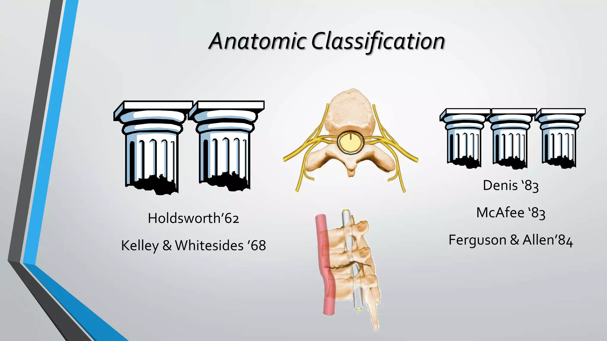
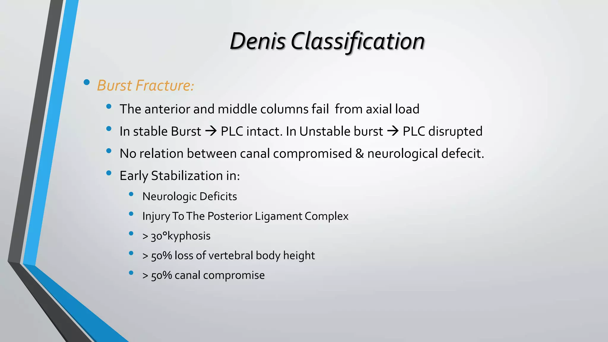
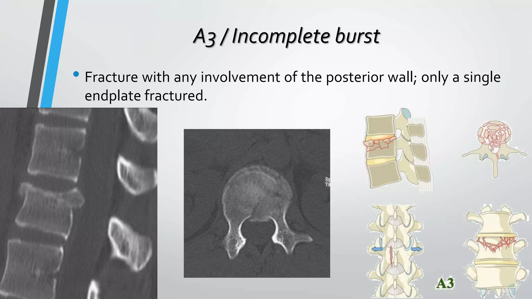
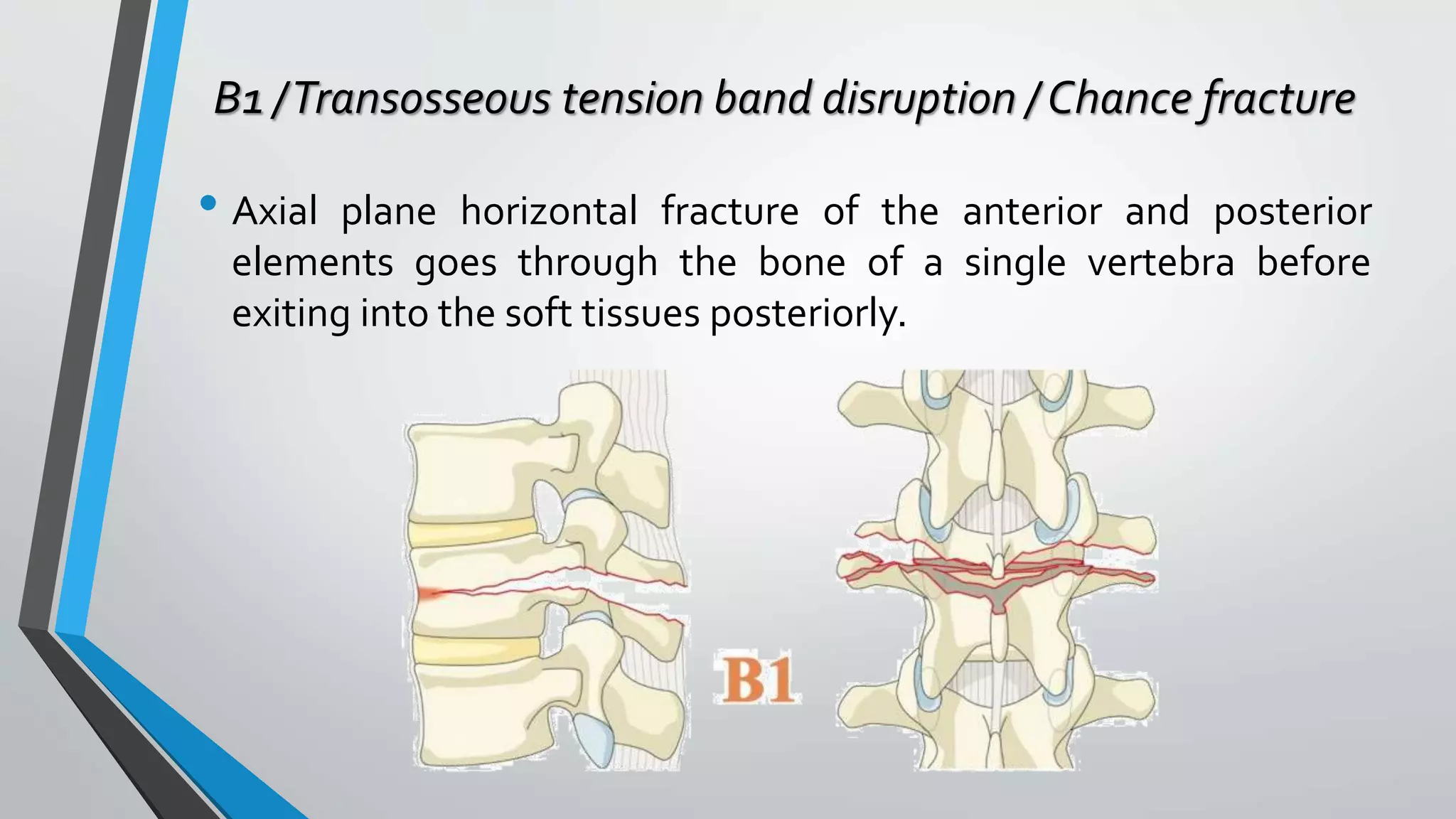
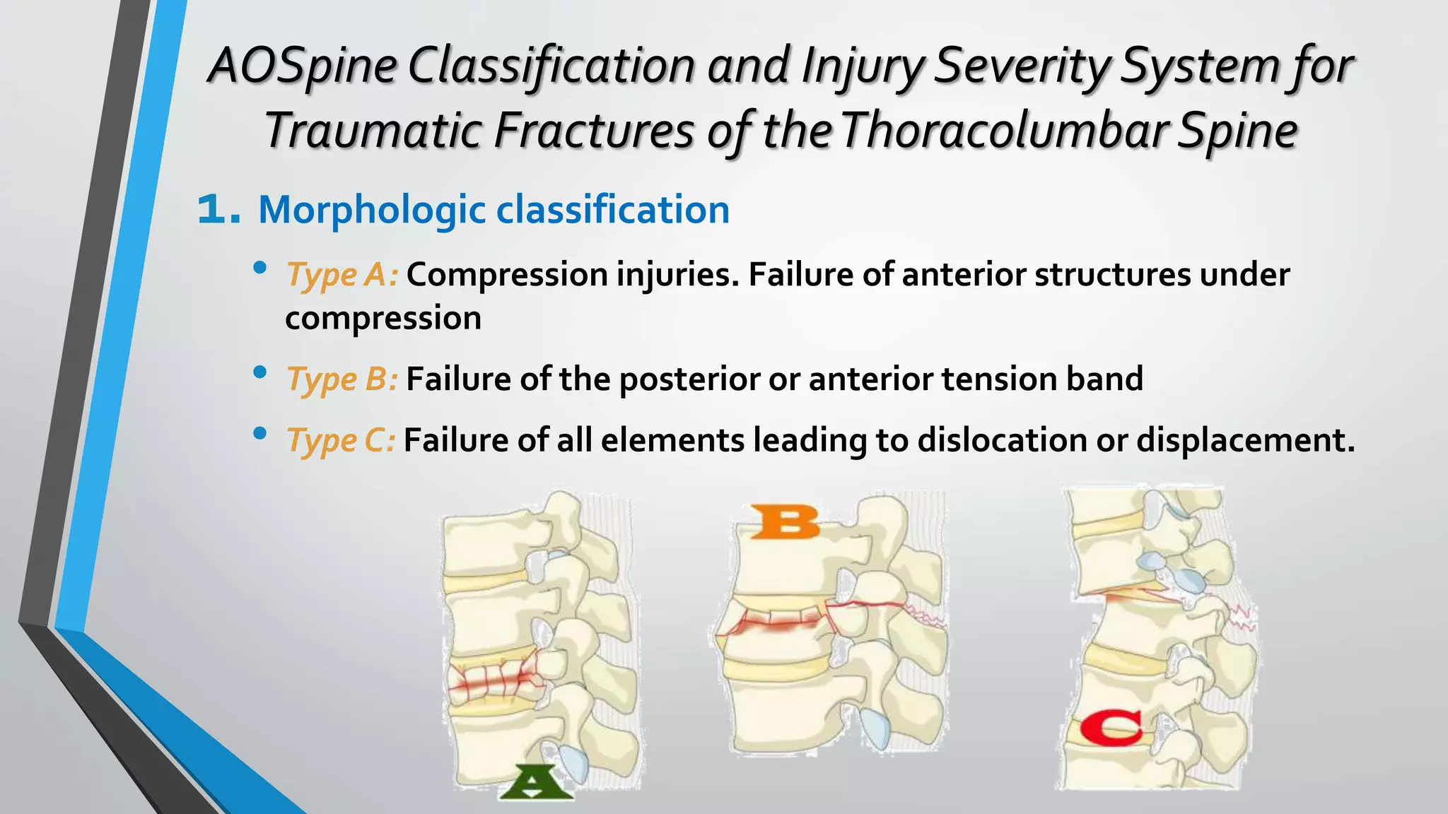
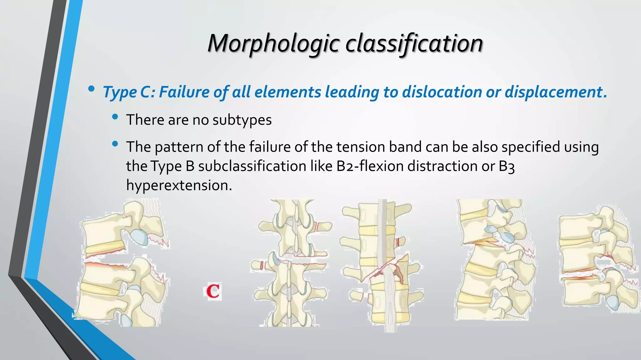
This document provides an overview of different classification systems for thoracolumbar fractures. It discusses early anatomic classifications including the 2-column theory and 3-column theory. It then summarizes the Denis classification and Load-Sharing classification. The AO classification and Thoracolumbar Injury Classification and Severity Score (TLICS) are also described. The document concludes by outlining a new AOSpine Classification and Injury Severity System for Traumatic Fractures of the Thoracolumbar Spine which is based on the morphological classification of the fracture, neurologic injury status, and clinical modifiers.
Pain from acute vertebral fracture appears to be due in part to instability (non-union or slow union at the fracture site), while more than 1/3 of patients become chronically painful.
Traditional treatment for patients with painful VCFs includes bed rest, narcotic analgesics and bracing, resulting in increased pain because of acceleration bone loss and muscle weakness.
Can read freely here
https://sethiortho.blogspot.com/
Challenges and Solutions in
Management of Distal Humerus Fractures
Epidemiology
Anatomy
Classification
Controversies and Recent studies
Approach
Implants selection
Plate configuration
Ulnar nerve transposition
Role of total elbow arthroplasty in DHF
Role of hemiarthroplasty in DHF
Metaphyseal comminution –
Anatomic complexity of the distal humerus
Positioning of the plates
TBW –
Skin closure
Osteoporotic nature of the bone –
Less BMD/Thin metaphysis
Screw Pullout strength is low
DHF account for 2% of all adult fractures
The common pattern of fracture
Intraarticular and involves both columns
Bimodal distribution
Peak incidence in young male and in older female patients
Young male – High-velocity injury
Older female - Osteoporosis
The distal humerus is flattened and expanded bony structure
It is composed of lateral and medial columns with the trochlea situated between these columns.
The location of the trochlea is central rather than medial
Formed by Medial SCR + M/Epicondyle
The distal end has 450 angulation with humeral shaft
M/ Epicondyle gives attachment for MCL & Common Flexor Origin
The MCL originates from the undersurface of the medial epicondyle where it is vulnerable to excessive dissection
Ulnar nerve
Formed by Lateral SCR and L/Epicondyle and Capitulum
Distal end has 200 with humeral shaft
L/ epicondyle gives attachment for LCL & common extensor origin
Its posterior surface is non articular and can be used as a site for a plate fixation
The lateral column curves anteriorly
Placement of a straight plate on the posterolateral surface of the humerus risks straightening of distal humerus.
The medial column including the medial epicondyle is in line with the humeral shaft.
It forms the center of the triangle
It has 30 - 80 – external rotation & 250 anterior divergent with the shaft
It forms a 40 - 80 degree valgus direction
X-ray -
Anterior-posterior view
lateral View
Traction View – This can help to define articular fragments and aid in pre-operative classification of the fracture.
NCCT – Elbow
Articular surfaces
Position of the fracture fragments
useful for identifying impacted fracture fragments that make reduction challenging
Olecranon Osteotomy Approach – 52-57%
Triceps sparing VS Olecranon osteotomy approach
The lateral column was often the first to fail as a result of excessive varus forces acting on the elbow during normal activities of daily living. Small anterior-posterior diameter
Smaller diameter of the humerus, permitting only one or two short screws for fixation.
Interruption of blood supply to the lateral column
blood supply to the lateral column is also derived from posterior segmental vessels. Sagittal plane plating has less risk of injuring these structures, which may improve the chances of union
Colorado spine surgeon, Dr. Donald Corenman, M.D., D.C. (http://neckandback.com), is an expert in treating spinal cord injuries associated with a traumatic fall, sports related injury or accident. Many spine fractures include a thoracolumbar fracture, which is a break in one or more of the thoracic and lumbar vertebrae. Spine fractures can be very serious but are also treatable in many cases. This presentation on spinal cord injuries, spine fractures and thoracolumbar fractures details events that can lead to this injury, symptoms and treatment options.
Dr. Corenman is a renowned Colorado spine surgeon and also is an expert at all spine conditions and disorders including scoliosis, degenerative disc disease, spinal stenosis, sciatica, herniated disc, slipped disc and spondylolythesis. He is also a sports medicine specialist and treats athletes with traumatic sports related injuries. He recently launched his own website (http://neckandback.com) to educate patients on spine disorders and to offer second opinions to physicians and colleagues who are seeking additional information on specific spine injuries and treatment options.
Anti ulcer drugs and their Advance pharmacology ||
Anti-ulcer drugs are medications used to prevent and treat ulcers in the stomach and upper part of the small intestine (duodenal ulcers). These ulcers are often caused by an imbalance between stomach acid and the mucosal lining, which protects the stomach lining.
||Scope: Overview of various classes of anti-ulcer drugs, their mechanisms of action, indications, side effects, and clinical considerations.
Title: Sense of Taste
Presenter: Dr. Faiza, Assistant Professor of Physiology
Qualifications:
MBBS (Best Graduate, AIMC Lahore)
FCPS Physiology
ICMT, CHPE, DHPE (STMU)
MPH (GC University, Faisalabad)
MBA (Virtual University of Pakistan)
Learning Objectives:
Describe the structure and function of taste buds.
Describe the relationship between the taste threshold and taste index of common substances.
Explain the chemical basis and signal transduction of taste perception for each type of primary taste sensation.
Recognize different abnormalities of taste perception and their causes.
Key Topics:
Significance of Taste Sensation:
Differentiation between pleasant and harmful food
Influence on behavior
Selection of food based on metabolic needs
Receptors of Taste:
Taste buds on the tongue
Influence of sense of smell, texture of food, and pain stimulation (e.g., by pepper)
Primary and Secondary Taste Sensations:
Primary taste sensations: Sweet, Sour, Salty, Bitter, Umami
Chemical basis and signal transduction mechanisms for each taste
Taste Threshold and Index:
Taste threshold values for Sweet (sucrose), Salty (NaCl), Sour (HCl), and Bitter (Quinine)
Taste index relationship: Inversely proportional to taste threshold
Taste Blindness:
Inability to taste certain substances, particularly thiourea compounds
Example: Phenylthiocarbamide
Structure and Function of Taste Buds:
Composition: Epithelial cells, Sustentacular/Supporting cells, Taste cells, Basal cells
Features: Taste pores, Taste hairs/microvilli, and Taste nerve fibers
Location of Taste Buds:
Found in papillae of the tongue (Fungiform, Circumvallate, Foliate)
Also present on the palate, tonsillar pillars, epiglottis, and proximal esophagus
Mechanism of Taste Stimulation:
Interaction of taste substances with receptors on microvilli
Signal transduction pathways for Umami, Sweet, Bitter, Sour, and Salty tastes
Taste Sensitivity and Adaptation:
Decrease in sensitivity with age
Rapid adaptation of taste sensation
Role of Saliva in Taste:
Dissolution of tastants to reach receptors
Washing away the stimulus
Taste Preferences and Aversions:
Mechanisms behind taste preference and aversion
Influence of receptors and neural pathways
Impact of Sensory Nerve Damage:
Degeneration of taste buds if the sensory nerve fiber is cut
Abnormalities of Taste Detection:
Conditions: Ageusia, Hypogeusia, Dysgeusia (parageusia)
Causes: Nerve damage, neurological disorders, infections, poor oral hygiene, adverse drug effects, deficiencies, aging, tobacco use, altered neurotransmitter levels
Neurotransmitters and Taste Threshold:
Effects of serotonin (5-HT) and norepinephrine (NE) on taste sensitivity
Supertasters:
25% of the population with heightened sensitivity to taste, especially bitterness
Increased number of fungiform papillae
The prostate is an exocrine gland of the male mammalian reproductive system
It is a walnut-sized gland that forms part of the male reproductive system and is located in front of the rectum and just below the urinary bladder
Function is to store and secrete a clear, slightly alkaline fluid that constitutes 10-30% of the volume of the seminal fluid that along with the spermatozoa, constitutes semen
A healthy human prostate measures (4cm-vertical, by 3cm-horizontal, 2cm ant-post ).
It surrounds the urethra just below the urinary bladder. It has anterior, median, posterior and two lateral lobes
It’s work is regulated by androgens which are responsible for male sex characteristics
Generalised disease of the prostate due to hormonal derangement which leads to non malignant enlargement of the gland (increase in the number of epithelial cells and stromal tissue)to cause compression of the urethra leading to symptoms (LUTS
micro teaching on communication m.sc nursing.pdfAnurag Sharma
Microteaching is a unique model of practice teaching. It is a viable instrument for the. desired change in the teaching behavior or the behavior potential which, in specified types of real. classroom situations, tends to facilitate the achievement of specified types of objectives.
Factory Supply Best Quality Pmk Oil CAS 28578–16–7 PMK Powder in Stockrebeccabio
Factory Supply Best Quality Pmk Oil CAS 28578–16–7 PMK Powder in Stock
Telegram: bmksupplier
signal: +85264872720
threema: TUD4A6YC
You can contact me on Telegram or Threema
Communicate promptly and reply
Free of customs clearance, Double Clearance 100% pass delivery to USA, Canada, Spain, Germany, Netherland, Poland, Italy, Sweden, UK, Czech Republic, Australia, Mexico, Russia, Ukraine, Kazakhstan.Door to door service
Hot Selling Organic intermediates
- Video recording of this lecture in English language: https://youtu.be/lK81BzxMqdo
- Video recording of this lecture in Arabic language: https://youtu.be/Ve4P0COk9OI
- Link to download the book free: https://nephrotube.blogspot.com/p/nephrotube-nephrology-books.html
- Link to NephroTube website: www.NephroTube.com
- Link to NephroTube social media accounts: https://nephrotube.blogspot.com/p/join-nephrotube-on-social-media.html
Couples presenting to the infertility clinic- Do they really have infertility...Sujoy Dasgupta
Dr Sujoy Dasgupta presented the study on "Couples presenting to the infertility clinic- Do they really have infertility? – The unexplored stories of non-consummation" in the 13th Congress of the Asia Pacific Initiative on Reproduction (ASPIRE 2024) at Manila on 24 May, 2024.
Tom Selleck Health: A Comprehensive Look at the Iconic Actor’s Wellness Journeygreendigital
Tom Selleck, an enduring figure in Hollywood. has captivated audiences for decades with his rugged charm, iconic moustache. and memorable roles in television and film. From his breakout role as Thomas Magnum in Magnum P.I. to his current portrayal of Frank Reagan in Blue Bloods. Selleck's career has spanned over 50 years. But beyond his professional achievements. fans have often been curious about Tom Selleck Health. especially as he has aged in the public eye.
Follow us on: Pinterest
Introduction
Many have been interested in Tom Selleck health. not only because of his enduring presence on screen but also because of the challenges. and lifestyle choices he has faced and made over the years. This article delves into the various aspects of Tom Selleck health. exploring his fitness regimen, diet, mental health. and the challenges he has encountered as he ages. We'll look at how he maintains his well-being. the health issues he has faced, and his approach to ageing .
Early Life and Career
Childhood and Athletic Beginnings
Tom Selleck was born on January 29, 1945, in Detroit, Michigan, and grew up in Sherman Oaks, California. From an early age, he was involved in sports, particularly basketball. which played a significant role in his physical development. His athletic pursuits continued into college. where he attended the University of Southern California (USC) on a basketball scholarship. This early involvement in sports laid a strong foundation for his physical health and disciplined lifestyle.
Transition to Acting
Selleck's transition from an athlete to an actor came with its physical demands. His first significant role in "Magnum P.I." required him to perform various stunts and maintain a fit appearance. This role, which he played from 1980 to 1988. necessitated a rigorous fitness routine to meet the show's demands. setting the stage for his long-term commitment to health and wellness.
Fitness Regimen
Workout Routine
Tom Selleck health and fitness regimen has evolved. adapting to his changing roles and age. During his "Magnum, P.I." days. Selleck's workouts were intense and focused on building and maintaining muscle mass. His routine included weightlifting, cardiovascular exercises. and specific training for the stunts he performed on the show.
Selleck adjusted his fitness routine as he aged to suit his body's needs. Today, his workouts focus on maintaining flexibility, strength, and cardiovascular health. He incorporates low-impact exercises such as swimming, walking, and light weightlifting. This balanced approach helps him stay fit without putting undue strain on his joints and muscles.
Importance of Flexibility and Mobility
In recent years, Selleck has emphasized the importance of flexibility and mobility in his fitness regimen. Understanding the natural decline in muscle mass and joint flexibility with age. he includes stretching and yoga in his routine. These practices help prevent injuries, improve posture, and maintain mobilit
Ozempic: Preoperative Management of Patients on GLP-1 Receptor Agonists Saeid Safari
Preoperative Management of Patients on GLP-1 Receptor Agonists like Ozempic and Semiglutide
ASA GUIDELINE
NYSORA Guideline
2 Case Reports of Gastric Ultrasound
These lecture slides, by Dr Sidra Arshad, offer a quick overview of physiological basis of a normal electrocardiogram.
Learning objectives:
1. Define an electrocardiogram (ECG) and electrocardiography
2. Describe how dipoles generated by the heart produce the waveforms of the ECG
3. Describe the components of a normal electrocardiogram of a typical bipolar leads (limb II)
4. Differentiate between intervals and segments
5. Enlist some common indications for obtaining an ECG
Study Resources:
1. Chapter 11, Guyton and Hall Textbook of Medical Physiology, 14th edition
2. Chapter 9, Human Physiology - From Cells to Systems, Lauralee Sherwood, 9th edition
3. Chapter 29, Ganong’s Review of Medical Physiology, 26th edition
4. Electrocardiogram, StatPearls - https://www.ncbi.nlm.nih.gov/books/NBK549803/
5. ECG in Medical Practice by ABM Abdullah, 4th edition
6. ECG Basics, http://www.nataliescasebook.com/tag/e-c-g-basics
New Directions in Targeted Therapeutic Approaches for Older Adults With Mantl...i3 Health
i3 Health is pleased to make the speaker slides from this activity available for use as a non-accredited self-study or teaching resource.
This slide deck presented by Dr. Kami Maddocks, Professor-Clinical in the Division of Hematology and
Associate Division Director for Ambulatory Operations
The Ohio State University Comprehensive Cancer Center, will provide insight into new directions in targeted therapeutic approaches for older adults with mantle cell lymphoma.
STATEMENT OF NEED
Mantle cell lymphoma (MCL) is a rare, aggressive B-cell non-Hodgkin lymphoma (NHL) accounting for 5% to 7% of all lymphomas. Its prognosis ranges from indolent disease that does not require treatment for years to very aggressive disease, which is associated with poor survival (Silkenstedt et al, 2021). Typically, MCL is diagnosed at advanced stage and in older patients who cannot tolerate intensive therapy (NCCN, 2022). Although recent advances have slightly increased remission rates, recurrence and relapse remain very common, leading to a median overall survival between 3 and 6 years (LLS, 2021). Though there are several effective options, progress is still needed towards establishing an accepted frontline approach for MCL (Castellino et al, 2022). Treatment selection and management of MCL are complicated by the heterogeneity of prognosis, advanced age and comorbidities of patients, and lack of an established standard approach for treatment, making it vital that clinicians be familiar with the latest research and advances in this area. In this activity chaired by Michael Wang, MD, Professor in the Department of Lymphoma & Myeloma at MD Anderson Cancer Center, expert faculty will discuss prognostic factors informing treatment, the promising results of recent trials in new therapeutic approaches, and the implications of treatment resistance in therapeutic selection for MCL.
Target Audience
Hematology/oncology fellows, attending faculty, and other health care professionals involved in the treatment of patients with mantle cell lymphoma (MCL).
Learning Objectives
1.) Identify clinical and biological prognostic factors that can guide treatment decision making for older adults with MCL
2.) Evaluate emerging data on targeted therapeutic approaches for treatment-naive and relapsed/refractory MCL and their applicability to older adults
3.) Assess mechanisms of resistance to targeted therapies for MCL and their implications for treatment selection
ARTIFICIAL INTELLIGENCE IN HEALTHCARE.pdfAnujkumaranit
Artificial intelligence (AI) refers to the simulation of human intelligence processes by machines, especially computer systems. It encompasses tasks such as learning, reasoning, problem-solving, perception, and language understanding. AI technologies are revolutionizing various fields, from healthcare to finance, by enabling machines to perform tasks that typically require human intelligence.
2. ?? Classification ??
• Indications for surgery
• Optimal time for surgery
• Best approach for surgery
• Should be:
• Comprehensive
• Reproducible
• Usable
• Accurate
5. Anatomic Classification
• 2 ColumnTheory: Holdsworth 1962
• Reviewed 1,000 patients: Separated spine into
anterior weight-bearing column (a) and
posterior tension-bearing column (b)
• Six types: simple wedge, dislocation, rotational
fracture-dislocation, extension, burst, and
shear.
• Stressed importance of posterior elements
• If destabilized, must consider surgery
6. Anatomic Classification
• 3 ColumnTheory: Denis 1983
• Based on radiographic review of 412 cases
• Anterior: ALL , anterior 2/3 body
• Middle: post 1/3 body, PLL
• Posterior: all structures posterior to PLL
• Middle column Injury was necessary & create instability.
• Posterior injury not sufficient to cause instability
7. Denis Classification
• Divides spinal fractures into minor and major injuries
• Minor injuries: fractures ofTransverse Process, Pars Interarticularis,
Spinous Process.
• Major injuries: Wedge Compression, Burst, Seat-belt type & Fracture-
Dislocation.
8. Denis Classification
• Wedge Compression Fracture:
• Isolated failure of the anterior column
• Result from forward flexion
• Rarely are associated with neurological deficit except in multiple adjacent
vertebral fracture
• PLC may be disrupted in tension if there is loss ofVB height >50%
9.
10. Denis Classification
• Burst Fracture:
• The anterior and middle columns fail from axial load
• In stable Burst PLC intact. In Unstable burst PLC disrupted
• No relation between canal compromised & neurological defecit.
• Early Stabilization in:
• Neurologic Deficits
• InjuryToThe Posterior Ligament Complex
• > 30°kyphosis
• > 50% loss of vertebral body height
• > 50% canal compromise
14. Denis Classification
• Fracture-dislocations
• All three columns have failed in compression, tension, rotation, shear
with translation deformity.
• At the affected level, one part of the spinal canal has been displaced in
the transverse plane
15.
16. Load-Sharing Classification
• McCormack Classification:
• Designed specifically for thoracolumbar burst fracture (1994)
• Devised method of predicting posterior failure
• 1-3 points assigned grades to amount ofVB comminution, displacement
of fracture fragments, degree of kyphosis
• Sum the points for a 3-9 scale
• < 6 points posterior only
• > 6 points anterior
<30% 30-60%
>60%
0-1mm 1-2mm >2mm
Comminution
Fragment Displacement
Kyphosis
Correction
<3° 4-9°
>10°
17. AO Classification
• Review of 1445 cases (Magerl,Gertzbein et al. European Spine Journal 1994)
• Based on direction of injury force
• 3 types, 53 injury patterns
18.
19. Thoracolumbar Injury Classification & Severity Score
(TLICS orTLISS)
• Introduced by the SpineTrauma Study
Group in 2005.
• Three major injury characteristics:
1. Injury morphology
2. Neurologic status
3. Integrity of the PLC
20.
21. TLICS
• The total score used to guide treatment:
• ≤ 3 points non-operatively
• ≥ 5 points surgical intervention
• = 4 points w/ or w/o surgery
22. Example (1)
• Dx: Compression Fx
• TLICS
• Morphology: Compression
• Neurology: Intact
• PLC: Intact
• 1 + 0 + 0 = 1 point Non-OP
27. Example (6)
• 18-year-old woman presented with severe mid back pain following a
rollover motor vehicle collision. Patient assessment revealed a normal
neurologic examination with a palpable, tender gap in the
thoracolumbar region.
28. Example (6)
• Dx:T11-12 fracture-
dislocation with a Chance
fracture atT12
• TLICS
• Morphology:Translation
• Neurology: Intact
• PLC: Injury
• 3 + 0 + 3 = 6 points OP
29. Example (7)
• A 63-year-old man sustained a 15- foot fall at work and reported
severe back pain. Assessment revealed a normal neurologic
examination with no posterior tenderness, gap, or step-off.
31. AOSpine Classification and Injury Severity System for
Traumatic Fractures of theThoracolumbar Spine
• This system is being subjected to a rigorous scientific assessment.
• Based on the evaluation of three basic parameters:
1. Morphologic classification of the fracture
2. Neurologic injury
3. Clinical modifiers
32. AOSpine Classification and Injury Severity System for
Traumatic Fractures of theThoracolumbar Spine
1. Morphologic classification
• Type A: Compression injuries. Failure of anterior structures under
compression
• Type B: Failure of the posterior or anterior tension band
• Type C: Failure of all elements leading to dislocation or displacement.
33. Morphologic classification
• Type A: Compression injuries.
• Five subtypes and no further sub-classification.
• A0 / minor, nonstructural fractures
• A1 /Wedge-compression
• A2 / Split
• A3 / Incomplete burst
• A4 / Complete burst
34. A0 / minor, nonstructural fractures
• Do not compromise the structural integrity of the spinal column
36. A2 / Split
• Fracture of both endplates without involvement of
the posterior wall of the vertebral body.
37. A3 / Incomplete burst
• Fracture with any involvement of the posterior wall; only a single
endplate fractured.
38. A4 / Complete burst
• Fracture with any involvement of the posterior wall and both
endplates.
39. AOSpine Classification and Injury Severity System for
Traumatic Fractures of theThoracolumbar Spine
1. Morphologic classification
• Type A: Compression injuries. Failure of anterior structures under
compression
• Type B: Failure of the posterior or anterior tension band
• Type C: Failure of all elements leading to dislocation or displacement.
40. Morphologic classification
• Type B: Failure of the posterior or anterior tension band
• There are three subtypes:
• B1 /Transosseous tension band disruption / Chance fracture
• B2 / Posterior tension band disruption
• B3 / Hyperextension
41. B1 /Transosseous tension band disruption / Chance fracture
• Axial plane horizontal fracture of the anterior and posterior
elements goes through the bone of a single vertebra before
exiting into the soft tissues posteriorly.
43. B2 / Posterior tension band disruption
• Bony and/or ligamentary failure of the posterior tension band
together with aType A fracture.
44. B3 / Hyperextension
• Injury through the disk or vertebral body leading to a
hyperextended position of the spinal column. Anterior structures,
especially the ALL are ruptured but there is a posterior hinge
preventing further displacement.
45. AOSpine Classification and Injury Severity System for
Traumatic Fractures of theThoracolumbar Spine
1. Morphologic classification
• Type A: Compression injuries. Failure of anterior structures under
compression
• Type B: Failure of the posterior or anterior tension band
• Type C: Failure of all elements leading to dislocation or displacement.
46. Morphologic classification
• Type C: Failure of all elements leading to dislocation or displacement.
• There are no subtypes
• The pattern of the failure of the tension band can be also specified using
theType B subclassification like B2-flexion distraction or B3
hyperextension.
49. AOSpine Classification and Injury Severity System for
Traumatic Fractures of theThoracolumbar Spine
• This system is being subjected to a rigorous scientific assessment.
• Based on the evaluation of three basic parameters:
1. Morphologic classification of the fracture
2. Neurologic injury
3. Clinical modifiers
50. AOSpine Classification and Injury Severity System for
Traumatic Fractures of theThoracolumbar Spine
2. Neurologic injury
• Neurologic status at the moment of admission should be scored
• N0: Neurologically intact
• N1:Transient neurologic deficit, which is no longer present
• N2: Radicular symptoms
• N3: Incomplete spinal cord injury or any degree of cauda equina injury
• N4: Complete spinal cord injury
• NX: Neurologic status is unknown due to sedation or head injury
51. AOSpine Classification and Injury Severity System for
Traumatic Fractures of theThoracolumbar Spine
• This system is being subjected to a rigorous scientific assessment.
• Based on the evaluation of three basic parameters:
1. Morphologic classification of the fracture
2. Neurologic injury
3. Clinical modifiers
52. AOSpine Classification and Injury Severity System for
Traumatic Fractures of theThoracolumbar Spine
3. Clinical modifiers
• There are two modifiers
• M1: is used to designate fractures with an indeterminate injury to the tension band
based on spinal imaging with or without MRI.This modifier is important for
designating those injuries with stable injuries from a bony standpoint for which
ligamentous insufficiency may help determine whether operative stabilization is a
consideration.
• M2: is used to designate a patient-specific comorbidity, which might argue either
for or against surgery for patients with relative surgical indications. Examples of an
M2 modifier include ankylosing spondylitis or burns affecting the skin overlying
the injured spine.
53.
54. References
• Thoracolumbar SpineTrauma Classification; J Am Acad Orthop Surg
2010;18:63-71
• A Review ofThoracolumbar Spine Fracture Classifications: Journal of
Orthopaedics andTraumaVol. 1 (2011),Article ID 235406, 5 pages.
• Thoracolumbar Spinal Injuries: http://www.springer.com/978-3-540-
40511-5
• Thoracic and Lumbar Spine Fractures and Dislocations: Assessment
and Classification: Christopher Bono, MD and Mitch Harris, MD; March
2004: Jim A.Youssef, MD; Revised January 2006 and May 2011; OTA
Editor's Notes
This should be classified as: T12-L1 Type B2 with T12 A4 according to the combination rules.