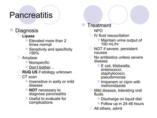
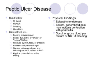
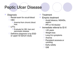
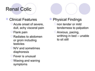
Acute Abdominal Pain
UNC Emergency Medicine
Medical Student Lecture Series
Case #1
24 yo healthy M with one day hx of abdominal pain. Pain
was generalized at first, now worse in right lower abd &
radiates to his right groin. He has vomited twice today.
Denies any diarrhea, fevers, dysuria or other complaints.
No appetite today. ROS otherwise negative.
PMHx: negative
PSurgHx: negative
Meds: none
NKDA
Social hx: no alcohol, tobacco or drug use
Family hx: non-contributory
Abdominal pain
What else do you want to know?
What is on your differential diagnosis so far?
(healthy male with RLQ abd pain….)
How do you approach the complaint of
abdominal pain in general?
Let’s review in this lecture:
 Types of pain
 History and physical examination
 Labs and imaging
 Abdominal pain in special populations (Elderly, HIV)
 Clinical pearls to help you in the ED
“Tell me more about your pain….”
Location
Quality
Severity
Onset
Duration
Modifying factors
Change over time
What kind of pain is it?
 Visceral
 Involves hollow or solid organs; midline pain due to bilateral innvervation
 Steady ache or vague discomfort to excruciating or colicky pain
 Poorly localized
 Epigastric region: stomach, duodenum, biliary tract
 Periumbilical: small bowel, appendix, cecum
 Suprapubic: colon, sigmoid, GU tract
 Parietal
 Involves parietal peritoneum
 Localized pain
 Causes tenderness and guarding which progress to rigidity and rebound as
peritonitis develops
 Referred
 Produces symptoms not signs
 Based on developmental embryology
 Ureteral obstruction → testicular pain
 Subdiaphragmatic irritation → ipsilateral shoulder or supraclavicular pain
 Gynecologic pathology → back or proximal lower extremity
 Biliary disease → right infrascapular pain
 MI → epigastric, neck, jaw or upper extremity pain
Ask about relevant ROS
GI symptoms
Nausea, vomiting, hematemesis, anorexia, diarrhea,
constipation, bloody stools, melena stools
GU symptoms
Dysuria, frequency, urgency, hematuria, incontinence
Gyn symptoms
Vaginal discharge, vaginal bleeding
General
Fever, lightheadedness
And don’t forget the history
 GI
 Past abdominal surgeries, h/o GB disease, ulcers; FamHx IBD
 GU
 Past surgeries, h/o kidney stones, pyelonephritis, UTI
 Gyn
 Last menses, sexual activity, contraception, h/o PID or STDs, h/o
ovarian cysts, past gynecological surgeries, pregnancies
 Vascular
 h/o MI, heart disease, a-fib, anticoagulation, CHF, PVD, Fam Hx of AAA
 Other medical history
 DM, organ transplant, HIV/AIDS, cancer
 Social
 Tobacco, drugs – Especially cocaine, alcohol
 Medications
 NSAIDs, H2 blockers, PPIs, immunosuppression, coumadin
Moving on to the Physical Exam
 General
 Pallor, diaphoresis, general appearance, level of distress or discomfort, is the patient lying
still or moving around in the bed
 Vital Signs
 Orthostatic VS when volume depletion is suspected
 Cardiac
 Arrhythmias
 Lungs
 Pneumonia
 Abdomen
 Look for distention, scars, masses
 Auscultate – hyperactive or obstructive BS increase likelihood of SBO fivefold – otherwise
not very helpful
 Palpate for tenderness, masses, aortic aneurysm, organomegaly, rebound, guarding, rigidity
 Percuss for tympany
 Look for hernias!
 rectal exam
 Back
 CVA tenderness
 Pelvic exam
 CMT
 Vaginal discharge – Culture
 Adenexal mass or fullness
Abdominal Findings
 Guarding
 Voluntary
Contraction of abdominal musculature in anticipation of palpation
Diminish by having patient flex knees
 Involuntary
Reflex spasm of abdominal muscles
aka: rigidity
Suggests peritoneal irritation
 Rebound
 Present in 1 of 4 patients without peritonitis
 Pain referred to the point of maximum tenderness when palpating
an adjacent quadrant is suggestive of peritonitis
 Rovsing’s sign in appendicitis
 Rectal exam
 Little evidence that tenderness adds any useful information beyond
abdominal examination
 Gross blood or melena indicates a GIB
Differential Diagnosis
It’s Huge!
 Use history and physical exam to narrow it down
 Rule out life-threatening pathology
 Half the time you will send the patient home with a diagnosis of nonspecific
abdominal pain (NSAP or Abdominal Pain – NOS)
 90% will be better or asymptomatic at 2-3 weeks
Differential Diagnosis
 Gastritis, ileitis, colitis, esophagitis
 Ulcers: gastric, peptic, esophageal
 Biliary disease: cholelithiasis, cholecystitis
 Hepatitis, pancreatitis, Cholangitis
 Splenic infarct, Splenic rupture
 Pancreatic psuedocyst
 Hollow viscous perforation
 Bowel obstruction, volvulus
 Diverticulitis
 Appendicitis
 Ovarian cyst
 Ovarian torsion
 Hernias: incarcerated, strangulated
 Kidney stones
 Pyelonephritis
 Hydronephrosis
 Inflammatory bowel disease: crohns, UC
 Gastroenteritis, enterocolitis
 pseudomembranous colitis, ischemia colitis
 Tumors: carcinomas, lipomas
 Meckels diverticulum
 Testicular torsion
 Epididymitis, prostatitis, orchitis, cystitis
 Constipation
 Abdominal aortic aneurysm, ruptures aneurysm
 Aortic dissection
 Mesenteric ischemia
 Organomegaly
 Hemilith infestation
 Porphyrias
 ACS
 Pneumonia
 Abdominal wall syndromes: muscle strain, hematomas,
trauma,
 Neuropathic causes: radicular pain
 Non-specific abdominal pain
 Group A beta-hemolytic streptococcal pharyngitis
 Rocky Mountain Spotted Fever
 Toxic Shock Syndrome
 Black widow envenomation
 Drugs: cocaine induced-ischemia, erythromycin, tetracyclines,
NSAIDs
 Mercury salts
 Acute inorganic lead poisoning
 Electrical injury
 Opioid withdrawal
 Mushroom toxicity
 AGA: DKA, AKA
 Adrenal crisis
 Thyroid storm
 Hypo- and hypercalcemia
 Sickle cell crisis
 Vasculitis
 Irritable bowel syndrome
 Ectopic pregnancy
 PID
 Urinary retention
 Ileus, Ogilvie syndrome
Most Common Causes in the ED
Non-specific abd pain 34%
Appendicitis 28%
Biliary tract dz 10%
SBO 4%
Gyn disease 4%
Pancreatitis 3%
Renal colic 3%
Perforated ulcer 3%
Cancer 2%
Diverticular dz 2%
Other 6%
What kind of tests should you order?
 Depends what you are looking
for!
 Abdominal series
 3 views: upright chest, flat view of
abdomen, upright view of abdomen
 Limited utility: restrict use to
patients with suspected obstruction
or free air
 Ultrasound
 Good for diagnosing AAA but not
ruptured AAA
 Good for pelvic pathology
 CT abdomen/pelvis
 Noncontrast for free air, renal colic,
ruptured AAA, (bowel obstruction)
 Contrast study for abscess,
infection, inflammation, unknown
cause
 MRI
 Most often used when unable to
obtain CT due to contrast issue
 Labs
 CBC: “What’s the white count?”
 Chemistries
 Liver function tests, Lipase
 Coagulation studies
 Urinalysis, urine culture
 GC/Chlamydia swabs
 Lactate
Disposition
Depends on the source
Non-specific abdominal pain
No source is identified
Vital signs are normal
Non specific abdominal exam, no evidence of peritonitis
or severe pain
Patient improves during ED visit
Patient able to take fluids
Have patient return to ED in 12-24 hours for re-
examination if not better or if they develop new
symptoms
Appendicitis: Obturator Sign
Passively flex
right hip and knee
then internally
rotate the hip
Appendicitis: CT findings
Abscess, fat
stranding
Cecum
Appendicitis
Diagnosis
 WBC
 Clinical appendicitis – call
your surgeon
 Maybe appendicitis - CT
scan
 Not likely appendicitis –
observe for 6-12 hours or
re-examination in 12
hours
Treatment
 NPO
 IVFs
 Preoperative antibiotics –
decrease the incidence of
postoperative wound
infections
Cover anaerobes, gram-
negative and enterococci
Zosyn 3.375 grams IV or
Unasyn 3 grams IV
 Analgesia
Diverticulitis
Risk factors
Diverticula
Increasing age
Clinical features
Steady, deep
discomfort in LLQ
Change in bowel habits
Urinary symptoms
Tenesmus
Paralytic ileus
SBO
Physical Exam
Low-grade fever
Localized tenderness
Rebound and guarding
Left-sided pain on
rectal exam
Occult blood
Peritoneal signs
Suggest perforation or
abscess rupture
Diverticulitis
Diagnosis
CT scan (IV and oral
contrast)
Pericolic fat stranding
Diverticula
Thickened bowel wall
Peridiverticular
abscess
Leukocytosis present in
only 36% of patients
Treatment
 Fluids
 Correct electrolyte
abnormalities
 NPO
 Abx: gentamicin AND
metronidazole OR
clindamycin OR
levaquin/flagyl
 For outpatients (non-toxic)
liquid diet x 48 hours
cipro and flagyl
Pancreatitis
 Risk Factors
 Alcohol
 Gallstones
 Drugs
Amiodarone, antivirals,
diuretics, NSAIDs,
antibiotics, more…..
 Severe hyperlipidemia
 Idiopathic
 Clinical Features
 Epigastric pain
 Constant, boring pain
 Radiates to back
 Severe
 N/V
 bloating
 Physical Findings
 Low-grade fevers
 Tachycardia, hypotension
 Respiratory symptoms
Atelectasis
Pleural effusion
 Peritonitis – a late finding
 Ileus
 Cullen sign*
Bluish discoloration around
the umbilicus
 Grey Turner sign*
Bluish discoloration of the
flanks
*Signs of hemorrhagic pancreatitis
Pancreatitis
Diagnosis
 Lipase
Elevated more than 2
times normal
Sensitivity and specificity
>90%
 Amylase
Nonspecific
Don’t bother…
 RUQ US if etiology unknown
 CT scan
Insensitive in early or mild
disease
NOT necessary to
diagnose pancreatitis
Useful to evaluate for
complications
Treatment
 NPO
 IV fluid resuscitation
Maintain urine output of
100 mL/hr
 NGT if severe, persistent
nausea
 No antibiotics unless severe
disease
E coli, Klebsiella,
enterococci,
staphylococci,
pseudomonas
Imipenem or cipro with
metronidazole
 Mild disease, tolerating oral
fluids
Discharge on liquid diet
Follow up in 24-48 hours
 All others, admit
Peptic Ulcer Disease
 Risk Factors
 H. pylori
 NSAIDs
 Smoking
 Hereditary
 Clinical Features
 Burning epigastric pain
 Sharp, dull, achy, or “empty” or
“hungry” feeling
 Relieved by milk, food, or antacids
 Awakens the patient at night
 Nausea, retrosternal pain and
belching are NOT related to PUD
 Atypical presentations in the
elderly
Physical Findings
 Epigastric tenderness
 Severe, generalized pain
may indicate perforation
with peritonitis
 Occult or gross blood per
rectum or NGT if bleeding
Peptic Ulcer Disease
 Diagnosis
 Rectal exam for occult blood
 CBC
Anemia from chronic blood
loss
 LFTs
Evaluate for GB, liver and
pancreatic disease
 Definitive diagnosis is by EGD
or upper GI barium study
 Treatment
 Empiric treatment
 Avoid tobacco, NSAIDs,
aspirin
 PPI or H2 blocker
 Immediate referral to GI if:
 >45 years
 Weight loss
 Long h/o symptoms
 Anemia
 Persistent anorexia or
vomiting
 Early satiety
 GIB
Perforated Peptic Ulcer
Abrupt onset of severe epigastric pain
followed by peritonitis
IV, oxygen, monitor
CBC, T&C, Lipase
Acute abdominal x-ray series
Lack of free air does NOT rule out perforation
Broad-spectrum antibiotics
Surgical consultation
Cholecystitis
 Diagnosis
 CBC, LFTs, Lipase
Elevated alkaline
phosphatase
Elevated lipase suggests
gallstone pancreatitis
 RUQ US
Thicken gallbladder wall
Pericholecystic fluid
Gallstones or sludge
Sonographic murphy sign
 HIDA scan
more sensitive & specific
than US
 H&P and laboratory findings
have a poor predictive value –
if you suspect it, get the US
 Treatment
 Surgical consult
 IV fluids
 Correct electrolyte
abnormalities
 Analgesia
 Antibiotics
Ceftriaxone 1 gram IV
If septic, broaden coverage
to zosyn, unasyn,
imipenem or add anaerobic
coverage to ceftriaxone
 NGT if intractable vomiting
Renal Colic
Clinical Features
Acute onset of severe,
dull, achy visceral pain
Flank pain
Radiates to abdomen
or groin including
testicles
N/V and sometimes
diaphoresis
Fever is unusual
Waxing and waning
symptoms
Physical Findings
non tender or mild
tenderness to palpation
Anxious, pacing,
writhing in bed – unable
to sit still
Renal Colic
 Diagnosis
 Urinalysis
RBCs
WBCs suggest infection or
other etiology for pain (ie
appendicitis)
 CBC
If infection suspected
 BUN/Creatinine
In older patients
If patient has single kidney
If severe obstruction is
suspected
 CT scan
In older patients or patients
with comorbidities (DM,
SCD)
Not necessary in young
patients or patients with h/o
stones that pass
spontaneously
 Treatment
 IV fluid boluses
 Analgesia
Narcotics
NSAIDS
• If no renal insufficiency
 Strain all urine
 Follow up with urology in 1-2
weeks
 If stone > 5mm, consider
admission and urology consult
 If toxic appearing or infection
found
IV antibiotics
Urologic consult
Just a few more to go….hang in
there
Ovarian torsion
Testicular torsion
GI bleeding
Abd pain in the Elderly
Abdominal Pain in the Elderly
Mortality rate for
abdominal pain in the
elderly is 11-14%
Perception of pain is
altered
Altered reporting of pain:
stoicism, fear,
communication problems
Most common causes:
 Cholecystitis
 Appendicitis
 Bowel obstruction
 Diverticulitis
 Perforated peptic ulcer
Don’t miss these:
 AAA, ruptured AAA
 Mesenteric ischemia
 Myocardial ischemia
 Aortic dissection
Appendicitis – do not exclude it because of prolonged
symptoms. Only 20% will have fever, N/V, RLQ pain and
↑WBC
Acute cholecystitis – most common surgical emergency
in the elderly.
Perforated peptic ulcer – only 50% report a sudden
onset of pain. In one series, missed diagnosis of PPU
was leading cause of death.
Mesenteric ischemia – we make the diagnosis only 25%
of the time. Early diagnosis improves chances of
survival. Overall survival is 30%.
Increased frequency of abdominal aortic aneurysms
AAA may look like renal colic in elderly patients
Abdominal Pain in the Elderly
Medical History
Common Presentation:
Hematemesis (source proximal to right colon)
Coffee-ground emesis
Melena
Hematochezia (distal colorectal source)
High level of suspicion with
 Hypotension
 Tachycardia
 Angina
 Syncope
 Weakness
 Confusion
 Cardiac arrest
Abdominal Pain Clinical Pearls
 Significant abdominal tenderness should never be attributed to
gastroenteritis
 Incidence of gastroenteritis in the elderly is very low
 Always perform genital examinations when lower abdominal pain is present
– in males and females, in young and old
 In older patients with renal colic symptoms, exclude AAA
 Severe pain should be taken as an indicator of serious disease
 Pain awakening the patient from sleep should always be considered
signficant
 Sudden, severe pain suggests serious disease
 Pain almost always precedes vomiting in surgical causes; converse is true
for most gastroenteritis and NSAP
 Acute cholecystitis is the most common surgical emergency in the elderly
 A lack of free air on a chest xray does NOT rule out perforation
 Signs and symptoms of PUD, gastritis, reflux and nonspecific dyspepsia
have significant overlap
 If the pain of biliary colic lasts more than 6 hours, suspect early cholecystitis

Acute abdominal pain ms lecture ppt

  • 1.
    Acute Abdominal Pain UNCEmergency Medicine Medical Student Lecture Series
  • 2.
    Case #1 24 yohealthy M with one day hx of abdominal pain. Pain was generalized at first, now worse in right lower abd & radiates to his right groin. He has vomited twice today. Denies any diarrhea, fevers, dysuria or other complaints. No appetite today. ROS otherwise negative. PMHx: negative PSurgHx: negative Meds: none NKDA Social hx: no alcohol, tobacco or drug use Family hx: non-contributory
  • 3.
    Abdominal pain What elsedo you want to know? What is on your differential diagnosis so far? (healthy male with RLQ abd pain….) How do you approach the complaint of abdominal pain in general? Let’s review in this lecture:  Types of pain  History and physical examination  Labs and imaging  Abdominal pain in special populations (Elderly, HIV)  Clinical pearls to help you in the ED
  • 4.
    “Tell me moreabout your pain….” Location Quality Severity Onset Duration Modifying factors Change over time
  • 5.
    What kind ofpain is it?  Visceral  Involves hollow or solid organs; midline pain due to bilateral innvervation  Steady ache or vague discomfort to excruciating or colicky pain  Poorly localized  Epigastric region: stomach, duodenum, biliary tract  Periumbilical: small bowel, appendix, cecum  Suprapubic: colon, sigmoid, GU tract  Parietal  Involves parietal peritoneum  Localized pain  Causes tenderness and guarding which progress to rigidity and rebound as peritonitis develops  Referred  Produces symptoms not signs  Based on developmental embryology  Ureteral obstruction → testicular pain  Subdiaphragmatic irritation → ipsilateral shoulder or supraclavicular pain  Gynecologic pathology → back or proximal lower extremity  Biliary disease → right infrascapular pain  MI → epigastric, neck, jaw or upper extremity pain
  • 6.
    Ask about relevantROS GI symptoms Nausea, vomiting, hematemesis, anorexia, diarrhea, constipation, bloody stools, melena stools GU symptoms Dysuria, frequency, urgency, hematuria, incontinence Gyn symptoms Vaginal discharge, vaginal bleeding General Fever, lightheadedness
  • 7.
    And don’t forgetthe history  GI  Past abdominal surgeries, h/o GB disease, ulcers; FamHx IBD  GU  Past surgeries, h/o kidney stones, pyelonephritis, UTI  Gyn  Last menses, sexual activity, contraception, h/o PID or STDs, h/o ovarian cysts, past gynecological surgeries, pregnancies  Vascular  h/o MI, heart disease, a-fib, anticoagulation, CHF, PVD, Fam Hx of AAA  Other medical history  DM, organ transplant, HIV/AIDS, cancer  Social  Tobacco, drugs – Especially cocaine, alcohol  Medications  NSAIDs, H2 blockers, PPIs, immunosuppression, coumadin
  • 8.
    Moving on tothe Physical Exam  General  Pallor, diaphoresis, general appearance, level of distress or discomfort, is the patient lying still or moving around in the bed  Vital Signs  Orthostatic VS when volume depletion is suspected  Cardiac  Arrhythmias  Lungs  Pneumonia  Abdomen  Look for distention, scars, masses  Auscultate – hyperactive or obstructive BS increase likelihood of SBO fivefold – otherwise not very helpful  Palpate for tenderness, masses, aortic aneurysm, organomegaly, rebound, guarding, rigidity  Percuss for tympany  Look for hernias!  rectal exam  Back  CVA tenderness  Pelvic exam  CMT  Vaginal discharge – Culture  Adenexal mass or fullness
  • 9.
    Abdominal Findings  Guarding Voluntary Contraction of abdominal musculature in anticipation of palpation Diminish by having patient flex knees  Involuntary Reflex spasm of abdominal muscles aka: rigidity Suggests peritoneal irritation  Rebound  Present in 1 of 4 patients without peritonitis  Pain referred to the point of maximum tenderness when palpating an adjacent quadrant is suggestive of peritonitis  Rovsing’s sign in appendicitis  Rectal exam  Little evidence that tenderness adds any useful information beyond abdominal examination  Gross blood or melena indicates a GIB
  • 10.
    Differential Diagnosis It’s Huge! Use history and physical exam to narrow it down  Rule out life-threatening pathology  Half the time you will send the patient home with a diagnosis of nonspecific abdominal pain (NSAP or Abdominal Pain – NOS)  90% will be better or asymptomatic at 2-3 weeks
  • 11.
    Differential Diagnosis  Gastritis,ileitis, colitis, esophagitis  Ulcers: gastric, peptic, esophageal  Biliary disease: cholelithiasis, cholecystitis  Hepatitis, pancreatitis, Cholangitis  Splenic infarct, Splenic rupture  Pancreatic psuedocyst  Hollow viscous perforation  Bowel obstruction, volvulus  Diverticulitis  Appendicitis  Ovarian cyst  Ovarian torsion  Hernias: incarcerated, strangulated  Kidney stones  Pyelonephritis  Hydronephrosis  Inflammatory bowel disease: crohns, UC  Gastroenteritis, enterocolitis  pseudomembranous colitis, ischemia colitis  Tumors: carcinomas, lipomas  Meckels diverticulum  Testicular torsion  Epididymitis, prostatitis, orchitis, cystitis  Constipation  Abdominal aortic aneurysm, ruptures aneurysm  Aortic dissection  Mesenteric ischemia  Organomegaly  Hemilith infestation  Porphyrias  ACS  Pneumonia  Abdominal wall syndromes: muscle strain, hematomas, trauma,  Neuropathic causes: radicular pain  Non-specific abdominal pain  Group A beta-hemolytic streptococcal pharyngitis  Rocky Mountain Spotted Fever  Toxic Shock Syndrome  Black widow envenomation  Drugs: cocaine induced-ischemia, erythromycin, tetracyclines, NSAIDs  Mercury salts  Acute inorganic lead poisoning  Electrical injury  Opioid withdrawal  Mushroom toxicity  AGA: DKA, AKA  Adrenal crisis  Thyroid storm  Hypo- and hypercalcemia  Sickle cell crisis  Vasculitis  Irritable bowel syndrome  Ectopic pregnancy  PID  Urinary retention  Ileus, Ogilvie syndrome
  • 12.
    Most Common Causesin the ED Non-specific abd pain 34% Appendicitis 28% Biliary tract dz 10% SBO 4% Gyn disease 4% Pancreatitis 3% Renal colic 3% Perforated ulcer 3% Cancer 2% Diverticular dz 2% Other 6%
  • 13.
    What kind oftests should you order?  Depends what you are looking for!  Abdominal series  3 views: upright chest, flat view of abdomen, upright view of abdomen  Limited utility: restrict use to patients with suspected obstruction or free air  Ultrasound  Good for diagnosing AAA but not ruptured AAA  Good for pelvic pathology  CT abdomen/pelvis  Noncontrast for free air, renal colic, ruptured AAA, (bowel obstruction)  Contrast study for abscess, infection, inflammation, unknown cause  MRI  Most often used when unable to obtain CT due to contrast issue  Labs  CBC: “What’s the white count?”  Chemistries  Liver function tests, Lipase  Coagulation studies  Urinalysis, urine culture  GC/Chlamydia swabs  Lactate
  • 14.
    Disposition Depends on thesource Non-specific abdominal pain No source is identified Vital signs are normal Non specific abdominal exam, no evidence of peritonitis or severe pain Patient improves during ED visit Patient able to take fluids Have patient return to ED in 12-24 hours for re- examination if not better or if they develop new symptoms
  • 15.
    Appendicitis: Obturator Sign Passivelyflex right hip and knee then internally rotate the hip
  • 16.
  • 17.
    Appendicitis Diagnosis  WBC  Clinicalappendicitis – call your surgeon  Maybe appendicitis - CT scan  Not likely appendicitis – observe for 6-12 hours or re-examination in 12 hours Treatment  NPO  IVFs  Preoperative antibiotics – decrease the incidence of postoperative wound infections Cover anaerobes, gram- negative and enterococci Zosyn 3.375 grams IV or Unasyn 3 grams IV  Analgesia
  • 18.
    Diverticulitis Risk factors Diverticula Increasing age Clinicalfeatures Steady, deep discomfort in LLQ Change in bowel habits Urinary symptoms Tenesmus Paralytic ileus SBO Physical Exam Low-grade fever Localized tenderness Rebound and guarding Left-sided pain on rectal exam Occult blood Peritoneal signs Suggest perforation or abscess rupture
  • 19.
    Diverticulitis Diagnosis CT scan (IVand oral contrast) Pericolic fat stranding Diverticula Thickened bowel wall Peridiverticular abscess Leukocytosis present in only 36% of patients Treatment  Fluids  Correct electrolyte abnormalities  NPO  Abx: gentamicin AND metronidazole OR clindamycin OR levaquin/flagyl  For outpatients (non-toxic) liquid diet x 48 hours cipro and flagyl
  • 20.
    Pancreatitis  Risk Factors Alcohol  Gallstones  Drugs Amiodarone, antivirals, diuretics, NSAIDs, antibiotics, more…..  Severe hyperlipidemia  Idiopathic  Clinical Features  Epigastric pain  Constant, boring pain  Radiates to back  Severe  N/V  bloating  Physical Findings  Low-grade fevers  Tachycardia, hypotension  Respiratory symptoms Atelectasis Pleural effusion  Peritonitis – a late finding  Ileus  Cullen sign* Bluish discoloration around the umbilicus  Grey Turner sign* Bluish discoloration of the flanks *Signs of hemorrhagic pancreatitis
  • 21.
    Pancreatitis Diagnosis  Lipase Elevated morethan 2 times normal Sensitivity and specificity >90%  Amylase Nonspecific Don’t bother…  RUQ US if etiology unknown  CT scan Insensitive in early or mild disease NOT necessary to diagnose pancreatitis Useful to evaluate for complications Treatment  NPO  IV fluid resuscitation Maintain urine output of 100 mL/hr  NGT if severe, persistent nausea  No antibiotics unless severe disease E coli, Klebsiella, enterococci, staphylococci, pseudomonas Imipenem or cipro with metronidazole  Mild disease, tolerating oral fluids Discharge on liquid diet Follow up in 24-48 hours  All others, admit
  • 22.
    Peptic Ulcer Disease Risk Factors  H. pylori  NSAIDs  Smoking  Hereditary  Clinical Features  Burning epigastric pain  Sharp, dull, achy, or “empty” or “hungry” feeling  Relieved by milk, food, or antacids  Awakens the patient at night  Nausea, retrosternal pain and belching are NOT related to PUD  Atypical presentations in the elderly Physical Findings  Epigastric tenderness  Severe, generalized pain may indicate perforation with peritonitis  Occult or gross blood per rectum or NGT if bleeding
  • 23.
    Peptic Ulcer Disease Diagnosis  Rectal exam for occult blood  CBC Anemia from chronic blood loss  LFTs Evaluate for GB, liver and pancreatic disease  Definitive diagnosis is by EGD or upper GI barium study  Treatment  Empiric treatment  Avoid tobacco, NSAIDs, aspirin  PPI or H2 blocker  Immediate referral to GI if:  >45 years  Weight loss  Long h/o symptoms  Anemia  Persistent anorexia or vomiting  Early satiety  GIB
  • 24.
    Perforated Peptic Ulcer Abruptonset of severe epigastric pain followed by peritonitis IV, oxygen, monitor CBC, T&C, Lipase Acute abdominal x-ray series Lack of free air does NOT rule out perforation Broad-spectrum antibiotics Surgical consultation
  • 25.
    Cholecystitis  Diagnosis  CBC,LFTs, Lipase Elevated alkaline phosphatase Elevated lipase suggests gallstone pancreatitis  RUQ US Thicken gallbladder wall Pericholecystic fluid Gallstones or sludge Sonographic murphy sign  HIDA scan more sensitive & specific than US  H&P and laboratory findings have a poor predictive value – if you suspect it, get the US  Treatment  Surgical consult  IV fluids  Correct electrolyte abnormalities  Analgesia  Antibiotics Ceftriaxone 1 gram IV If septic, broaden coverage to zosyn, unasyn, imipenem or add anaerobic coverage to ceftriaxone  NGT if intractable vomiting
  • 26.
    Renal Colic Clinical Features Acuteonset of severe, dull, achy visceral pain Flank pain Radiates to abdomen or groin including testicles N/V and sometimes diaphoresis Fever is unusual Waxing and waning symptoms Physical Findings non tender or mild tenderness to palpation Anxious, pacing, writhing in bed – unable to sit still
  • 27.
    Renal Colic  Diagnosis Urinalysis RBCs WBCs suggest infection or other etiology for pain (ie appendicitis)  CBC If infection suspected  BUN/Creatinine In older patients If patient has single kidney If severe obstruction is suspected  CT scan In older patients or patients with comorbidities (DM, SCD) Not necessary in young patients or patients with h/o stones that pass spontaneously  Treatment  IV fluid boluses  Analgesia Narcotics NSAIDS • If no renal insufficiency  Strain all urine  Follow up with urology in 1-2 weeks  If stone > 5mm, consider admission and urology consult  If toxic appearing or infection found IV antibiotics Urologic consult
  • 28.
    Just a fewmore to go….hang in there Ovarian torsion Testicular torsion GI bleeding Abd pain in the Elderly
  • 29.
    Abdominal Pain inthe Elderly Mortality rate for abdominal pain in the elderly is 11-14% Perception of pain is altered Altered reporting of pain: stoicism, fear, communication problems Most common causes:  Cholecystitis  Appendicitis  Bowel obstruction  Diverticulitis  Perforated peptic ulcer Don’t miss these:  AAA, ruptured AAA  Mesenteric ischemia  Myocardial ischemia  Aortic dissection
  • 30.
    Appendicitis – donot exclude it because of prolonged symptoms. Only 20% will have fever, N/V, RLQ pain and ↑WBC Acute cholecystitis – most common surgical emergency in the elderly. Perforated peptic ulcer – only 50% report a sudden onset of pain. In one series, missed diagnosis of PPU was leading cause of death. Mesenteric ischemia – we make the diagnosis only 25% of the time. Early diagnosis improves chances of survival. Overall survival is 30%. Increased frequency of abdominal aortic aneurysms AAA may look like renal colic in elderly patients Abdominal Pain in the Elderly
  • 31.
    Medical History Common Presentation: Hematemesis(source proximal to right colon) Coffee-ground emesis Melena Hematochezia (distal colorectal source) High level of suspicion with  Hypotension  Tachycardia  Angina  Syncope  Weakness  Confusion  Cardiac arrest
  • 32.
    Abdominal Pain ClinicalPearls  Significant abdominal tenderness should never be attributed to gastroenteritis  Incidence of gastroenteritis in the elderly is very low  Always perform genital examinations when lower abdominal pain is present – in males and females, in young and old  In older patients with renal colic symptoms, exclude AAA  Severe pain should be taken as an indicator of serious disease  Pain awakening the patient from sleep should always be considered signficant  Sudden, severe pain suggests serious disease  Pain almost always precedes vomiting in surgical causes; converse is true for most gastroenteritis and NSAP  Acute cholecystitis is the most common surgical emergency in the elderly  A lack of free air on a chest xray does NOT rule out perforation  Signs and symptoms of PUD, gastritis, reflux and nonspecific dyspepsia have significant overlap  If the pain of biliary colic lasts more than 6 hours, suspect early cholecystitis

Editor's Notes

  • #3 Basic cases to go through the most common abd pain complaints we see in the ED
  • #9 Orthostatic VS are less reliable in the diabetic, elderly, those on beta-blocker. Pulse increase of 30 or presyncope on standing are highly sensitive for loss of 1 L of blood or 3L of fluid. BP changes are less reliable. Patient must be standing at least one minute before measurements are taken.
  • #10 Heme positive stool – 10% of people over the age of 50 sent home with diagnosis of NSAP and heme positive stools were found to have cancer within a year. Heme positive stool in the setting of suspected PUD should elicit more urgent referral for further evaluation
  • #33 For free air – instill 500cc of air into stomach via NGT and repeat xray or do noncontrast CT scan