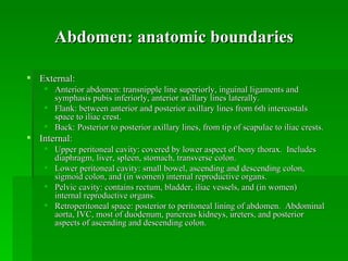
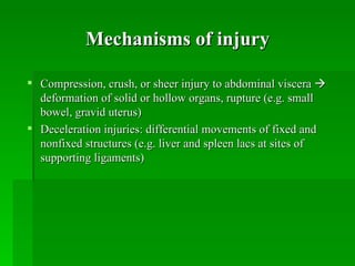
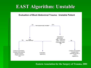
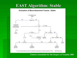
Blunt Abdominal Trauma:
       Evaluation
          Trauma Conference
           January 9th, 2006

           Greg Feldman, MD
    PGY1, General Surgery Department
        Stanford Medical Center
Outline

   Anatomic definition of abdomen
   Mechanisms of injury in blunt trauma
   Typical injury patterns
   Assessment of blunt abdominal trauma
   Diagnostic algorithms
Abdomen: anatomic boundaries

 External:
     Anterior abdomen: transnipple line superiorly, inguinal ligaments and
      symphasis pubis inferiorly, anterior axillary lines laterally.
     Flank: between anterior and posterior axillary lines from 6th intercostals
      space to iliac crest.
     Back: Posterior to posterior axillary lines, from tip of scapulae to iliac crests.
 Internal:
     Upper peritoneal cavity: covered by lower aspect of bony thorax. Includes
      diaphragm, liver, spleen, stomach, transverse colon.
     Lower peritoneal cavity: small bowel, ascending and descending colon,
      sigmoid colon, and (in women) internal reproductive organs.
     Pelvic cavity: contains rectum, bladder, iliac vessels, and (in women)
      internal reproductive organs.
     Retroperitoneal space: posterior to peritoneal lining of abdomen. Abdominal
      aorta, IVC, most of duodenum, pancreas kidneys, ureters, and posterior
      aspects of ascending and descending colon.
Mechanisms of injury

 Compression, crush, or sheer injury to abdominal viscera 
  deformation of solid or hollow organs, rupture (e.g. small
  bowel, gravid uterus)
 Deceleration injuries: differential movements of fixed and
  nonfixed structures (e.g. liver and spleen lacs at sites of
  supporting ligaments)
Common injury patterns
 In patients undergoing laparotomy for blunt trauma, most frequently
  injured organs are spleen (40-55%), liver (35-45%), and small bowel
  (5-10%). (ATLS, 2001)
 Duodenum:
     Classically, frontal-impact MVC with unrestrained driver; or direct blow to
      abdomen.
     Bloody gastric aspirate, retroperitoneal air on XR or CT
     Confirmed with upper GI series or double contrast CT
 Small bowel injury:
     Generally from sudden deceleration with subsequent tearing near fixed
      points of attachment.
     Often associated with seat belt sign, lumbar distraction fracture (Chance
      fracture)
     DPL superior to FAST or CT for diagnosis.
Common injury patterns (2)
 Pancreas:
    Direct epigastric blow compressing pancreas against vertebral column.
    Early normal serum amylase does NOT exclude major pancreatic trauma.
    CT with PO/IV contrast – NOT particularly sensitive in immediate post-
     injury period.

 Diaphragm:
    Most commonly, 5-10 cm rupture involving posterolateral hemidiaphragm.
    Noted on CXR: blurred or elevated hemidiaphragm, hemothorax, GT in
     chest

 Genitourinary:
    Anterior injuries (below UG diaphragm): usually from straddle impact.
    Posterior injuries (above UG diaphragm): in patient with multisystem
     injuries and pelvic fractures.
Common injury patterns (3)

 Solid organ injury
    Laceration to liver, spleen, or kidney
    Injury to one of these three + hemodynamic instability: considered
     indication for urgent laparotomy
    Isolated solid organ injury in hemodynamically stable patient: can
     often be managed nonoperatively.

 Pelvic fractures:
      Suggest major force applied to patient.
      Usually auto-ped, MVC, or motorcycle
      Significant association with intraperitoneal and retroperitoneal
       organs and vascular structures.
Restraining devices
   Lap seat belt
        Mesenteric tear or avulsion
        Rupture of small bowel or colon
        Iliac artery or abdominal aorta thrombosis
        Chance fracture of lumbar vertebrae (hyperflexion)
   Shoulder Harness
        Rupture of upper abdominal viscera
        Intimal tear or thrombosis in innominate, carotid, subclavian, or vertebral arteries
        Fracture or dislocation of C-spine
        Rib fractures
        Pulmonary contusion
   Air Bag
        Corneal abrasions, keratitis
        Abrasions of face, neck, chest
        Cardiac rupture
        C or T-spine fracture
Assessment: History

   Mechanism
   Symptoms, events, PMH, Meds, EtOH/drugs
   MVC:
     Speed
     Type of collision (frontal, lateral, sideswipe, rear,
      rollover)
     Vehicle intrusion into passenger compartment
     Types of restraints
     Deployment of air bag
     Patient's position in vehicle
Assessment: Physical Exam

 Inspection, auscultation, percussion, palpation
    Inspection: abrasions, contusions, lacerations, deformity
       Grey-Turner, Kehr, Balance, Cullen
    Auscultation: careful exam advised by ATLS.
     (Controversial utility in trauma setting.)
    Percussion: subtle signs of peritonitis; tympany in gastric
     dilatation or free air; dullness with hemoperitoneum
    Palpation: elicit superficial, deep, or rebound tenderness;
     involuntary muscle guarding
Physical Exam: Eponyms

 Grey-Turner sign:
    Bluish discoloration of lower flanks, lower back; associated with
     retroperitoneal bleeding of pancreas, kidney, or pelvic fracture.
 Cullen sign:
    Bluish discoloration around umbilicus, indicates peritoneal bleeding,
     often pancreatic hemorrhage.
 Kehr sign:
    L shoulder pain while supine; caused by diaphragmatic irritation
     (splenic injury, free air, intra-abd bleeding)
 Balance sign:
    Dull percussion in LUQ. Sign of splenic injury; blood accumulating
     in subcapsular or extracapsular spleen.
Diagnostic adjuncts

 Labs: BMP, CBC, coags, b-HCG, amy/lip, U/A, tox screen,
  T&C
 Plain films: CXR, pelvis; abd films generally lower priority
 DPL
 FAST
 CT
Diagnostic Peritoneal Lavage
 98% sensitive for intraperitoneal bleeding (ATLS)
 Open or closed (Seldinger); usually infraumbilical, but may be
  supraumbilical in pelvic frxs or advanced pregnancy.
 Free aspiration of blood, GI contents, or bile in demodynamically
  abnormal pt: indication for laparotomy
 If gross blood (> 10 mL) or GI contents not aspirated, perform lavage
  with 1000 mL warmed LR. Allow to mix, compress abdomen and
  logross paient, the sent to lab. + test: >100,000 RBC/mm3, >500 WBC/
  mm3, Gram stain with bacteria.
 Alters subsequent examination of patient

 Has been somewhat superceded by FAST in common use; now generally
  performed in unstable patients with intermediate FAST exams, or with
  suspicion for small bowel injury.
FAST: Strengths and Limitations
Strengths                               Limitations
 Rapid (~2 mins)                        Does not typically identify source of
 Portable                                 bleeding, or detect injuries that do
 Inexpensive                              not cause hemoperitoneum
 Technically simple, easy to train      Requires extensive training to assess
                                           parenchyma reliably
   (studies show competence can be
   achieved after ~30 studies)           Limited in detecting <250 cc
 Can be performed serially                intraperitoneal fluid
 Useful for guiding triage decisions    Particularly poor at detecting bowel
                                           and mesentery damage (44%
   in trauma patients                      sensitivity)
                                         Difficult to assess retroperitoneum
                                         Limited by habitus in obese patients
FAST: Accuracy

For identifying hemoperitoneum in blunt abdominal trauma:
    Sensitivity 76 - 90%
    Specificity 95 - 100%
The larger the hemoperitoneum, the higher the sensitivity. So
     sensitivity increases for clinically significant
     hemoperitoneum.

How much fluid can FAST detect?
  250 cc total
  100 cc in Morison’s pouch
Does FAST replace CT?

Only at the extremes.
 Unstable patient, (+) FAST  OR
 Stable patient, low force injury, (-) FAST  consider
   observing patient.

CT is far more sensitive than FAST for detecting and
   characterizing abdominal injury in trauma. The gold
   standard for characterizing intraparenchymal injury.

“Death begins with a CT.” Never send an unstable patient to
   CT. FAST, however, can be performed during
   resuscitation.
CT
EAST level I recommendations (2001):
 CT is recommended for evaluation of hemodynamically
  stable patients with equivocal findings on physical
  examination, associated neurologic injury, or multiple
  extra-abdominal injuries.

   CT is the diagnostic modality of choice for nonoperative
    management of solid visceral injuries.
EAST Algorithm: Unstable


        Eastern Association for the Surgery of Trauma, 2001
EAST Algorithm: Stable


      Eastern Association for the Surgery of Trauma, 2001
References
   Hoff et al. EAST Practice Management Guidelines Work Group.
    Practice Management Guidelines for the Evaluation of Blunt
    Abdominal Trauma, 2001. www.east.org.
   American College of Surgeons Committee on Trauma.
    Advanced Trauma Life Support for Doctors; Student Course
    Manual, 7th edition, 2004.
   Scalea TM, Rodriquez A, Chiu WC. Focused Assessment with
    Sonography for Trauma (FAST): Results from an International
    Consensus Conference. J. Trauma 1999;46:466-472.
   Yoshii H, Sato M, Yamamoto S. Usefulness and Limitations of
    Ultrasonography in the Initial Evaluation of Blunt Abdominal
    Trauma. J. Trauma 1998;45:45-51.
Acknowledgements
   Dr. Shelly Erford
   Dr. Denny Jenkins
   Carol Thomson
   Dr. Natalie Kirilchik
   Dr. Subarna Biswas
   Drs. Brundage, Spain, and Gregg
   Stanford Medical Center ACS/Trauma Service
   Noah Feinstein
   Dr. Gillian Lieberman
   Dr. Jason Tracy

Abdominal trauma

  • 1.
    Blunt Abdominal Trauma: Evaluation Trauma Conference January 9th, 2006 Greg Feldman, MD PGY1, General Surgery Department Stanford Medical Center
  • 2.
    Outline  Anatomic definition of abdomen  Mechanisms of injury in blunt trauma  Typical injury patterns  Assessment of blunt abdominal trauma  Diagnostic algorithms
  • 3.
    Abdomen: anatomic boundaries External:  Anterior abdomen: transnipple line superiorly, inguinal ligaments and symphasis pubis inferiorly, anterior axillary lines laterally.  Flank: between anterior and posterior axillary lines from 6th intercostals space to iliac crest.  Back: Posterior to posterior axillary lines, from tip of scapulae to iliac crests.  Internal:  Upper peritoneal cavity: covered by lower aspect of bony thorax. Includes diaphragm, liver, spleen, stomach, transverse colon.  Lower peritoneal cavity: small bowel, ascending and descending colon, sigmoid colon, and (in women) internal reproductive organs.  Pelvic cavity: contains rectum, bladder, iliac vessels, and (in women) internal reproductive organs.  Retroperitoneal space: posterior to peritoneal lining of abdomen. Abdominal aorta, IVC, most of duodenum, pancreas kidneys, ureters, and posterior aspects of ascending and descending colon.
  • 4.
    Mechanisms of injury Compression, crush, or sheer injury to abdominal viscera  deformation of solid or hollow organs, rupture (e.g. small bowel, gravid uterus)  Deceleration injuries: differential movements of fixed and nonfixed structures (e.g. liver and spleen lacs at sites of supporting ligaments)
  • 5.
    Common injury patterns In patients undergoing laparotomy for blunt trauma, most frequently injured organs are spleen (40-55%), liver (35-45%), and small bowel (5-10%). (ATLS, 2001)  Duodenum:  Classically, frontal-impact MVC with unrestrained driver; or direct blow to abdomen.  Bloody gastric aspirate, retroperitoneal air on XR or CT  Confirmed with upper GI series or double contrast CT  Small bowel injury:  Generally from sudden deceleration with subsequent tearing near fixed points of attachment.  Often associated with seat belt sign, lumbar distraction fracture (Chance fracture)  DPL superior to FAST or CT for diagnosis.
  • 6.
    Common injury patterns(2)  Pancreas:  Direct epigastric blow compressing pancreas against vertebral column.  Early normal serum amylase does NOT exclude major pancreatic trauma.  CT with PO/IV contrast – NOT particularly sensitive in immediate post- injury period.  Diaphragm:  Most commonly, 5-10 cm rupture involving posterolateral hemidiaphragm.  Noted on CXR: blurred or elevated hemidiaphragm, hemothorax, GT in chest  Genitourinary:  Anterior injuries (below UG diaphragm): usually from straddle impact.  Posterior injuries (above UG diaphragm): in patient with multisystem injuries and pelvic fractures.
  • 7.
    Common injury patterns(3)  Solid organ injury  Laceration to liver, spleen, or kidney  Injury to one of these three + hemodynamic instability: considered indication for urgent laparotomy  Isolated solid organ injury in hemodynamically stable patient: can often be managed nonoperatively.  Pelvic fractures:  Suggest major force applied to patient.  Usually auto-ped, MVC, or motorcycle  Significant association with intraperitoneal and retroperitoneal organs and vascular structures.
  • 8.
    Restraining devices  Lap seat belt  Mesenteric tear or avulsion  Rupture of small bowel or colon  Iliac artery or abdominal aorta thrombosis  Chance fracture of lumbar vertebrae (hyperflexion)  Shoulder Harness  Rupture of upper abdominal viscera  Intimal tear or thrombosis in innominate, carotid, subclavian, or vertebral arteries  Fracture or dislocation of C-spine  Rib fractures  Pulmonary contusion  Air Bag  Corneal abrasions, keratitis  Abrasions of face, neck, chest  Cardiac rupture  C or T-spine fracture
  • 9.
    Assessment: History  Mechanism  Symptoms, events, PMH, Meds, EtOH/drugs  MVC:  Speed  Type of collision (frontal, lateral, sideswipe, rear, rollover)  Vehicle intrusion into passenger compartment  Types of restraints  Deployment of air bag  Patient's position in vehicle
  • 10.
    Assessment: Physical Exam Inspection, auscultation, percussion, palpation  Inspection: abrasions, contusions, lacerations, deformity  Grey-Turner, Kehr, Balance, Cullen  Auscultation: careful exam advised by ATLS. (Controversial utility in trauma setting.)  Percussion: subtle signs of peritonitis; tympany in gastric dilatation or free air; dullness with hemoperitoneum  Palpation: elicit superficial, deep, or rebound tenderness; involuntary muscle guarding
  • 11.
    Physical Exam: Eponyms Grey-Turner sign:  Bluish discoloration of lower flanks, lower back; associated with retroperitoneal bleeding of pancreas, kidney, or pelvic fracture.  Cullen sign:  Bluish discoloration around umbilicus, indicates peritoneal bleeding, often pancreatic hemorrhage.  Kehr sign:  L shoulder pain while supine; caused by diaphragmatic irritation (splenic injury, free air, intra-abd bleeding)  Balance sign:  Dull percussion in LUQ. Sign of splenic injury; blood accumulating in subcapsular or extracapsular spleen.
  • 12.
    Diagnostic adjuncts  Labs:BMP, CBC, coags, b-HCG, amy/lip, U/A, tox screen, T&C  Plain films: CXR, pelvis; abd films generally lower priority  DPL  FAST  CT
  • 13.
    Diagnostic Peritoneal Lavage 98% sensitive for intraperitoneal bleeding (ATLS)  Open or closed (Seldinger); usually infraumbilical, but may be supraumbilical in pelvic frxs or advanced pregnancy.  Free aspiration of blood, GI contents, or bile in demodynamically abnormal pt: indication for laparotomy  If gross blood (> 10 mL) or GI contents not aspirated, perform lavage with 1000 mL warmed LR. Allow to mix, compress abdomen and logross paient, the sent to lab. + test: >100,000 RBC/mm3, >500 WBC/ mm3, Gram stain with bacteria.  Alters subsequent examination of patient  Has been somewhat superceded by FAST in common use; now generally performed in unstable patients with intermediate FAST exams, or with suspicion for small bowel injury.
  • 14.
    FAST: Strengths andLimitations Strengths Limitations  Rapid (~2 mins)  Does not typically identify source of  Portable bleeding, or detect injuries that do  Inexpensive not cause hemoperitoneum  Technically simple, easy to train  Requires extensive training to assess parenchyma reliably (studies show competence can be achieved after ~30 studies)  Limited in detecting <250 cc  Can be performed serially intraperitoneal fluid  Useful for guiding triage decisions  Particularly poor at detecting bowel and mesentery damage (44% in trauma patients sensitivity)  Difficult to assess retroperitoneum  Limited by habitus in obese patients
  • 15.
    FAST: Accuracy For identifyinghemoperitoneum in blunt abdominal trauma:  Sensitivity 76 - 90%  Specificity 95 - 100% The larger the hemoperitoneum, the higher the sensitivity. So sensitivity increases for clinically significant hemoperitoneum. How much fluid can FAST detect?  250 cc total  100 cc in Morison’s pouch
  • 16.
    Does FAST replaceCT? Only at the extremes.  Unstable patient, (+) FAST  OR  Stable patient, low force injury, (-) FAST  consider observing patient. CT is far more sensitive than FAST for detecting and characterizing abdominal injury in trauma. The gold standard for characterizing intraparenchymal injury. “Death begins with a CT.” Never send an unstable patient to CT. FAST, however, can be performed during resuscitation.
  • 17.
    CT EAST level Irecommendations (2001):  CT is recommended for evaluation of hemodynamically stable patients with equivocal findings on physical examination, associated neurologic injury, or multiple extra-abdominal injuries.  CT is the diagnostic modality of choice for nonoperative management of solid visceral injuries.
  • 18.
    EAST Algorithm: Unstable Eastern Association for the Surgery of Trauma, 2001
  • 19.
    EAST Algorithm: Stable Eastern Association for the Surgery of Trauma, 2001
  • 20.
    References  Hoff et al. EAST Practice Management Guidelines Work Group. Practice Management Guidelines for the Evaluation of Blunt Abdominal Trauma, 2001. www.east.org.  American College of Surgeons Committee on Trauma. Advanced Trauma Life Support for Doctors; Student Course Manual, 7th edition, 2004.  Scalea TM, Rodriquez A, Chiu WC. Focused Assessment with Sonography for Trauma (FAST): Results from an International Consensus Conference. J. Trauma 1999;46:466-472.  Yoshii H, Sato M, Yamamoto S. Usefulness and Limitations of Ultrasonography in the Initial Evaluation of Blunt Abdominal Trauma. J. Trauma 1998;45:45-51.
  • 21.
    Acknowledgements  Dr. Shelly Erford  Dr. Denny Jenkins  Carol Thomson  Dr. Natalie Kirilchik  Dr. Subarna Biswas  Drs. Brundage, Spain, and Gregg  Stanford Medical Center ACS/Trauma Service  Noah Feinstein  Dr. Gillian Lieberman  Dr. Jason Tracy