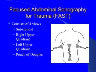
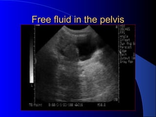
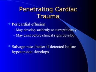
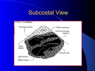
Emergency UltrasoundEmergency Ultrasound
inin
TraumaTrauma
Anthony J Weekes MD,Anthony J Weekes MD,
RDMSRDMS
Janet G. Alteveer, MDJanet G. Alteveer, MD
Sarah Stahmer, MD
Clinical CaseClinical Case
GR is a 62 y male who hit his right torso
when he slipped on an icy sidewalk. He
denies head trauma, and can walk
without a limp. Two hours later the pain
in his lower chest has increased he
comes to the ED.
Clinical CaseClinical Case
PE: BP116/72, pulse109, RR 24.
There is a minor abrasion to right lateral
chest, which is tender to palpation.
Diffuse mild abdominal tenderness.
Meds: Coumadin for irregular heartbeat
Clinical CaseClinical Case
 2 large IV’s placed,
CXR done. Blood
tests sent.
 Bedside ultrasound
done.
 CXR revealed lower
rib fractures, no HTX
or PTX
Clinical CaseClinical Case
FFP ordered and OR notified.
He is found to have a liver laceration
and 500 cc of blood in the peritoneal
cavity.
Diagnostic Modalities in BluntDiagnostic Modalities in Blunt
Abdominal TraumaAbdominal Trauma
Diagnostic Peritoneal Lavage (DPL)
CAT Scan
Ultrasound (FAST exam)
Diagnostic Peritoneal LavageDiagnostic Peritoneal Lavage
 Advantages
– Very sensitive for
identifying intra-
peritoneal blood
– 106
RBC/mm3
approx.
20 ml blood in 1L
lavage fluid
– Can be done at the
bedside
– Can be done in 10-15
minutes
 Disadvantages
– Overly sensitive, may
result in too high a
laparotomy rate
– Invasive
– Difficult in pregnancy,
or with many prior
surgeries
– Can not be repeated
CT ScanCT Scan
 Advantages
– Identifies specific
injuries
– Good for hollow viscus
and retroperitoneal
injury
– High sensitivity and
specificity
 Disadvantages
– Expensive equipment
– 30-60 minutes to
complete study
– Only for stable
patients
– Not for pregnant
patients
FAST
Focused Abdominal
Sonography in Trauma
FASTFAST
 Advantages
– Can be performed in 5
minutes at the bedside
– Non-invasive
– Repeat exams
– Sensitivity and
specificity for free
fluid equal to DPL and
CT
 Disadvantages
– Operator dependent
– May not identify
specific injury
– Poor for hollow viscus
or retroperitoneal
injury
– Obesity, subcutaneous
air may interfere with
exam
FAST PrinciplesFAST Principles
 Detects free
intraperitoneal fluid
 Blood/fluid pools in
dependent areas
 Pelvis
– Most dependent
 Hepatorenal fossa
– Most dependent area in
supramesocolic region
FAST PrinciplesFAST Principles
 Pelvis and Supra-
mesocolic areas
communicate
– Phrenicolic ligament
prevents flow
 Liver/spleen injury
– Represents 2/3 of cases of
blunt abdominal trauma
FAST- principlesFAST- principles
Intraperitoneal fluid may be
– Blood
– Preexisting ascites
– Urine
– Intestinal contents
FAST – limitationsFAST – limitations
US relatively insensitive for detecting
traumatic abdominal organ injury
Fluid may pool at variable rates
– Minimum volume for US detection
– Multiple views at multiple sites
– Serial exams: repeat exam if there is a change
in clinical picture
Operator dependent
Evidence supporting use ofEvidence supporting use of
FASTFAST
Multiple studies in USA by EM and trauma
surgeons
Studies from Europe and Japan
Policy statements by specialty organizations
Emergency department ultrasound in the
evaluation of blunt abdominal trauma.
Jehle, D., et al, Am J Emerg Med, 1993
– Single view of Morison’s pouch in 44 patients
– Performed by physicians after 2 weeks training
– US compared to DPL and laparotomy
– Sensitivity 81.8%
– Specificity 93.9%
Trauma surgical studyTrauma surgical study
A prospective study of surgeon-
performed ultrasound as the primary
adjuvant modality of injured patient
assessment. 1994 Rozycki et al.
N=358 patients
Outcomes used: US detection of
hemoperitoneum/pericardial effusion
ResultsResults
53/358 (15%) patients w/ free fluid on
“gold standard”
All patients: Sens 81.5%, spec 99.7%
Blunt trauma: Sens 78.6%, spec 100%
PPV 98.1%, NPV 96.2%
Overall accuracy was 96.5% for detection
of hemoperitoneum or pericardium
Trauma StudyTrauma Study
Rozycki G, et al 1998 Surgeon-performed
ultrasound for the assessment of truncal
injuries. Lessons learned from 1540 patients
FAST exam on patients with precordial or
transthoracic wounds or blunt abdominal
trauma
Protocol:
+ Pericardial fluid OR
Stable CT
+IP fluid
Unstable OR
 Results
– N= 1540 pts, 80/1540 (5%) with FF
– Overall: Sens 83.3%, Spec 99.7%
– PPV 95%, NPV 99%
– Precordial/Transthor : Sens 100%, Spec 99.3%
– Hypotensive BAT: Sens 100%, Spec 100%
FAST – Specialty SocietiesFAST – Specialty Societies
 Established clinical role in Europe, Australia,
Japan, Israel
 German Surgical Society requires candidates’
proficiency in ultrasound
 United States
– US in ATLS
– US policies by frontline specialties
 American College of Surgeons
 ACEP,SAEM & AAEM
FASTFAST
Perform during
– Resuscitation
– Physical exam
– Stabilization
EquipmentEquipment
Curved array
 Various “footprints”
– Small footprint for thorax
– Large for abdomen
 Variable frequencies
– 5.0 MHz: thin, child
– 3.5 MHz: versatile
– 2.0 MHz: cardiac, large
pts
Time to Complete ScanTime to Complete Scan
Each view: 30-60 seconds
Number of views dependent on clinical
question and findings on initial views
Total exam time usually < 3-5 minutes
1988 Armenian earthquake
– 400 trauma US scans in 72 hrs
Focused Abdominal SonographyFocused Abdominal Sonography
for Trauma (FAST)for Trauma (FAST)
 Consists of 4 views
– Subxiphoid
– Right Upper
Quadrant
– Left Upper
Quadrant
– Pouch of Douglas
FASTFAST
 Increased sensitivity with
increased number of views
 Will identify pleural
effusions
 Reliably detects as little as
50-100cc in the thorax
 Sensitivity >96%,
specificity 99-100%
Clinical experience with FASTClinical experience with FAST
Intraperitoneal fluid
– Sensitivity 82-98%, specificity 88-100%
Morison’s pouch alone 36-82% sensitivity
Increased sensitivity with
– Increasing number of views
– Trendelenberg
– Serial examinations
Can detect as little as 250cc of free fluid
Clinical ExperienceClinical Experience
Solid organ disruption
– 40% sensitivity for all organs
– 33-94% for splenic injury
Hollow viscus injury
– Sensitivity 57%
Retroperitoneal injury
– Sensitivity for identification of hemorrhage
<60%
RUQRUQ
 Probe at right thoraco-
abdominal junction
 Liver : large acoustic
window
 Probe marker cephalad
 Rib interference?
– Rotate 30°
counterclockwise
Scan PlaneScan Plane
 Same image if probe
positioned
– Anterior
– Mid axillary
– Posterior
RUQRUQ
 Image on screen:
– Liver cephalad
– Kidney inferiorly
– Morison’s Pouch*:
space between
Glisson’s capsule and
Gerota’s fascia
*
*
*
*
Normal RUQNormal RUQ
 Image kidney
– Longitudinally
– Transversely
 Two toned structure
– Cortex/medulla
– Renal sinus
Appearance of bloodAppearance of blood
 Fresh blood
– Anechoic (black)
 Coagulating blood
– First hypoechoic
– Later hyperechoic
Normal
Morison’s Pouch
Free fluid in
Morison’s
Pouch
Branney, S.W. et al: Quantitative sensitivity of
ultrasound in detecting free intraperitoneal
fluid J Trauma:1995: 39
Peritoneal lavage fluid infused in 100 patients
Simultaneous scan of Morison’s pouch
– By physicians ( Surgery,EM, Radiology)
– Blinded to volume and rate of infusion
– Mean volume of detection: 619cc
– Sensitivity at 1 liter: 97%
– 10% physicians detected less than 400cc
Caveat to Branney study:
– Artificial condition: infused fluid
– Fluid in Morison’s after pelvis overflow
Tiling et al :
– 200 -250ml detected by US
– Collection >0.5cm suggests over 500ml
Transvaginal/rectal
– 15ml of free intraperitoneal fluid
Volume Assessment by US
Detection of Fluid byDetection of Fluid by
UltrasoundUltrasound
 Affected by
positioning
 Location of bleed
 Rate of bleeding
 Operator Experience
 Value of sensitivity of
Ultrasound:
– Detects clinically
injuries
– Non-detection of fluid
 May indicate self-
limited bleeding
All Fluid is not BloodAll Fluid is not Blood
Ascites
Ruptured Ovarian Cyst
Lavage fluid
Urine from ruptured bladder
Mimics of Fluid in RUQMimics of Fluid in RUQ
Perinephric fat
– May be hypoechoic like blood
– Usually evenly layered along kidney
– If in doubt, compare to left kidney
Abdominal inflammation
– Widened extra-renal space
– Echogenicity of kidney becomes more like the
liver parenchyma
PitfallsPitfalls
RUQ
– Not attempting multiple probe placements
– Not placing the probe cephalad enough to use the
acoustic window of the liver
 Scanning too soon before enough blood has
accumulated
 Not repeating the scan
LUQLUQ
 Probe at left posterior
axillary line
 Near ribs 9 and 10
 Angle probe obliquely
(avoid ribs)
LUQ Scan PlaneLUQ Scan Plane
 More difficult
– Acoustic window
(spleen) is smaller than
liver
– Mild inspiration will
optimize image
– Bowel interference is
common
LUQ ScanLUQ Scan
spleen
kidney
*Splenorenal fossa – a potential space
*
*
*
*
Normal
Spleno-renal
view
Free fluid
around spleen
To Evaluate the ThoraxTo Evaluate the Thorax
 Move probe
– cephalad
– longitudinal
 Image
Liver
Diaphragm
Pleural space
HemothoraxHemothorax
liver
diaphragmfluid
Small Pleural EffusionSmall Pleural Effusion
Large Pleural Effusion
Ma O John, Mateer J, Trauma Ultrasound
Examination Versus Chest Radiography in the
Detection of Hemothorax
Ann Emerg Med: March 1997
 240 trauma US study patients
 26 had hemothorax ( CT or chest tube)
 CXR and US
– 0 false positive
– 1 false negative
– 25 true positive
– 214 true negative
Pelvic ViewPelvic View
 Probe should be
placed in the
suprapubic position
 Either can be
transverse or
longitudinal
 Helpful to image
before placement of
a Foley catheter
Pelvis (Long View)Pelvis (Long View)
Pelvis: TransversePelvis: Transverse
Normal
Transverse
pelvic
Fluid in pelvis
Pelvic View – SagittalPelvic View – Sagittal
 Fluid in front of the
bladder
 If bladder is empty
or Foley already
placed:
Trick of trade
– IV bag on abdomen
– Scan through bag
clot bladder
Blood in the PelvisBlood in the Pelvis
Free fluid in the pelvisFree fluid in the pelvis
FAST AlgorithmFAST Algorithm
Normal
Hemodynamic
Status
AlteredMS
ConfoundingInjury
GrossHematuria
HCT< 35%
Repeat U/S30’
HCTat 4h
Observe8h
US:
Freefluid?
Nonoperative
or
cirrhosis?
LAPAROTOMY
DPL
Abdominal CT
Peritoneal
Irritation?
DPL
NO
NO NO
US
Freefluid?
NO
Branney, et. al.
J Trauma, 1997
YES
YES
YES
YES
YES
NO NO
YES
Ultrasound in the Detection of Injury
From Blunt or Penetrating Thoracic
Trauma
Penetrating ThoracicPenetrating Thoracic
InjuryInjury
Clinical challenge
– Where is the penetration?
– What was the weapon?
– What was the trajectory?
– What organ(s) have been injured?
– Improved outcomes in patients with normal or
near-normal vital signs
Penetrating CardiacPenetrating Cardiac
TraumaTrauma
Pericardial effusion
– May develop suddenly or surreptitiously
– May exist before clinical signs develop
Salvage rates better if detected before
hypotension develops
Clinical CaseClinical Case
QD is 37 year old male brought in by EMS
for ingesting entire bottle of unidentified
red and white pills. In the ambulance bay he
pulls out a knife and stabs himself in the left
nipple.
Clinical CaseClinical Case
Initial BP 116/72, pulse 109 RR 24. IV’s
placed.
No JVD, Clear breath sounds, non tender
abdomen
As CXR is about to be done, pulse increases
to 134.
Bedside ultrasound is done while cartridge
is developed.
Clinical CaseClinical Case
Patient is taken to the OR
Penetrating cardiac wound is repaired
Subcostal ViewSubcostal View
Most practical in
trauma setting
Away from airway
and neck/chest
procedures
Also called Sub-
Xyphoid view
Subcostal ViewSubcostal View
Subcostal View
Pericardial FluidPericardial Fluid
fluid
Occult Penetrating CardiacOccult Penetrating Cardiac
TraumaTrauma
Observation unreliable
Subxiphoid window
– Invasive
– 100% sensitive, 92% specific
– Negative exploration rates (as high as 80%)
Ultrasound reliable indicator of even small
pericardial effusion
Trauma StudyTrauma Study
The role of ultrasound in patients with
possible penetrating cardiac wounds: a
prospective multicenter study.
Rozycki GS: J Trauma. 1999
 Pericardial scans performed in 261 patients
 Sensitivity 100%, specificity 96.9%
 PPV: 81% NPV:100%
 Time interval BUS to OR: 12.1 +/- 5.9 min
Avoid PitfallsAvoid Pitfalls
Normal echo does not definitively rule out
major pericardial injury
Repeat echo with ∆ clinical picture
Epicardial fat pad may easily be
misinterpreted as “clot”
Hemothorax may be confused with
pericardial effusion
Blunt Cardiac TraumaBlunt Cardiac Trauma
 Basic Assessments
– Pericardial effusion
– Assess for wall motion
abnormality
– RV:
 closest to anterior chest
wall
 Most likely to be
injured
 Advanced
Assessments
– Assess thoracic aorta –
may need TEE to see
all of thoracic aorta
 Hematoma
 Intimal flap
 Abnormal contour
– Valvular dysfunction
or septal rupture
Blunt cardiac traumaBlunt cardiac trauma
Injuries difficult to assess by FAST
– Valvular incompetence
– Myocardial rupture
– Intracardiac thrombosis
– Ventricular aneurysm
– Coronary Thrombosis
– Intra-cardiac Thrombosis
“ The most important preoperative objective in
the management of the patient with trauma is
to ascertain whether or not laparotomy is
needed, and not the diagnosis of a specific
organ injury”

29 us trauma

  • 1.
    Emergency UltrasoundEmergency Ultrasound inin TraumaTrauma AnthonyJ Weekes MD,Anthony J Weekes MD, RDMSRDMS Janet G. Alteveer, MDJanet G. Alteveer, MD Sarah Stahmer, MD
  • 2.
    Clinical CaseClinical Case GRis a 62 y male who hit his right torso when he slipped on an icy sidewalk. He denies head trauma, and can walk without a limp. Two hours later the pain in his lower chest has increased he comes to the ED.
  • 3.
    Clinical CaseClinical Case PE:BP116/72, pulse109, RR 24. There is a minor abrasion to right lateral chest, which is tender to palpation. Diffuse mild abdominal tenderness. Meds: Coumadin for irregular heartbeat
  • 4.
    Clinical CaseClinical Case 2 large IV’s placed, CXR done. Blood tests sent.  Bedside ultrasound done.  CXR revealed lower rib fractures, no HTX or PTX
  • 5.
    Clinical CaseClinical Case FFPordered and OR notified. He is found to have a liver laceration and 500 cc of blood in the peritoneal cavity.
  • 6.
    Diagnostic Modalities inBluntDiagnostic Modalities in Blunt Abdominal TraumaAbdominal Trauma Diagnostic Peritoneal Lavage (DPL) CAT Scan Ultrasound (FAST exam)
  • 7.
    Diagnostic Peritoneal LavageDiagnosticPeritoneal Lavage  Advantages – Very sensitive for identifying intra- peritoneal blood – 106 RBC/mm3 approx. 20 ml blood in 1L lavage fluid – Can be done at the bedside – Can be done in 10-15 minutes  Disadvantages – Overly sensitive, may result in too high a laparotomy rate – Invasive – Difficult in pregnancy, or with many prior surgeries – Can not be repeated
  • 8.
    CT ScanCT Scan Advantages – Identifies specific injuries – Good for hollow viscus and retroperitoneal injury – High sensitivity and specificity  Disadvantages – Expensive equipment – 30-60 minutes to complete study – Only for stable patients – Not for pregnant patients
  • 9.
  • 10.
    FASTFAST  Advantages – Canbe performed in 5 minutes at the bedside – Non-invasive – Repeat exams – Sensitivity and specificity for free fluid equal to DPL and CT  Disadvantages – Operator dependent – May not identify specific injury – Poor for hollow viscus or retroperitoneal injury – Obesity, subcutaneous air may interfere with exam
  • 11.
    FAST PrinciplesFAST Principles Detects free intraperitoneal fluid  Blood/fluid pools in dependent areas  Pelvis – Most dependent  Hepatorenal fossa – Most dependent area in supramesocolic region
  • 12.
    FAST PrinciplesFAST Principles Pelvis and Supra- mesocolic areas communicate – Phrenicolic ligament prevents flow  Liver/spleen injury – Represents 2/3 of cases of blunt abdominal trauma
  • 13.
    FAST- principlesFAST- principles Intraperitonealfluid may be – Blood – Preexisting ascites – Urine – Intestinal contents
  • 14.
    FAST – limitationsFAST– limitations US relatively insensitive for detecting traumatic abdominal organ injury Fluid may pool at variable rates – Minimum volume for US detection – Multiple views at multiple sites – Serial exams: repeat exam if there is a change in clinical picture Operator dependent
  • 15.
    Evidence supporting useofEvidence supporting use of FASTFAST Multiple studies in USA by EM and trauma surgeons Studies from Europe and Japan Policy statements by specialty organizations
  • 16.
    Emergency department ultrasoundin the evaluation of blunt abdominal trauma. Jehle, D., et al, Am J Emerg Med, 1993 – Single view of Morison’s pouch in 44 patients – Performed by physicians after 2 weeks training – US compared to DPL and laparotomy – Sensitivity 81.8% – Specificity 93.9%
  • 17.
    Trauma surgical studyTraumasurgical study A prospective study of surgeon- performed ultrasound as the primary adjuvant modality of injured patient assessment. 1994 Rozycki et al. N=358 patients Outcomes used: US detection of hemoperitoneum/pericardial effusion
  • 18.
    ResultsResults 53/358 (15%) patientsw/ free fluid on “gold standard” All patients: Sens 81.5%, spec 99.7% Blunt trauma: Sens 78.6%, spec 100% PPV 98.1%, NPV 96.2% Overall accuracy was 96.5% for detection of hemoperitoneum or pericardium
  • 19.
    Trauma StudyTrauma Study RozyckiG, et al 1998 Surgeon-performed ultrasound for the assessment of truncal injuries. Lessons learned from 1540 patients FAST exam on patients with precordial or transthoracic wounds or blunt abdominal trauma
  • 20.
    Protocol: + Pericardial fluidOR Stable CT +IP fluid Unstable OR  Results – N= 1540 pts, 80/1540 (5%) with FF – Overall: Sens 83.3%, Spec 99.7% – PPV 95%, NPV 99% – Precordial/Transthor : Sens 100%, Spec 99.3% – Hypotensive BAT: Sens 100%, Spec 100%
  • 21.
    FAST – SpecialtySocietiesFAST – Specialty Societies  Established clinical role in Europe, Australia, Japan, Israel  German Surgical Society requires candidates’ proficiency in ultrasound  United States – US in ATLS – US policies by frontline specialties  American College of Surgeons  ACEP,SAEM & AAEM
  • 22.
    FASTFAST Perform during – Resuscitation –Physical exam – Stabilization
  • 23.
    EquipmentEquipment Curved array  Various“footprints” – Small footprint for thorax – Large for abdomen  Variable frequencies – 5.0 MHz: thin, child – 3.5 MHz: versatile – 2.0 MHz: cardiac, large pts
  • 24.
    Time to CompleteScanTime to Complete Scan Each view: 30-60 seconds Number of views dependent on clinical question and findings on initial views Total exam time usually < 3-5 minutes 1988 Armenian earthquake – 400 trauma US scans in 72 hrs
  • 25.
    Focused Abdominal SonographyFocusedAbdominal Sonography for Trauma (FAST)for Trauma (FAST)  Consists of 4 views – Subxiphoid – Right Upper Quadrant – Left Upper Quadrant – Pouch of Douglas
  • 26.
    FASTFAST  Increased sensitivitywith increased number of views  Will identify pleural effusions  Reliably detects as little as 50-100cc in the thorax  Sensitivity >96%, specificity 99-100%
  • 27.
    Clinical experience withFASTClinical experience with FAST Intraperitoneal fluid – Sensitivity 82-98%, specificity 88-100% Morison’s pouch alone 36-82% sensitivity Increased sensitivity with – Increasing number of views – Trendelenberg – Serial examinations Can detect as little as 250cc of free fluid
  • 28.
    Clinical ExperienceClinical Experience Solidorgan disruption – 40% sensitivity for all organs – 33-94% for splenic injury Hollow viscus injury – Sensitivity 57% Retroperitoneal injury – Sensitivity for identification of hemorrhage <60%
  • 29.
    RUQRUQ  Probe atright thoraco- abdominal junction  Liver : large acoustic window  Probe marker cephalad  Rib interference? – Rotate 30° counterclockwise
  • 30.
    Scan PlaneScan Plane Same image if probe positioned – Anterior – Mid axillary – Posterior
  • 31.
    RUQRUQ  Image onscreen: – Liver cephalad – Kidney inferiorly – Morison’s Pouch*: space between Glisson’s capsule and Gerota’s fascia * * * *
  • 32.
    Normal RUQNormal RUQ Image kidney – Longitudinally – Transversely  Two toned structure – Cortex/medulla – Renal sinus
  • 33.
    Appearance of bloodAppearanceof blood  Fresh blood – Anechoic (black)  Coagulating blood – First hypoechoic – Later hyperechoic
  • 34.
  • 41.
    Branney, S.W. etal: Quantitative sensitivity of ultrasound in detecting free intraperitoneal fluid J Trauma:1995: 39 Peritoneal lavage fluid infused in 100 patients Simultaneous scan of Morison’s pouch – By physicians ( Surgery,EM, Radiology) – Blinded to volume and rate of infusion – Mean volume of detection: 619cc – Sensitivity at 1 liter: 97% – 10% physicians detected less than 400cc
  • 42.
    Caveat to Branneystudy: – Artificial condition: infused fluid – Fluid in Morison’s after pelvis overflow Tiling et al : – 200 -250ml detected by US – Collection >0.5cm suggests over 500ml Transvaginal/rectal – 15ml of free intraperitoneal fluid Volume Assessment by US
  • 43.
    Detection of FluidbyDetection of Fluid by UltrasoundUltrasound  Affected by positioning  Location of bleed  Rate of bleeding  Operator Experience  Value of sensitivity of Ultrasound: – Detects clinically injuries – Non-detection of fluid  May indicate self- limited bleeding
  • 44.
    All Fluid isnot BloodAll Fluid is not Blood Ascites Ruptured Ovarian Cyst Lavage fluid Urine from ruptured bladder
  • 45.
    Mimics of Fluidin RUQMimics of Fluid in RUQ Perinephric fat – May be hypoechoic like blood – Usually evenly layered along kidney – If in doubt, compare to left kidney Abdominal inflammation – Widened extra-renal space – Echogenicity of kidney becomes more like the liver parenchyma
  • 46.
    PitfallsPitfalls RUQ – Not attemptingmultiple probe placements – Not placing the probe cephalad enough to use the acoustic window of the liver  Scanning too soon before enough blood has accumulated  Not repeating the scan
  • 47.
    LUQLUQ  Probe atleft posterior axillary line  Near ribs 9 and 10  Angle probe obliquely (avoid ribs)
  • 48.
    LUQ Scan PlaneLUQScan Plane  More difficult – Acoustic window (spleen) is smaller than liver – Mild inspiration will optimize image – Bowel interference is common
  • 49.
    LUQ ScanLUQ Scan spleen kidney *Splenorenalfossa – a potential space * * * *
  • 50.
  • 56.
    To Evaluate theThoraxTo Evaluate the Thorax  Move probe – cephalad – longitudinal  Image Liver Diaphragm Pleural space
  • 57.
  • 58.
    Small Pleural EffusionSmallPleural Effusion Large Pleural Effusion
  • 59.
    Ma O John,Mateer J, Trauma Ultrasound Examination Versus Chest Radiography in the Detection of Hemothorax Ann Emerg Med: March 1997  240 trauma US study patients  26 had hemothorax ( CT or chest tube)  CXR and US – 0 false positive – 1 false negative – 25 true positive – 214 true negative
  • 60.
    Pelvic ViewPelvic View Probe should be placed in the suprapubic position  Either can be transverse or longitudinal  Helpful to image before placement of a Foley catheter
  • 61.
  • 62.
  • 63.
  • 64.
    Pelvic View –SagittalPelvic View – Sagittal  Fluid in front of the bladder  If bladder is empty or Foley already placed: Trick of trade – IV bag on abdomen – Scan through bag clot bladder
  • 65.
    Blood in thePelvisBlood in the Pelvis
  • 68.
    Free fluid inthe pelvisFree fluid in the pelvis
  • 69.
    FAST AlgorithmFAST Algorithm Normal Hemodynamic Status AlteredMS ConfoundingInjury GrossHematuria HCT<35% Repeat U/S30’ HCTat 4h Observe8h US: Freefluid? Nonoperative or cirrhosis? LAPAROTOMY DPL Abdominal CT Peritoneal Irritation? DPL NO NO NO US Freefluid? NO Branney, et. al. J Trauma, 1997 YES YES YES YES YES NO NO YES
  • 70.
    Ultrasound in theDetection of Injury From Blunt or Penetrating Thoracic Trauma
  • 71.
    Penetrating ThoracicPenetrating Thoracic InjuryInjury Clinicalchallenge – Where is the penetration? – What was the weapon? – What was the trajectory? – What organ(s) have been injured? – Improved outcomes in patients with normal or near-normal vital signs
  • 72.
    Penetrating CardiacPenetrating Cardiac TraumaTrauma Pericardialeffusion – May develop suddenly or surreptitiously – May exist before clinical signs develop Salvage rates better if detected before hypotension develops
  • 73.
    Clinical CaseClinical Case QDis 37 year old male brought in by EMS for ingesting entire bottle of unidentified red and white pills. In the ambulance bay he pulls out a knife and stabs himself in the left nipple.
  • 74.
    Clinical CaseClinical Case InitialBP 116/72, pulse 109 RR 24. IV’s placed. No JVD, Clear breath sounds, non tender abdomen As CXR is about to be done, pulse increases to 134. Bedside ultrasound is done while cartridge is developed.
  • 75.
  • 76.
    Clinical CaseClinical Case Patientis taken to the OR Penetrating cardiac wound is repaired
  • 77.
    Subcostal ViewSubcostal View Mostpractical in trauma setting Away from airway and neck/chest procedures Also called Sub- Xyphoid view
  • 78.
  • 79.
  • 80.
  • 86.
    Occult Penetrating CardiacOccultPenetrating Cardiac TraumaTrauma Observation unreliable Subxiphoid window – Invasive – 100% sensitive, 92% specific – Negative exploration rates (as high as 80%) Ultrasound reliable indicator of even small pericardial effusion
  • 87.
    Trauma StudyTrauma Study Therole of ultrasound in patients with possible penetrating cardiac wounds: a prospective multicenter study. Rozycki GS: J Trauma. 1999  Pericardial scans performed in 261 patients  Sensitivity 100%, specificity 96.9%  PPV: 81% NPV:100%  Time interval BUS to OR: 12.1 +/- 5.9 min
  • 88.
    Avoid PitfallsAvoid Pitfalls Normalecho does not definitively rule out major pericardial injury Repeat echo with ∆ clinical picture Epicardial fat pad may easily be misinterpreted as “clot” Hemothorax may be confused with pericardial effusion
  • 89.
    Blunt Cardiac TraumaBluntCardiac Trauma  Basic Assessments – Pericardial effusion – Assess for wall motion abnormality – RV:  closest to anterior chest wall  Most likely to be injured  Advanced Assessments – Assess thoracic aorta – may need TEE to see all of thoracic aorta  Hematoma  Intimal flap  Abnormal contour – Valvular dysfunction or septal rupture
  • 90.
    Blunt cardiac traumaBluntcardiac trauma Injuries difficult to assess by FAST – Valvular incompetence – Myocardial rupture – Intracardiac thrombosis – Ventricular aneurysm – Coronary Thrombosis – Intra-cardiac Thrombosis
  • 91.
    “ The mostimportant preoperative objective in the management of the patient with trauma is to ascertain whether or not laparotomy is needed, and not the diagnosis of a specific organ injury”