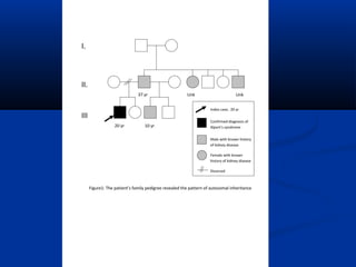
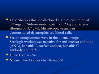
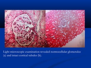
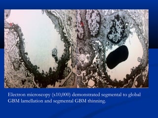
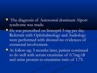
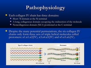
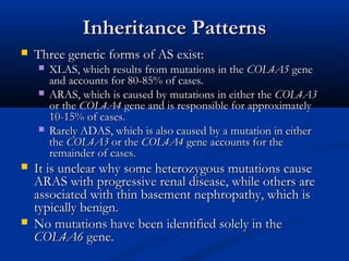
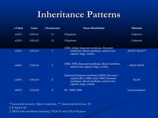
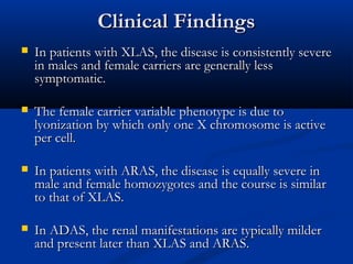
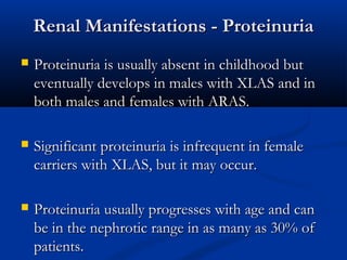
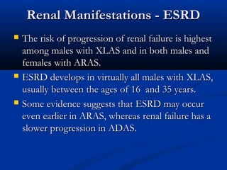
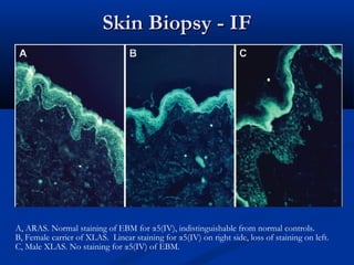
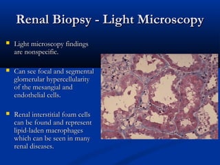
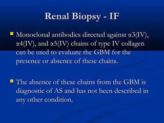
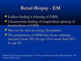
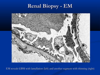
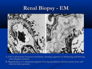
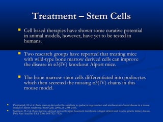
A Challenging Case:A Challenging Case:
Hematuria andHematuria and
ProteinuriaProteinuria
Wisit CheungpasitpornWisit Cheungpasitporn
Case Presentation
 A 19-year-old Caucasian male presented to
Nephrology Clinic for evaluation of proteinuria
and microscopic hematuria.
 He was in the process of undergoing a physical
for the Marine Corps and was found to have
proteinuria.
 PMH
 asymptomatic microscopic hematuria since
childhood.
 Medication
 Ibuprofen 400 mg PRN Q8 hours for muscle pain.
Use occasionally.
Case Presentation
 Social History
 Drinks beer occasionally
 Denies tobacco uses
 Family history
 Kidney problem in his father, paternal uncle and
paternal aunts.
 His half brother who shared the same father also
has a “red blood cell presented in his urine”.
 Physical examination revealed a blood pressure
of 136/60 mmHg with no peripheral edema.
 Laboratory evaluation disclosed a serum creatinine of
0.7 mg/dl, 24-hour urine protein of 2.4 g and serum
albumin of 3.7 g/dl. Microscopic urianalysis
demonstrated dysmorphic red blood cells.
 Serum complements were in the normal range.
Serologic workup was negative for anti-nuclear antibody
(ANA), hepatitis B surface antigen, hepatitis C
antibody, and HIV.
 HbA1C of 4.7 %
 Normal sized kidneys by ultrasound
Renal biopsy
Light microscopic examination revealed normocellular glomerulus
(a) and intact cortical tubules (b).
IF: negative for IgM, IgG, C3, and C4.
Electron microscopy (x10,000) demonstrated segmental to global
GBM lamellation and segmental GBM thinning.
 The diagnosis of Autosomal dominant Alport
syndrome was made.
 He was prescribed on lisinopril 5 mg per day.
Referrals with Ophthalmology and Audiology
were performed with showed no evidences of
extrarenal involvement.
 At follow-up, 3 months later, patient continued
to do well with serum creatinine of 0.7mg/dl
and urine protein-to-creatinine ratio of 1.75.
Alport SyndromeAlport Syndrome
HistoryHistory
HistoryHistory
 In 1927, Dr. Cecil Alport followed 3 later generationsIn 1927, Dr. Cecil Alport followed 3 later generations
of the same family and he recognized that deafness wasof the same family and he recognized that deafness was
a syndromic component and that the disorder tended toa syndromic component and that the disorder tended to
be more severe in males than females, that affectedbe more severe in males than females, that affected
males died of uremia, while females lived to old age.males died of uremia, while females lived to old age.
 Subsequently, many more families were described andSubsequently, many more families were described and
the disease was named Alport Syndrome (AS) in 1961.the disease was named Alport Syndrome (AS) in 1961.
LB GuthrieLB Guthrie”Idiopathic,” or congenital, hereditary and familial haematuria.”Idiopathic,” or congenital, hereditary and familial haematuria.Lancet, London, 1902, 1: 1243-1246.Lancet, London, 1902, 1: 1243-1246.
AF Hurst:AF Hurst:Hereditary familial congenital haemorrhagic nephritis occurring in sixteen individuals in three generations.Hereditary familial congenital haemorrhagic nephritis occurring in sixteen individuals in three generations. Guy’s Hosp Rec, 1923, 3: 368-370Guy’s Hosp Rec, 1923, 3: 368-370
C Alport:C Alport: Hereditary familial congenital haemorrhagic nephritis.Hereditary familial congenital haemorrhagic nephritis. British Medical Journal, London, 1927, I: 504-506.British Medical Journal, London, 1927, I: 504-506.
BackgroundBackground
 The incidence of AS is approximately 1 in 5000The incidence of AS is approximately 1 in 5000
births.births.
 In the US, accounts for approximately 3% ofIn the US, accounts for approximately 3% of
children and 0.2% of adults with ESRD.children and 0.2% of adults with ESRD.
 In Europe, the incidence AS is greater andIn Europe, the incidence AS is greater and
accounts for 0.6% of patients with ESRD.accounts for 0.6% of patients with ESRD.
PathophysiologyPathophysiology
 AS is a primary basement membrane disorder arisingAS is a primary basement membrane disorder arising
from mutations in genes encoding several members offrom mutations in genes encoding several members of
the type IV collagen family.the type IV collagen family.
 Basement membranes are assembled through anBasement membranes are assembled through an
interweaving of type IV collagen with laminins andinterweaving of type IV collagen with laminins and
sulfated proteoglycans.sulfated proteoglycans.
 Six genes,Six genes, COL4A1, COL4A2, COL4A3, COL4A4,COL4A1, COL4A2, COL4A3, COL4A4,
COL4A5COL4A5 andand COL4A6COL4A6 encode the six chains ofencode the six chains of
collagen IV, α1(IV) through α6(IV), respectively.collagen IV, α1(IV) through α6(IV), respectively.
PathophysiologyPathophysiology
 Each collagen IV chain has three domains:Each collagen IV chain has three domains:
 Short 7S domain at the N-terminalShort 7S domain at the N-terminal
 A long, collagenous domain occupying the midsection of the moleculeA long, collagenous domain occupying the midsection of the molecule
 Noncollagenous domain (NC1) positioned at the C terminalNoncollagenous domain (NC1) positioned at the C terminal
 Despite the many potential permutations, the six collagen IVDespite the many potential permutations, the six collagen IV
chains only form three sets of triple helical molecules calledchains only form three sets of triple helical molecules called
protomers: α1.α1.α2(IV), α3.α4.α5(IV) and α5.α5.α6(IV).protomers: α1.α1.α2(IV), α3.α4.α5(IV) and α5.α5.α6(IV).
PathophysiologyPathophysiology
 Two NC1 trimers unite toTwo NC1 trimers unite to
form a hexamer.form a hexamer.
 Four 7S domains formFour 7S domains form
tetramers with othertetramers with other
protomersprotomers
 The three protomers onlyThe three protomers only
form three sets of hexamersform three sets of hexamers
to form collagenousto form collagenous
networks:networks:
 α1.α1.α2(α1.α1.α2(IVIV) - α1.α1.α2() - α1.α1.α2(IVIV))
 α3.α4.α5(α3.α4.α5(IVIV) – α3.α4.α5() – α3.α4.α5(IVIV))
 α1.α1.α2(α1.α1.α2(IVIV) – α5.α5.α6() – α5.α5.α6(IVIV))
Inheritance PatternsInheritance Patterns
 Three genetic forms of AS exist:Three genetic forms of AS exist:
 XLAS, which results from mutations in theXLAS, which results from mutations in the COL4A5COL4A5 genegene
and accounts for 80-85% of cases.and accounts for 80-85% of cases.
 ARAS, which is caused by mutations in either theARAS, which is caused by mutations in either the COL4A3COL4A3
or theor the COL4A4COL4A4 gene and is responsible for approximatelygene and is responsible for approximately
10-15% of cases.10-15% of cases.
 Rarely ADAS, which is also caused by a mutation in eitherRarely ADAS, which is also caused by a mutation in either
thethe COL4A3COL4A3 or theor the COL4A4COL4A4 gene accounts for thegene accounts for the
remainder of cases.remainder of cases.
 It is unclear why some heterozygous mutations causeIt is unclear why some heterozygous mutations cause
ARAS with progressive renal disease, while others areARAS with progressive renal disease, while others are
associated with thin basement nephropathy, which isassociated with thin basement nephropathy, which is
typically benign.typically benign.
 No mutations have been identified solely in theNo mutations have been identified solely in the
COL4A6COL4A6 gene.gene.
Inheritance PatternsInheritance Patterns
α Chain Genes Chromosome Tissue Distribution Mutation
α1(IV) COL4A1 13 Ubiquitous Unknown
α2(IV) COL4A2 13 Ubiquitous Unknown
α3(IV) COL4A3 2
GBM, tubular basement membrane, Descemet
membrane, Bruch membrane, anterior lens
capsule, lungs, cochlea
ARAS*/ADAS**
α4(IV) COL4A4 2
GBM, TBM, Descemet membrane, Bruch membrane,
anterior lens capsule, lungs, cochlea
ARAS/ADAS
α5(IV) COL4A5 X
Epidermal basement membrane (EBM), Bowman’s
capsule (BC), GBM, distal TBM, Descemet
membrane, Bruch membrane, anterior lens
capsule, lungs, cochlea
XLAS†
α6(IV) COL4A6 X BC, TBM, EBM Leiomyomatosis‡
*Autosomal recessive Alport syndrome, ** Autosomal dominant AS
† X-linked AS
‡ ARAS with mutations spanning COL4A5 and COL4A6 genes
X Linked MutationsX Linked Mutations
 In theIn the COL4A5COL4A5 genes from the families with XLAS, more thangenes from the families with XLAS, more than
300 gene mutations have been reported.300 gene mutations have been reported.
 MostMost COL4A5COL4A5 mutations are small and include missensemutations are small and include missense
mutations, splice-site mutations, and small deletions where renalmutations, splice-site mutations, and small deletions where renal
failure and deafness occur after 30 years of age (adult form).failure and deafness occur after 30 years of age (adult form).
 Approximately 20% of the mutations are major rearrangementsApproximately 20% of the mutations are major rearrangements
at theat the COL4A5COL4A5 locus (i.e., large deletions, reading frame shifts,locus (i.e., large deletions, reading frame shifts,
etc) in which patients are symptomatic before the age of 30etc) in which patients are symptomatic before the age of 30
(juvenile form).(juvenile form).
 A rare of deletion spanningA rare of deletion spanning COL4A5COL4A5 andand COL4A6COL4A6 genes isgenes is
associated with a combination of XLAS and diffuseassociated with a combination of XLAS and diffuse
leiomyomatosis.leiomyomatosis.
Autosomal MutationsAutosomal Mutations
 To date, only 6 mutations in theTo date, only 6 mutations in the COL4A3COL4A3 gene and 12gene and 12
mutations in themutations in the COL4A4COL4A4 gene have been identified ingene have been identified in
patients with ARAS.patients with ARAS.
 ARAS patients are either homozygous or compoundARAS patients are either homozygous or compound
heterozygous for their mutations, and their parents areheterozygous for their mutations, and their parents are
usually asymptomatic carriers.usually asymptomatic carriers.
 ADAS is more rare than XLAS or ARAS and is a resultADAS is more rare than XLAS or ARAS and is a result
of a dominant negative mutation of theof a dominant negative mutation of the COL4A3COL4A3 oror
COL4A4COL4A4 genes whose gene product actsgenes whose gene product acts
antagonistically to the wild-type allele.antagonistically to the wild-type allele.
Clinical FindingsClinical Findings
 In patients with XLAS, the disease is consistently severeIn patients with XLAS, the disease is consistently severe
in males and female carriers are generally lessin males and female carriers are generally less
symptomatic.symptomatic.
 The female carrier variable phenotype is due toThe female carrier variable phenotype is due to
lyonization by which only one X chromosome is activelyonization by which only one X chromosome is active
per cell.per cell.
 In patients with ARAS, the disease is equally severe inIn patients with ARAS, the disease is equally severe in
male and female homozygotes and the course is similarmale and female homozygotes and the course is similar
to that of XLAS.to that of XLAS.
 In ADAS, the renal manifestations are typically milderIn ADAS, the renal manifestations are typically milder
and present later than XLAS and ARAS.and present later than XLAS and ARAS.
Renal Manifestations - HematuriaRenal Manifestations - Hematuria
 Gross or microscopic hematuria is the most commonGross or microscopic hematuria is the most common
and earliest manifestation.and earliest manifestation.
 Microscopic hematuria is observed usually in the firstMicroscopic hematuria is observed usually in the first
few years of life in all males and in 95% of females.few years of life in all males and in 95% of females.
 Hematuria is usually persistent in males, whereas it canHematuria is usually persistent in males, whereas it can
be intermittent in females.be intermittent in females.
 Like IgA nephropathy, approximately 60-70% ofLike IgA nephropathy, approximately 60-70% of
patients experience episodes of gross hematuria, oftenpatients experience episodes of gross hematuria, often
precipitated by upper respiratory infection, during theprecipitated by upper respiratory infection, during the
first 2 decades of life.first 2 decades of life.
Renal Manifestations - ProteinuriaRenal Manifestations - Proteinuria
 Proteinuria is usually absent in childhood butProteinuria is usually absent in childhood but
eventually develops in males with XLAS and ineventually develops in males with XLAS and in
both males and females with ARAS.both males and females with ARAS.
 Significant proteinuria is infrequent in femaleSignificant proteinuria is infrequent in female
carriers with XLAS, but it may occur.carriers with XLAS, but it may occur.
 Proteinuria usually progresses with age and canProteinuria usually progresses with age and can
be in the nephrotic range in as many as 30% ofbe in the nephrotic range in as many as 30% of
patients.patients.
Renal Manifestations - ESRDRenal Manifestations - ESRD
 The risk of progression of renal failure is highestThe risk of progression of renal failure is highest
among males with XLAS and in both males andamong males with XLAS and in both males and
females with ARAS.females with ARAS.
 ESRD develops in virtually all males with XLAS,ESRD develops in virtually all males with XLAS,
usually between the ages of 16 and 35 years.usually between the ages of 16 and 35 years.
 Some evidence suggests that ESRD may occurSome evidence suggests that ESRD may occur
even earlier in ARAS, whereas renal failure has aeven earlier in ARAS, whereas renal failure has a
slower progression in ADAS.slower progression in ADAS.
Hearing DeficitsHearing Deficits
 Bilateral sensorineural hearing loss is a characteristicBilateral sensorineural hearing loss is a characteristic
feature observed frequently, but not universally.feature observed frequently, but not universally.
 May reflect impaired adhesion of the Organ of CortiMay reflect impaired adhesion of the Organ of Corti
(which contain auditory sensory cells) to the basilar(which contain auditory sensory cells) to the basilar
membrane of the inner ear.membrane of the inner ear.
 About 50% of male patients with XLAS showAbout 50% of male patients with XLAS show
sensorineural deafness by age 25 years, and about 90% aresensorineural deafness by age 25 years, and about 90% are
deaf by age 40 years.deaf by age 40 years.
Ocular Findings – Anterior LenticonusOcular Findings – Anterior Lenticonus
 Conical protrusion of the central portion of theConical protrusion of the central portion of the
lens into the anterior chamber.lens into the anterior chamber.
 It is most marked anteriorly because it is theIt is most marked anteriorly because it is the
region where the capsule is thinnest, the stressesregion where the capsule is thinnest, the stresses
of accommodation are greatest, and the lens isof accommodation are greatest, and the lens is
least supported.least supported.
 Occurs in approximately 15-20% of AS patients.Occurs in approximately 15-20% of AS patients.
Ocular Findings – Anterior LenticonusOcular Findings – Anterior Lenticonus
LeiomyomatosisLeiomyomatosis
 Diffuse leiomyomatosis of the gastrointestinal, respiratoryDiffuse leiomyomatosis of the gastrointestinal, respiratory
and female genital tracts has been reported in someand female genital tracts has been reported in some
families with AS (particularly esophagus andfamilies with AS (particularly esophagus and
tracheobronchial tree).tracheobronchial tree).
 Seen in 2-5% of patients and carriers of XLAS who haveSeen in 2-5% of patients and carriers of XLAS who have
deletions that involvedeletions that involve COL4A5COL4A5 and extend to the secondand extend to the second
intron of the adjacentintron of the adjacent COL4A6COL4A6 gene.gene.
 Symptoms usually appear in late childhood and includeSymptoms usually appear in late childhood and include
dysphagia, postprandial vomiting, substernal or epigastricdysphagia, postprandial vomiting, substernal or epigastric
pain, recurrent bronchitis, dyspnea, cough, and stridor.pain, recurrent bronchitis, dyspnea, cough, and stridor.
DiagnosisDiagnosis
DiagnosisDiagnosis
 Historical information (family history, hearing loss,Historical information (family history, hearing loss,
visual disturbances, gross hematuria)visual disturbances, gross hematuria)
 Tissue biopsy often reveals ultrastructural abnormalitiesTissue biopsy often reveals ultrastructural abnormalities
and confirm diagnosis.and confirm diagnosis.
 Skin biopsy is less invasive than renal biopsy andSkin biopsy is less invasive than renal biopsy and
should be obtained first.should be obtained first.
 Molecular genetic testing in equivocal biopsy cases,Molecular genetic testing in equivocal biopsy cases,
patients in whom biopsy is contraindicated andpatients in whom biopsy is contraindicated and
prenatal testing.prenatal testing.
Skin BiopsySkin Biopsy
 The absence ofThe absence of αα5(IV) chains in the epidermal basement membrane5(IV) chains in the epidermal basement membrane
on skin biopsy is diagnostic of XLAS.on skin biopsy is diagnostic of XLAS.
 However, the absence ofHowever, the absence of αα5(IV) chains in the epidermal basement5(IV) chains in the epidermal basement
membrane is observed in only 80% of males with XLAS.membrane is observed in only 80% of males with XLAS.
 Therefore, the presence ofTherefore, the presence of αα5(IV) chains in the epidermal basement5(IV) chains in the epidermal basement
membrane does not rule out the diagnosis of XLAS.membrane does not rule out the diagnosis of XLAS.
 Furthermore,Furthermore, αα3(IV) and3(IV) and αα4(IV) chains are not found in the4(IV) chains are not found in the
epidermal basement membrane so skin biopsy can not be used forepidermal basement membrane so skin biopsy can not be used for
the diagnosis of ARAS and ADAS.the diagnosis of ARAS and ADAS.
Skin Biopsy - IFSkin Biopsy - IF
A, ARAS. Normal staining of EBM for α5(IV), indistinguishable from normal controls.
B, Female carrier of XLAS. Linear staining for α5(IV) on right side, loss of staining on left.
C, Male XLAS. No staining for α5(IV) of EBM.
Renal Biopsy - Light MicroscopyRenal Biopsy - Light Microscopy
 Light microscopy findingsLight microscopy findings
are nonspecific.are nonspecific.
 Can see focal and segmentalCan see focal and segmental
glomerular hypercellularityglomerular hypercellularity
of the mesangial andof the mesangial and
endothelial cells.endothelial cells.
 Renal interstitial foam cellsRenal interstitial foam cells
can be found and representcan be found and represent
lipid-laden macrophageslipid-laden macrophages
which can be seen in manywhich can be seen in many
renal diseases.renal diseases.
Renal Biopsy - IFRenal Biopsy - IF
 Monoclonal antibodies directed againstMonoclonal antibodies directed against αα3(IV),3(IV),
αα4(IV), and4(IV), and αα5(IV) chains of type IV collagen5(IV) chains of type IV collagen
can be used to evaluate the GBM for thecan be used to evaluate the GBM for the
presence or absence of these chains.presence or absence of these chains.
 The absence of these chains from the GBM isThe absence of these chains from the GBM is
diagnostic of AS and has not been described indiagnostic of AS and has not been described in
any other condition.any other condition.
Renal Biopsy - EMRenal Biopsy - EM
 Earliest finding is thinning of GBM.Earliest finding is thinning of GBM.
 Characteristic finding of longitudinal splitting ofCharacteristic finding of longitudinal splitting of
lamina densa of GBM.lamina densa of GBM.
 May not be seen in young AS patients.May not be seen in young AS patients.
 The proportion of GBM that shows splittingThe proportion of GBM that shows splitting
increases from 30% by age 10 to more than 90%increases from 30% by age 10 to more than 90%
by age 30.by age 30.
Rumpelt, HJ. Hereditary nephropathy: Correlation of clinical data with GBM alterations. Clin
Nephrol 1980; 13:203.
Renal Biopsy - EMRenal Biopsy - EM
EM of patient with AS, arrows are pointing to the splitting and lamellation of the GBM.EM of patient with AS, arrows are pointing to the splitting and lamellation of the GBM.
Renal Biopsy - EMRenal Biopsy - EM
EM reveals GBM with lamellation (left) and another segment with thinning (right)EM reveals GBM with lamellation (left) and another segment with thinning (right)
Renal Biopsy - EMRenal Biopsy - EM
A, EM of glomerular basement membrane, showing segments of thickening and thinningA, EM of glomerular basement membrane, showing segments of thickening and thinning
with irregular contours.with irregular contours.
B, Magnification of a thickened segment showing lamellation, electron-lucent areas andB, Magnification of a thickened segment showing lamellation, electron-lucent areas and
electron-dense granules.electron-dense granules.
TreatmentTreatment
Treatment – Angiotensin BlockadeTreatment – Angiotensin Blockade
 It has been proposed, although unproven, that angiotensinIt has been proposed, although unproven, that angiotensin
blockade may diminish the rate of proteinuria leading toblockade may diminish the rate of proteinuria leading to
glomerulosclerosis and thereby disease progression.glomerulosclerosis and thereby disease progression.
 To date, only small uncontrolled trials have demonstrated theTo date, only small uncontrolled trials have demonstrated the
effect of ACE inhibitors on reducing proteinuria in humans.effect of ACE inhibitors on reducing proteinuria in humans.
 Preemptive therapy with ACE inhibitors in anPreemptive therapy with ACE inhibitors in an αα3(IV) knockout3(IV) knockout
Alport mouse model prolonged lifespan until death from renalAlport mouse model prolonged lifespan until death from renal
failure by more than 100%.failure by more than 100%.
 In the absence of more data, the use of ACE inhibitors isIn the absence of more data, the use of ACE inhibitors is
reasonable in patients with Alport syndrome.reasonable in patients with Alport syndrome.
 Cohen, EP. In hereditary nephritis ACE inihibition decreases proteinuria and may slow the rate of progression. Am J Kidney Dis,Cohen, EP. In hereditary nephritis ACE inihibition decreases proteinuria and may slow the rate of progression. Am J Kidney Dis,
1996; 27:199.1996; 27:199.
 Gross, O et al. Preemptive ramipril therapy delays renal failure and reduces renal fibrosis inGross, O et al. Preemptive ramipril therapy delays renal failure and reduces renal fibrosis in COL4A3COL4A3-knockout mice with Alport-knockout mice with Alport
syndrome. KI 2003; 63: 438-446.syndrome. KI 2003; 63: 438-446.
Treatment - CyclosporineTreatment - Cyclosporine
 Cyclosporine has also been studied in smallCyclosporine has also been studied in small
uncontrolled trials as well.uncontrolled trials as well.
 One study of eight Alport males who receivedOne study of eight Alport males who received
cyclosporine for a mean duration of 8.4 years suggestedcyclosporine for a mean duration of 8.4 years suggested
a slower progression to ESRD as compared to relateda slower progression to ESRD as compared to related
effected males.effected males.
 Another study demonstrated reduction in proteinuria,Another study demonstrated reduction in proteinuria,
however, 4 of 9 patients exhibited cyclosporinehowever, 4 of 9 patients exhibited cyclosporine
nephrotoxicity.nephrotoxicity.
 Callis, L et al. Long-term effects of cyclosporine A in Alport’s syndrome, KI 1999; 55: 1051-1056Callis, L et al. Long-term effects of cyclosporine A in Alport’s syndrome, KI 1999; 55: 1051-1056
 Charbit, M et al. Cyclosporine therapy in patients with Alport syndrome. Pediatric Nephrology 2007; 22:57-Charbit, M et al. Cyclosporine therapy in patients with Alport syndrome. Pediatric Nephrology 2007; 22:57-
63.63.
Treatment – Stem CellsTreatment – Stem Cells
 Cell based therapies have shown some curative potentialCell based therapies have shown some curative potential
in animal models, however, have yet to be tested inin animal models, however, have yet to be tested in
humans.humans.
 Two research groups have reported that treating miceTwo research groups have reported that treating mice
with wild-type bone marrow derived cells can improvewith wild-type bone marrow derived cells can improve
the disease inthe disease in αα3(IV) knockout Alport mice.3(IV) knockout Alport mice.
 The bone marrow stem cells differentiated into podocytesThe bone marrow stem cells differentiated into podocytes
which then secreted the missingwhich then secreted the missing αα3(IV) chains in this3(IV) chains in this
mouse model.mouse model.
 Prodromidi, EI et al. Bone marrow-derived cells contribute to podocyte regeneration and amelioration of renal disease in a mouseProdromidi, EI et al. Bone marrow-derived cells contribute to podocyte regeneration and amelioration of renal disease in a mouse
model of Alport syndrome. Stem Cells. 2006; 24: 2448-2455.model of Alport syndrome. Stem Cells. 2006; 24: 2448-2455.
 Sugimoto H et al. Bone marrow–derived stem cells repair basement membrane collagen defects and reverse genetic kidney disease.Sugimoto H et al. Bone marrow–derived stem cells repair basement membrane collagen defects and reverse genetic kidney disease.
Proc Natl Acad Sci USA 2006; 103:7321-7326.Proc Natl Acad Sci USA 2006; 103:7321-7326.
Treatment – Renal TransplantTreatment – Renal Transplant
 AS is essentially cured with renal transplantation, and asAS is essentially cured with renal transplantation, and as
one would suspect unless the donor has the disease, ASone would suspect unless the donor has the disease, AS
will not occur in the transplanted organ.will not occur in the transplanted organ.
 The most significant and devastating, albeit rare,The most significant and devastating, albeit rare,
complication of transplantation iscomplication of transplantation is antiglomerularantiglomerular
basement membrane nephritis.basement membrane nephritis.
 ApproximatelyApproximately 3-5%3-5% of patients with Alport syndromeof patients with Alport syndrome
who receive a transplant develop anti-GBM antibody towho receive a transplant develop anti-GBM antibody to
the NC1 component of the α3(IV) chain.the NC1 component of the α3(IV) chain.
 Post-transplant anti-GBM nephritis usually developsPost-transplant anti-GBM nephritis usually develops
within thewithin the first yearfirst year of the transplant.of the transplant.
Thank you.Thank you.

Alport syndrome

  • 1.
    A Challenging Case:AChallenging Case: Hematuria andHematuria and ProteinuriaProteinuria Wisit CheungpasitpornWisit Cheungpasitporn
  • 2.
    Case Presentation  A19-year-old Caucasian male presented to Nephrology Clinic for evaluation of proteinuria and microscopic hematuria.  He was in the process of undergoing a physical for the Marine Corps and was found to have proteinuria.
  • 3.
     PMH  asymptomaticmicroscopic hematuria since childhood.  Medication  Ibuprofen 400 mg PRN Q8 hours for muscle pain. Use occasionally. Case Presentation
  • 4.
     Social History Drinks beer occasionally  Denies tobacco uses  Family history  Kidney problem in his father, paternal uncle and paternal aunts.  His half brother who shared the same father also has a “red blood cell presented in his urine”.
  • 6.
     Physical examinationrevealed a blood pressure of 136/60 mmHg with no peripheral edema.
  • 7.
     Laboratory evaluationdisclosed a serum creatinine of 0.7 mg/dl, 24-hour urine protein of 2.4 g and serum albumin of 3.7 g/dl. Microscopic urianalysis demonstrated dysmorphic red blood cells.  Serum complements were in the normal range. Serologic workup was negative for anti-nuclear antibody (ANA), hepatitis B surface antigen, hepatitis C antibody, and HIV.  HbA1C of 4.7 %  Normal sized kidneys by ultrasound
  • 8.
  • 9.
    Light microscopic examinationrevealed normocellular glomerulus (a) and intact cortical tubules (b).
  • 10.
    IF: negative forIgM, IgG, C3, and C4.
  • 11.
    Electron microscopy (x10,000)demonstrated segmental to global GBM lamellation and segmental GBM thinning.
  • 12.
     The diagnosisof Autosomal dominant Alport syndrome was made.  He was prescribed on lisinopril 5 mg per day. Referrals with Ophthalmology and Audiology were performed with showed no evidences of extrarenal involvement.  At follow-up, 3 months later, patient continued to do well with serum creatinine of 0.7mg/dl and urine protein-to-creatinine ratio of 1.75.
  • 13.
  • 14.
  • 15.
    HistoryHistory  In 1927,Dr. Cecil Alport followed 3 later generationsIn 1927, Dr. Cecil Alport followed 3 later generations of the same family and he recognized that deafness wasof the same family and he recognized that deafness was a syndromic component and that the disorder tended toa syndromic component and that the disorder tended to be more severe in males than females, that affectedbe more severe in males than females, that affected males died of uremia, while females lived to old age.males died of uremia, while females lived to old age.  Subsequently, many more families were described andSubsequently, many more families were described and the disease was named Alport Syndrome (AS) in 1961.the disease was named Alport Syndrome (AS) in 1961. LB GuthrieLB Guthrie”Idiopathic,” or congenital, hereditary and familial haematuria.”Idiopathic,” or congenital, hereditary and familial haematuria.Lancet, London, 1902, 1: 1243-1246.Lancet, London, 1902, 1: 1243-1246. AF Hurst:AF Hurst:Hereditary familial congenital haemorrhagic nephritis occurring in sixteen individuals in three generations.Hereditary familial congenital haemorrhagic nephritis occurring in sixteen individuals in three generations. Guy’s Hosp Rec, 1923, 3: 368-370Guy’s Hosp Rec, 1923, 3: 368-370 C Alport:C Alport: Hereditary familial congenital haemorrhagic nephritis.Hereditary familial congenital haemorrhagic nephritis. British Medical Journal, London, 1927, I: 504-506.British Medical Journal, London, 1927, I: 504-506.
  • 16.
    BackgroundBackground  The incidenceof AS is approximately 1 in 5000The incidence of AS is approximately 1 in 5000 births.births.  In the US, accounts for approximately 3% ofIn the US, accounts for approximately 3% of children and 0.2% of adults with ESRD.children and 0.2% of adults with ESRD.  In Europe, the incidence AS is greater andIn Europe, the incidence AS is greater and accounts for 0.6% of patients with ESRD.accounts for 0.6% of patients with ESRD.
  • 17.
  • 18.
    PathophysiologyPathophysiology  AS isa primary basement membrane disorder arisingAS is a primary basement membrane disorder arising from mutations in genes encoding several members offrom mutations in genes encoding several members of the type IV collagen family.the type IV collagen family.  Basement membranes are assembled through anBasement membranes are assembled through an interweaving of type IV collagen with laminins andinterweaving of type IV collagen with laminins and sulfated proteoglycans.sulfated proteoglycans.  Six genes,Six genes, COL4A1, COL4A2, COL4A3, COL4A4,COL4A1, COL4A2, COL4A3, COL4A4, COL4A5COL4A5 andand COL4A6COL4A6 encode the six chains ofencode the six chains of collagen IV, α1(IV) through α6(IV), respectively.collagen IV, α1(IV) through α6(IV), respectively.
  • 19.
    PathophysiologyPathophysiology  Each collagenIV chain has three domains:Each collagen IV chain has three domains:  Short 7S domain at the N-terminalShort 7S domain at the N-terminal  A long, collagenous domain occupying the midsection of the moleculeA long, collagenous domain occupying the midsection of the molecule  Noncollagenous domain (NC1) positioned at the C terminalNoncollagenous domain (NC1) positioned at the C terminal  Despite the many potential permutations, the six collagen IVDespite the many potential permutations, the six collagen IV chains only form three sets of triple helical molecules calledchains only form three sets of triple helical molecules called protomers: α1.α1.α2(IV), α3.α4.α5(IV) and α5.α5.α6(IV).protomers: α1.α1.α2(IV), α3.α4.α5(IV) and α5.α5.α6(IV).
  • 20.
    PathophysiologyPathophysiology  Two NC1trimers unite toTwo NC1 trimers unite to form a hexamer.form a hexamer.  Four 7S domains formFour 7S domains form tetramers with othertetramers with other protomersprotomers  The three protomers onlyThe three protomers only form three sets of hexamersform three sets of hexamers to form collagenousto form collagenous networks:networks:  α1.α1.α2(α1.α1.α2(IVIV) - α1.α1.α2() - α1.α1.α2(IVIV))  α3.α4.α5(α3.α4.α5(IVIV) – α3.α4.α5() – α3.α4.α5(IVIV))  α1.α1.α2(α1.α1.α2(IVIV) – α5.α5.α6() – α5.α5.α6(IVIV))
  • 21.
  • 22.
    Inheritance PatternsInheritance Patterns Three genetic forms of AS exist:Three genetic forms of AS exist:  XLAS, which results from mutations in theXLAS, which results from mutations in the COL4A5COL4A5 genegene and accounts for 80-85% of cases.and accounts for 80-85% of cases.  ARAS, which is caused by mutations in either theARAS, which is caused by mutations in either the COL4A3COL4A3 or theor the COL4A4COL4A4 gene and is responsible for approximatelygene and is responsible for approximately 10-15% of cases.10-15% of cases.  Rarely ADAS, which is also caused by a mutation in eitherRarely ADAS, which is also caused by a mutation in either thethe COL4A3COL4A3 or theor the COL4A4COL4A4 gene accounts for thegene accounts for the remainder of cases.remainder of cases.  It is unclear why some heterozygous mutations causeIt is unclear why some heterozygous mutations cause ARAS with progressive renal disease, while others areARAS with progressive renal disease, while others are associated with thin basement nephropathy, which isassociated with thin basement nephropathy, which is typically benign.typically benign.  No mutations have been identified solely in theNo mutations have been identified solely in the COL4A6COL4A6 gene.gene.
  • 23.
    Inheritance PatternsInheritance Patterns αChain Genes Chromosome Tissue Distribution Mutation α1(IV) COL4A1 13 Ubiquitous Unknown α2(IV) COL4A2 13 Ubiquitous Unknown α3(IV) COL4A3 2 GBM, tubular basement membrane, Descemet membrane, Bruch membrane, anterior lens capsule, lungs, cochlea ARAS*/ADAS** α4(IV) COL4A4 2 GBM, TBM, Descemet membrane, Bruch membrane, anterior lens capsule, lungs, cochlea ARAS/ADAS α5(IV) COL4A5 X Epidermal basement membrane (EBM), Bowman’s capsule (BC), GBM, distal TBM, Descemet membrane, Bruch membrane, anterior lens capsule, lungs, cochlea XLAS† α6(IV) COL4A6 X BC, TBM, EBM Leiomyomatosis‡ *Autosomal recessive Alport syndrome, ** Autosomal dominant AS † X-linked AS ‡ ARAS with mutations spanning COL4A5 and COL4A6 genes
  • 24.
    X Linked MutationsXLinked Mutations  In theIn the COL4A5COL4A5 genes from the families with XLAS, more thangenes from the families with XLAS, more than 300 gene mutations have been reported.300 gene mutations have been reported.  MostMost COL4A5COL4A5 mutations are small and include missensemutations are small and include missense mutations, splice-site mutations, and small deletions where renalmutations, splice-site mutations, and small deletions where renal failure and deafness occur after 30 years of age (adult form).failure and deafness occur after 30 years of age (adult form).  Approximately 20% of the mutations are major rearrangementsApproximately 20% of the mutations are major rearrangements at theat the COL4A5COL4A5 locus (i.e., large deletions, reading frame shifts,locus (i.e., large deletions, reading frame shifts, etc) in which patients are symptomatic before the age of 30etc) in which patients are symptomatic before the age of 30 (juvenile form).(juvenile form).  A rare of deletion spanningA rare of deletion spanning COL4A5COL4A5 andand COL4A6COL4A6 genes isgenes is associated with a combination of XLAS and diffuseassociated with a combination of XLAS and diffuse leiomyomatosis.leiomyomatosis.
  • 25.
    Autosomal MutationsAutosomal Mutations To date, only 6 mutations in theTo date, only 6 mutations in the COL4A3COL4A3 gene and 12gene and 12 mutations in themutations in the COL4A4COL4A4 gene have been identified ingene have been identified in patients with ARAS.patients with ARAS.  ARAS patients are either homozygous or compoundARAS patients are either homozygous or compound heterozygous for their mutations, and their parents areheterozygous for their mutations, and their parents are usually asymptomatic carriers.usually asymptomatic carriers.  ADAS is more rare than XLAS or ARAS and is a resultADAS is more rare than XLAS or ARAS and is a result of a dominant negative mutation of theof a dominant negative mutation of the COL4A3COL4A3 oror COL4A4COL4A4 genes whose gene product actsgenes whose gene product acts antagonistically to the wild-type allele.antagonistically to the wild-type allele.
  • 26.
  • 27.
    Clinical FindingsClinical Findings In patients with XLAS, the disease is consistently severeIn patients with XLAS, the disease is consistently severe in males and female carriers are generally lessin males and female carriers are generally less symptomatic.symptomatic.  The female carrier variable phenotype is due toThe female carrier variable phenotype is due to lyonization by which only one X chromosome is activelyonization by which only one X chromosome is active per cell.per cell.  In patients with ARAS, the disease is equally severe inIn patients with ARAS, the disease is equally severe in male and female homozygotes and the course is similarmale and female homozygotes and the course is similar to that of XLAS.to that of XLAS.  In ADAS, the renal manifestations are typically milderIn ADAS, the renal manifestations are typically milder and present later than XLAS and ARAS.and present later than XLAS and ARAS.
  • 28.
    Renal Manifestations -HematuriaRenal Manifestations - Hematuria  Gross or microscopic hematuria is the most commonGross or microscopic hematuria is the most common and earliest manifestation.and earliest manifestation.  Microscopic hematuria is observed usually in the firstMicroscopic hematuria is observed usually in the first few years of life in all males and in 95% of females.few years of life in all males and in 95% of females.  Hematuria is usually persistent in males, whereas it canHematuria is usually persistent in males, whereas it can be intermittent in females.be intermittent in females.  Like IgA nephropathy, approximately 60-70% ofLike IgA nephropathy, approximately 60-70% of patients experience episodes of gross hematuria, oftenpatients experience episodes of gross hematuria, often precipitated by upper respiratory infection, during theprecipitated by upper respiratory infection, during the first 2 decades of life.first 2 decades of life.
  • 29.
    Renal Manifestations -ProteinuriaRenal Manifestations - Proteinuria  Proteinuria is usually absent in childhood butProteinuria is usually absent in childhood but eventually develops in males with XLAS and ineventually develops in males with XLAS and in both males and females with ARAS.both males and females with ARAS.  Significant proteinuria is infrequent in femaleSignificant proteinuria is infrequent in female carriers with XLAS, but it may occur.carriers with XLAS, but it may occur.  Proteinuria usually progresses with age and canProteinuria usually progresses with age and can be in the nephrotic range in as many as 30% ofbe in the nephrotic range in as many as 30% of patients.patients.
  • 30.
    Renal Manifestations -ESRDRenal Manifestations - ESRD  The risk of progression of renal failure is highestThe risk of progression of renal failure is highest among males with XLAS and in both males andamong males with XLAS and in both males and females with ARAS.females with ARAS.  ESRD develops in virtually all males with XLAS,ESRD develops in virtually all males with XLAS, usually between the ages of 16 and 35 years.usually between the ages of 16 and 35 years.  Some evidence suggests that ESRD may occurSome evidence suggests that ESRD may occur even earlier in ARAS, whereas renal failure has aeven earlier in ARAS, whereas renal failure has a slower progression in ADAS.slower progression in ADAS.
  • 31.
    Hearing DeficitsHearing Deficits Bilateral sensorineural hearing loss is a characteristicBilateral sensorineural hearing loss is a characteristic feature observed frequently, but not universally.feature observed frequently, but not universally.  May reflect impaired adhesion of the Organ of CortiMay reflect impaired adhesion of the Organ of Corti (which contain auditory sensory cells) to the basilar(which contain auditory sensory cells) to the basilar membrane of the inner ear.membrane of the inner ear.  About 50% of male patients with XLAS showAbout 50% of male patients with XLAS show sensorineural deafness by age 25 years, and about 90% aresensorineural deafness by age 25 years, and about 90% are deaf by age 40 years.deaf by age 40 years.
  • 32.
    Ocular Findings –Anterior LenticonusOcular Findings – Anterior Lenticonus  Conical protrusion of the central portion of theConical protrusion of the central portion of the lens into the anterior chamber.lens into the anterior chamber.  It is most marked anteriorly because it is theIt is most marked anteriorly because it is the region where the capsule is thinnest, the stressesregion where the capsule is thinnest, the stresses of accommodation are greatest, and the lens isof accommodation are greatest, and the lens is least supported.least supported.  Occurs in approximately 15-20% of AS patients.Occurs in approximately 15-20% of AS patients.
  • 33.
    Ocular Findings –Anterior LenticonusOcular Findings – Anterior Lenticonus
  • 34.
    LeiomyomatosisLeiomyomatosis  Diffuse leiomyomatosisof the gastrointestinal, respiratoryDiffuse leiomyomatosis of the gastrointestinal, respiratory and female genital tracts has been reported in someand female genital tracts has been reported in some families with AS (particularly esophagus andfamilies with AS (particularly esophagus and tracheobronchial tree).tracheobronchial tree).  Seen in 2-5% of patients and carriers of XLAS who haveSeen in 2-5% of patients and carriers of XLAS who have deletions that involvedeletions that involve COL4A5COL4A5 and extend to the secondand extend to the second intron of the adjacentintron of the adjacent COL4A6COL4A6 gene.gene.  Symptoms usually appear in late childhood and includeSymptoms usually appear in late childhood and include dysphagia, postprandial vomiting, substernal or epigastricdysphagia, postprandial vomiting, substernal or epigastric pain, recurrent bronchitis, dyspnea, cough, and stridor.pain, recurrent bronchitis, dyspnea, cough, and stridor.
  • 35.
  • 36.
    DiagnosisDiagnosis  Historical information(family history, hearing loss,Historical information (family history, hearing loss, visual disturbances, gross hematuria)visual disturbances, gross hematuria)  Tissue biopsy often reveals ultrastructural abnormalitiesTissue biopsy often reveals ultrastructural abnormalities and confirm diagnosis.and confirm diagnosis.  Skin biopsy is less invasive than renal biopsy andSkin biopsy is less invasive than renal biopsy and should be obtained first.should be obtained first.  Molecular genetic testing in equivocal biopsy cases,Molecular genetic testing in equivocal biopsy cases, patients in whom biopsy is contraindicated andpatients in whom biopsy is contraindicated and prenatal testing.prenatal testing.
  • 37.
    Skin BiopsySkin Biopsy The absence ofThe absence of αα5(IV) chains in the epidermal basement membrane5(IV) chains in the epidermal basement membrane on skin biopsy is diagnostic of XLAS.on skin biopsy is diagnostic of XLAS.  However, the absence ofHowever, the absence of αα5(IV) chains in the epidermal basement5(IV) chains in the epidermal basement membrane is observed in only 80% of males with XLAS.membrane is observed in only 80% of males with XLAS.  Therefore, the presence ofTherefore, the presence of αα5(IV) chains in the epidermal basement5(IV) chains in the epidermal basement membrane does not rule out the diagnosis of XLAS.membrane does not rule out the diagnosis of XLAS.  Furthermore,Furthermore, αα3(IV) and3(IV) and αα4(IV) chains are not found in the4(IV) chains are not found in the epidermal basement membrane so skin biopsy can not be used forepidermal basement membrane so skin biopsy can not be used for the diagnosis of ARAS and ADAS.the diagnosis of ARAS and ADAS.
  • 38.
    Skin Biopsy -IFSkin Biopsy - IF A, ARAS. Normal staining of EBM for α5(IV), indistinguishable from normal controls. B, Female carrier of XLAS. Linear staining for α5(IV) on right side, loss of staining on left. C, Male XLAS. No staining for α5(IV) of EBM.
  • 39.
    Renal Biopsy -Light MicroscopyRenal Biopsy - Light Microscopy  Light microscopy findingsLight microscopy findings are nonspecific.are nonspecific.  Can see focal and segmentalCan see focal and segmental glomerular hypercellularityglomerular hypercellularity of the mesangial andof the mesangial and endothelial cells.endothelial cells.  Renal interstitial foam cellsRenal interstitial foam cells can be found and representcan be found and represent lipid-laden macrophageslipid-laden macrophages which can be seen in manywhich can be seen in many renal diseases.renal diseases.
  • 40.
    Renal Biopsy -IFRenal Biopsy - IF  Monoclonal antibodies directed againstMonoclonal antibodies directed against αα3(IV),3(IV), αα4(IV), and4(IV), and αα5(IV) chains of type IV collagen5(IV) chains of type IV collagen can be used to evaluate the GBM for thecan be used to evaluate the GBM for the presence or absence of these chains.presence or absence of these chains.  The absence of these chains from the GBM isThe absence of these chains from the GBM is diagnostic of AS and has not been described indiagnostic of AS and has not been described in any other condition.any other condition.
  • 41.
    Renal Biopsy -EMRenal Biopsy - EM  Earliest finding is thinning of GBM.Earliest finding is thinning of GBM.  Characteristic finding of longitudinal splitting ofCharacteristic finding of longitudinal splitting of lamina densa of GBM.lamina densa of GBM.  May not be seen in young AS patients.May not be seen in young AS patients.  The proportion of GBM that shows splittingThe proportion of GBM that shows splitting increases from 30% by age 10 to more than 90%increases from 30% by age 10 to more than 90% by age 30.by age 30. Rumpelt, HJ. Hereditary nephropathy: Correlation of clinical data with GBM alterations. Clin Nephrol 1980; 13:203.
  • 42.
    Renal Biopsy -EMRenal Biopsy - EM EM of patient with AS, arrows are pointing to the splitting and lamellation of the GBM.EM of patient with AS, arrows are pointing to the splitting and lamellation of the GBM.
  • 43.
    Renal Biopsy -EMRenal Biopsy - EM EM reveals GBM with lamellation (left) and another segment with thinning (right)EM reveals GBM with lamellation (left) and another segment with thinning (right)
  • 44.
    Renal Biopsy -EMRenal Biopsy - EM A, EM of glomerular basement membrane, showing segments of thickening and thinningA, EM of glomerular basement membrane, showing segments of thickening and thinning with irregular contours.with irregular contours. B, Magnification of a thickened segment showing lamellation, electron-lucent areas andB, Magnification of a thickened segment showing lamellation, electron-lucent areas and electron-dense granules.electron-dense granules.
  • 45.
  • 46.
    Treatment – AngiotensinBlockadeTreatment – Angiotensin Blockade  It has been proposed, although unproven, that angiotensinIt has been proposed, although unproven, that angiotensin blockade may diminish the rate of proteinuria leading toblockade may diminish the rate of proteinuria leading to glomerulosclerosis and thereby disease progression.glomerulosclerosis and thereby disease progression.  To date, only small uncontrolled trials have demonstrated theTo date, only small uncontrolled trials have demonstrated the effect of ACE inhibitors on reducing proteinuria in humans.effect of ACE inhibitors on reducing proteinuria in humans.  Preemptive therapy with ACE inhibitors in anPreemptive therapy with ACE inhibitors in an αα3(IV) knockout3(IV) knockout Alport mouse model prolonged lifespan until death from renalAlport mouse model prolonged lifespan until death from renal failure by more than 100%.failure by more than 100%.  In the absence of more data, the use of ACE inhibitors isIn the absence of more data, the use of ACE inhibitors is reasonable in patients with Alport syndrome.reasonable in patients with Alport syndrome.  Cohen, EP. In hereditary nephritis ACE inihibition decreases proteinuria and may slow the rate of progression. Am J Kidney Dis,Cohen, EP. In hereditary nephritis ACE inihibition decreases proteinuria and may slow the rate of progression. Am J Kidney Dis, 1996; 27:199.1996; 27:199.  Gross, O et al. Preemptive ramipril therapy delays renal failure and reduces renal fibrosis inGross, O et al. Preemptive ramipril therapy delays renal failure and reduces renal fibrosis in COL4A3COL4A3-knockout mice with Alport-knockout mice with Alport syndrome. KI 2003; 63: 438-446.syndrome. KI 2003; 63: 438-446.
  • 47.
    Treatment - CyclosporineTreatment- Cyclosporine  Cyclosporine has also been studied in smallCyclosporine has also been studied in small uncontrolled trials as well.uncontrolled trials as well.  One study of eight Alport males who receivedOne study of eight Alport males who received cyclosporine for a mean duration of 8.4 years suggestedcyclosporine for a mean duration of 8.4 years suggested a slower progression to ESRD as compared to relateda slower progression to ESRD as compared to related effected males.effected males.  Another study demonstrated reduction in proteinuria,Another study demonstrated reduction in proteinuria, however, 4 of 9 patients exhibited cyclosporinehowever, 4 of 9 patients exhibited cyclosporine nephrotoxicity.nephrotoxicity.  Callis, L et al. Long-term effects of cyclosporine A in Alport’s syndrome, KI 1999; 55: 1051-1056Callis, L et al. Long-term effects of cyclosporine A in Alport’s syndrome, KI 1999; 55: 1051-1056  Charbit, M et al. Cyclosporine therapy in patients with Alport syndrome. Pediatric Nephrology 2007; 22:57-Charbit, M et al. Cyclosporine therapy in patients with Alport syndrome. Pediatric Nephrology 2007; 22:57- 63.63.
  • 48.
    Treatment – StemCellsTreatment – Stem Cells  Cell based therapies have shown some curative potentialCell based therapies have shown some curative potential in animal models, however, have yet to be tested inin animal models, however, have yet to be tested in humans.humans.  Two research groups have reported that treating miceTwo research groups have reported that treating mice with wild-type bone marrow derived cells can improvewith wild-type bone marrow derived cells can improve the disease inthe disease in αα3(IV) knockout Alport mice.3(IV) knockout Alport mice.  The bone marrow stem cells differentiated into podocytesThe bone marrow stem cells differentiated into podocytes which then secreted the missingwhich then secreted the missing αα3(IV) chains in this3(IV) chains in this mouse model.mouse model.  Prodromidi, EI et al. Bone marrow-derived cells contribute to podocyte regeneration and amelioration of renal disease in a mouseProdromidi, EI et al. Bone marrow-derived cells contribute to podocyte regeneration and amelioration of renal disease in a mouse model of Alport syndrome. Stem Cells. 2006; 24: 2448-2455.model of Alport syndrome. Stem Cells. 2006; 24: 2448-2455.  Sugimoto H et al. Bone marrow–derived stem cells repair basement membrane collagen defects and reverse genetic kidney disease.Sugimoto H et al. Bone marrow–derived stem cells repair basement membrane collagen defects and reverse genetic kidney disease. Proc Natl Acad Sci USA 2006; 103:7321-7326.Proc Natl Acad Sci USA 2006; 103:7321-7326.
  • 49.
    Treatment – RenalTransplantTreatment – Renal Transplant  AS is essentially cured with renal transplantation, and asAS is essentially cured with renal transplantation, and as one would suspect unless the donor has the disease, ASone would suspect unless the donor has the disease, AS will not occur in the transplanted organ.will not occur in the transplanted organ.  The most significant and devastating, albeit rare,The most significant and devastating, albeit rare, complication of transplantation iscomplication of transplantation is antiglomerularantiglomerular basement membrane nephritis.basement membrane nephritis.  ApproximatelyApproximately 3-5%3-5% of patients with Alport syndromeof patients with Alport syndrome who receive a transplant develop anti-GBM antibody towho receive a transplant develop anti-GBM antibody to the NC1 component of the α3(IV) chain.the NC1 component of the α3(IV) chain.  Post-transplant anti-GBM nephritis usually developsPost-transplant anti-GBM nephritis usually develops within thewithin the first yearfirst year of the transplant.of the transplant.
  • 50.