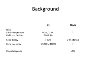
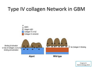
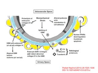
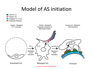
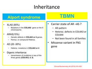
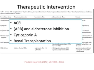
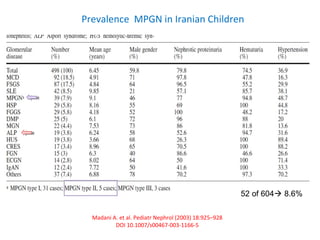
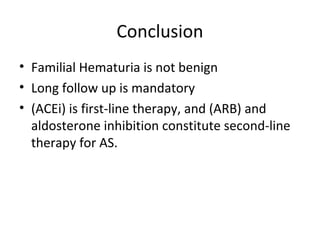
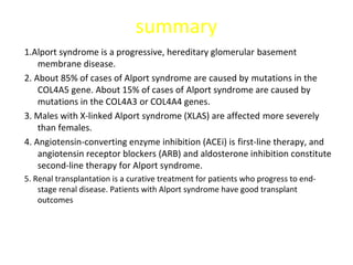
Familial Hematuria
Nakysa Hooman
Professor of Pediatric Nephrology
Iran University of Medical Sciences
2018
Abūbakr Mohammad-e Zakariyyā-ye Rāzī
854 CE -925 CE (Ray, Iran)
Rhazes describes the
discrimination between
vesical and renal hematuria in a
very scientific and up-to-date
manner
Changizi Ashtiyani et al, IJKD 2011;5:300-8
History
Cecil Alport
In 1927
FH+ Deafness+
severity in
male
Cecil Alport
In 1927
FH+ Deafness+
severity in
male
Arthur Frederick
Hurst
In 1923
FH+ uremia
Arthur Frederick
Hurst
In 1923
FH+ uremia
Leonard Guthrie
in 1902
FH
Leonard Guthrie
in 1902
FH
McConville
McAdams
In 1966
FBH
McConville
McAdams
In 1966
FBH
LB, Guthrie”.Lancet London, 1902, 1: 1243-1246.
AF Hurst. Guy’s Hosp Rec, 1923, 3: 368-370
C Alport:. British Medical Journal, London, 1927, I: 504-506.
Classification
• Heritable collagen IV nephropathies
– Alport syndrome
– Thin basement membrane disease
• Glomerulopathy with fibronectin deposits ( FN1)
• C3 glomerulopathy
• IgAN
Etiologies for Idiopathic Hematuria (+/- proteinuria)
n= 1092
Clark M, et al 2015, DOI 10.1515/dx-2015-0020
Background
AS TBMD
ESRD-
Adult- USA/Europe
Children-USA/Iran
0.2% / 0.6%
3% /2.3%
?
Renal biopsy 1-12% 5-9% (donor)
Gene frequency 1:5000 to 10000 ?
Clinical diagnosis <1%
Pathophysiology
collagen IV nephropathies
biology,2016
Type IV collagen of Basement membrane
• Two NC1 trimers unite to
form a hexamer.
• Four 7S domains form
tetramers with other
protomers
• The three protomers only
form three sets of hexamers
to form collagenous
networks:
– α1.α1.α2(IV) - α1.α1.α2(IV)
– α3.α4.α5(IV) – α3.α4.α5(IV)
– α1.α1.α2(IV) – α5.α5.α6(IV)
Type IV collagen Network in GBM
Cosgrov D.
Matrix biology,2016
Pediatr Nephrol (2013) 28:1025–1036
DOI 10.1007/s00467-012-2272-z
Model of AS initiation
Cosgrov D. Matrix biology,2016
Pediatr Nephrol (2013) 28:1025–1036
DOI 10.1007/s00467-012-2272-z
Alport syndrome
• XLAS (85%)
– Mutations in the COL4A5 gene on the X
chromosome.
• ARAS(15%)
– Genetic defects in COL4A3 or 4 genes.
– Homoz. or compound Heteroz.
• AD (20 -30%)
– Heteroz. mutations in COL4A3 or 4
• Digenic inheritance
– transmission of mutations in two of the
three genes (COL4A3, 4, 5).
TBMN
• Carrier state of AR –AS ?
– AD pattern
– Heteroz. defects in COL4A3 or
COL4A4
– Not been found in all families
• Missense variant in FN1
gene
Clinical Biochemistry 49 (2016) 816–820
Inheritance
TBMN
< 30 yr Hematuria , >30 yr Proteinuria+ CRF< 30 yr Hematuria , >30 yr Proteinuria+ CRF
Deltas C,et al. Pediatr Nephrol (2012) 27:1221–1231
AS- Clinical findings
• Renal manifestation
– Asymptomatic Hematuria >> Proteinuria >> HTN >>ESRD
– >> 16 /35/ 45-60 yr
• XLAS/ ARAS/ ADAS
• EAR
– SNHL>> High freq
• EYE
– Lens >>Ant. Lenticonus
– Retina >> dot and fleck or fleck retinopathy
• Macular hole
– Cornea >> posterio polymorphous dystrophy and recurrent corneal
erosion
• Leiomyomatosis
Clinical Progression of ADAS in European population
• Severe clinical
presentation
• High phenotype
variability
• Real prevalence
is higher
Rosado C, et al. Kidney Blood Press Res 2015;40:435-442
Pediatr Nephrol (2013) 28:1025–1036
• ACEI
• (ARB) and aldosterone inhibition
• Cyclosporin A
• Renal Transplantation
Therapeutic Intervention
RAAS blockade delayed ESRD in heterozygous Alport patients
Temme, et al. Kidney international. 81:779-83, 2012
Potential Therapy
• Stem cell therapy
– Bone marrow stem cells
– Amniotic fluid stem cells
– Mesenchymal stem cells
• Gene therapy
• Anti-inflammatory therapy
– DDR1 inhibitors
– EGFR inhibitors
– Integrin inhibitors
– Endothelin receptor antagonists
– Anti-miR-21
It appears effective in Alport
mice, despite controversial
results.
Further research on stem cell
therapy in Alport mice is required.
Gene transfer has worked well in Alport
pigs, but failed in Alport dogs.
Appropriate lentiviral vectors and good
animal models is crucial to improving
gene therapy for Alport syndrome in the
future.
All these anti-inflammatory
agents mentioned above are
very likely to be used clinically
to treat Alport syndrome in the
future.
Pediatr Nephrol .DOI 10.1007/s00467-017-3784-3
F1000Research 2017, 6(F1000 Faculty Rev):50 Last updated: 17 JAN 2017
J Nephrol Ther 6:256. doi:10.4172/2161-0959.1000256
Glomerulopathy with fibronectin
deposits
• FN1 (fibronectin) gene mutations
• AD trait.
• It is characterized by
– Microscopic hematuria, proteinuria, hypertension
– Progress to ESRD, during 2nd
to 6th
decade of life
Am J Kidney Dis. 2017;70(5):e21-e22
Madani A. et al. Pediatr Nephrol (2003) 18:925–928
DOI 10.1007/s00467-003-1166-5
Prevalence MPGN in Iranian Children
52 of 604 8.6%
C3 Glomerulopathy
• Three clinical conditions
– Dense Deposit Disease
– C3 glomerulonephritis
– Complement factor H-related 5 (CFHR5)
nephropathy
• Present with proteinuria, hematuria, hypertension,
and/or renal failure.
• leads to ESRD, within 10 years of the diagnosis, in
36%–50% of patients
• variants in CFHR5 may contribute to the genetic
susceptibility to IgAN
doi:10.1681/ASN.2015010012
https://doi.org/10.1159/000348654
CFHR5- Clinical Presentation
< 30 yr Hematuria , >30 yr CRF + Proteinuria< 30 yr Hematuria , >30 yr CRF + Proteinuria
Deltas C,et al. Pediatr Nephrol (2012) 27:1221–1231
Pathophysiology
Conclusion
• Familial Hematuria is not benign
• Long follow up is mandatory
• (ACEi) is first-line therapy, and (ARB) and
aldosterone inhibition constitute second-line
therapy for AS.
summary
1.Alport syndrome is a progressive, hereditary glomerular basement
membrane disease.
2. About 85% of cases of Alport syndrome are caused by mutations in the
COL4A5 gene. About 15% of cases of Alport syndrome are caused by
mutations in the COL4A3 or COL4A4 genes.
3. Males with X-linked Alport syndrome (XLAS) are affected more severely
than females.
4. Angiotensin-converting enzyme inhibition (ACEi) is first-line therapy, and
angiotensin receptor blockers (ARB) and aldosterone inhibition constitute
second-line therapy for Alport syndrome.
5. Renal transplantation is a curative treatment for patients who progress to end-
stage renal disease. Patients with Alport syndrome have good transplant
outcomes
The statue of Razi in the "Scholars Pavilion" in United Nations Office in Vienna
Thank
You
Thank
You

Familial hematuria

  • 1.
    Familial Hematuria Nakysa Hooman Professorof Pediatric Nephrology Iran University of Medical Sciences 2018
  • 2.
    Abūbakr Mohammad-e Zakariyyā-yeRāzī 854 CE -925 CE (Ray, Iran) Rhazes describes the discrimination between vesical and renal hematuria in a very scientific and up-to-date manner Changizi Ashtiyani et al, IJKD 2011;5:300-8 History
  • 3.
    Cecil Alport In 1927 FH+Deafness+ severity in male Cecil Alport In 1927 FH+ Deafness+ severity in male Arthur Frederick Hurst In 1923 FH+ uremia Arthur Frederick Hurst In 1923 FH+ uremia Leonard Guthrie in 1902 FH Leonard Guthrie in 1902 FH McConville McAdams In 1966 FBH McConville McAdams In 1966 FBH LB, Guthrie”.Lancet London, 1902, 1: 1243-1246. AF Hurst. Guy’s Hosp Rec, 1923, 3: 368-370 C Alport:. British Medical Journal, London, 1927, I: 504-506.
  • 4.
    Classification • Heritable collagenIV nephropathies – Alport syndrome – Thin basement membrane disease • Glomerulopathy with fibronectin deposits ( FN1) • C3 glomerulopathy • IgAN
  • 5.
    Etiologies for IdiopathicHematuria (+/- proteinuria) n= 1092 Clark M, et al 2015, DOI 10.1515/dx-2015-0020
  • 6.
    Background AS TBMD ESRD- Adult- USA/Europe Children-USA/Iran 0.2%/ 0.6% 3% /2.3% ? Renal biopsy 1-12% 5-9% (donor) Gene frequency 1:5000 to 10000 ? Clinical diagnosis <1%
  • 7.
  • 8.
    biology,2016 Type IV collagenof Basement membrane
  • 9.
    • Two NC1trimers unite to form a hexamer. • Four 7S domains form tetramers with other protomers • The three protomers only form three sets of hexamers to form collagenous networks: – α1.α1.α2(IV) - α1.α1.α2(IV) – α3.α4.α5(IV) – α3.α4.α5(IV) – α1.α1.α2(IV) – α5.α5.α6(IV)
  • 10.
    Type IV collagenNetwork in GBM Cosgrov D. Matrix biology,2016
  • 11.
    Pediatr Nephrol (2013)28:1025–1036 DOI 10.1007/s00467-012-2272-z
  • 12.
    Model of ASinitiation Cosgrov D. Matrix biology,2016
  • 13.
    Pediatr Nephrol (2013)28:1025–1036 DOI 10.1007/s00467-012-2272-z
  • 14.
    Alport syndrome • XLAS(85%) – Mutations in the COL4A5 gene on the X chromosome. • ARAS(15%) – Genetic defects in COL4A3 or 4 genes. – Homoz. or compound Heteroz. • AD (20 -30%) – Heteroz. mutations in COL4A3 or 4 • Digenic inheritance – transmission of mutations in two of the three genes (COL4A3, 4, 5). TBMN • Carrier state of AR –AS ? – AD pattern – Heteroz. defects in COL4A3 or COL4A4 – Not been found in all families • Missense variant in FN1 gene Clinical Biochemistry 49 (2016) 816–820 Inheritance
  • 15.
    TBMN < 30 yrHematuria , >30 yr Proteinuria+ CRF< 30 yr Hematuria , >30 yr Proteinuria+ CRF Deltas C,et al. Pediatr Nephrol (2012) 27:1221–1231
  • 16.
    AS- Clinical findings •Renal manifestation – Asymptomatic Hematuria >> Proteinuria >> HTN >>ESRD – >> 16 /35/ 45-60 yr • XLAS/ ARAS/ ADAS • EAR – SNHL>> High freq • EYE – Lens >>Ant. Lenticonus – Retina >> dot and fleck or fleck retinopathy • Macular hole – Cornea >> posterio polymorphous dystrophy and recurrent corneal erosion • Leiomyomatosis
  • 17.
    Clinical Progression ofADAS in European population • Severe clinical presentation • High phenotype variability • Real prevalence is higher Rosado C, et al. Kidney Blood Press Res 2015;40:435-442
  • 18.
    Pediatr Nephrol (2013)28:1025–1036 • ACEI • (ARB) and aldosterone inhibition • Cyclosporin A • Renal Transplantation Therapeutic Intervention
  • 19.
    RAAS blockade delayedESRD in heterozygous Alport patients Temme, et al. Kidney international. 81:779-83, 2012
  • 20.
    Potential Therapy • Stemcell therapy – Bone marrow stem cells – Amniotic fluid stem cells – Mesenchymal stem cells • Gene therapy • Anti-inflammatory therapy – DDR1 inhibitors – EGFR inhibitors – Integrin inhibitors – Endothelin receptor antagonists – Anti-miR-21 It appears effective in Alport mice, despite controversial results. Further research on stem cell therapy in Alport mice is required. Gene transfer has worked well in Alport pigs, but failed in Alport dogs. Appropriate lentiviral vectors and good animal models is crucial to improving gene therapy for Alport syndrome in the future. All these anti-inflammatory agents mentioned above are very likely to be used clinically to treat Alport syndrome in the future. Pediatr Nephrol .DOI 10.1007/s00467-017-3784-3 F1000Research 2017, 6(F1000 Faculty Rev):50 Last updated: 17 JAN 2017
  • 22.
    J Nephrol Ther6:256. doi:10.4172/2161-0959.1000256
  • 23.
    Glomerulopathy with fibronectin deposits •FN1 (fibronectin) gene mutations • AD trait. • It is characterized by – Microscopic hematuria, proteinuria, hypertension – Progress to ESRD, during 2nd to 6th decade of life Am J Kidney Dis. 2017;70(5):e21-e22
  • 24.
    Madani A. etal. Pediatr Nephrol (2003) 18:925–928 DOI 10.1007/s00467-003-1166-5 Prevalence MPGN in Iranian Children 52 of 604 8.6%
  • 25.
    C3 Glomerulopathy • Threeclinical conditions – Dense Deposit Disease – C3 glomerulonephritis – Complement factor H-related 5 (CFHR5) nephropathy • Present with proteinuria, hematuria, hypertension, and/or renal failure. • leads to ESRD, within 10 years of the diagnosis, in 36%–50% of patients • variants in CFHR5 may contribute to the genetic susceptibility to IgAN doi:10.1681/ASN.2015010012 https://doi.org/10.1159/000348654
  • 26.
    CFHR5- Clinical Presentation <30 yr Hematuria , >30 yr CRF + Proteinuria< 30 yr Hematuria , >30 yr CRF + Proteinuria Deltas C,et al. Pediatr Nephrol (2012) 27:1221–1231
  • 27.
  • 28.
    Conclusion • Familial Hematuriais not benign • Long follow up is mandatory • (ACEi) is first-line therapy, and (ARB) and aldosterone inhibition constitute second-line therapy for AS.
  • 30.
    summary 1.Alport syndrome isa progressive, hereditary glomerular basement membrane disease. 2. About 85% of cases of Alport syndrome are caused by mutations in the COL4A5 gene. About 15% of cases of Alport syndrome are caused by mutations in the COL4A3 or COL4A4 genes. 3. Males with X-linked Alport syndrome (XLAS) are affected more severely than females. 4. Angiotensin-converting enzyme inhibition (ACEi) is first-line therapy, and angiotensin receptor blockers (ARB) and aldosterone inhibition constitute second-line therapy for Alport syndrome. 5. Renal transplantation is a curative treatment for patients who progress to end- stage renal disease. Patients with Alport syndrome have good transplant outcomes
  • 31.
    The statue ofRazi in the "Scholars Pavilion" in United Nations Office in Vienna Thank You Thank You

Editor's Notes

  • #3 Sudden hematuria is due to a ruptured renal vessel but this cannot be the case in the bladder because it cannot be for a vesical vessel to rupture due to plenty of blood coming to it as it happens in the kidney. This is because blood is not filtered in the vessels of the bladder as it does in the vessels of the kidney. But, the amount of blood that comes to the bladder is only enough for its nutrition, while in the kidney, because blood is filtered in it, and then, large blood vessels and plenty of blood comes to it, far more than its need for nutrition. Also the vessels in the bladder are not close to the interior and unsupported as the vessels which enter deep into the kidney
  • #4 Dr. Leonard Guthrie in 1902, described a family with members who had hematuria that “may vary in extent, liable to paroxysmal exacerbations with influenza-like symptoms, and not marked by edema.” He called the syndrome congenital hereditary family hematuria, none of the affected individuals exhibited evidence of chronic renal damage at the time. Arthur Frederick Hurst in 1923 described the development of uremia in several members of this family. In 1927, Dr. Cecil Alport followed 3 later generations of the same family and he recognized that deafness was a syndromic component and that the disorder tended to be more severe in males than females, that affected males died of uremia, while females lived to old age.
  • #9 Each collagen IV chain has three domains: Short 7S domain at the N-terminal A long, collagenous domain occupying the midsection of the molecule Noncollagenous domain (NC1) positioned at the C terminal Despite the many potential permutations, the six collagen IV chains only form three sets of triple helical molecules called protomers: α1.α1.α2(IV), α3.α4.α5(IV) and α5.α5.α6(IV). ---------------------------------------------------------- Two NC1 trimers unite to form a hexamer. Four 7S domains form tetramers with other protomers The three protomers only form three sets of hexamers to form collagenous networks: α1.α1.α2(IV) - α1.α1.α2(IV) α3.α4.α5(IV) – α3.α4.α5(IV) α1.α1.α2(IV) – α5.α5.α6(IV)
  • #12 Outline of the glomerular basement membrane pathology of Alport syndrome. The embryonic (α1.α2.α2) type IV collagen is less resistant to biomechanical strain and subject to proteolysis by MMPs (particularly MMP-12 secreted by podocytes following binding of MCP-1 secreted by mesangial cells to the CCR2 receptor). It is thought that the podocyte might detect altered collagen protomers via DDR1–tyrosine kinase transmembrane receptors expressed at the lateral base of podocyte foot processes between the GBM and slit diaphragm known to be important for cell adhesion and repair. There is evidence for PPARγ down regulation with decrease slit diaphragm protein gene transcription and podocyte apoptosis.
  • #13 Model for Alport glomerular disease initiation. The changes in GBM composition result in elevated biomechanical strain on all glomerular cells. In the endothelial cells, this results in elevated expression of endothelin-1, which activates the endothelin A receptors on mesangial cells. ETAR signaling in mesangial cells results in the activation of the small GTPase CDC42 which induces the formation of filopodia (shown in red) at the mesangial angles. These filopodia invade the sub-endothelial aspect of the GBM and deposit mesangial proteins in the GBM, including laminin α2 (shown in green). Laminin α2 activates focal adhesion kinase (*FAK) in the podocyte pedicles, which in turn activates NFkappaB, resulting in nuclear translocation and activation of pro-inflammatory genes.
  • #14 Recent experimental evidence has shown that when angiotensin II binds to its receptor on the PTECs, it results in activation of the epidermal growth factor receptor (EGFR) and increases TGF-β1 expression by two means, either by phosphorylation of extracellular regulated kinase (ERK) or the non-receptor tyrosine kinase Src, the latter being ligand-independent, induced by ROS, and more persistent -------------------------------------------------------------------- was purified and discovered to be a split product of complement protein C3 -------------------------------------------------------------------------- intraluminal complement activation, and in particular the anaphylatoxin C3a (not MAC/C5b-9), is important for epithelial-to-mesenchymal transition (EMT) via activation of TGF-β1, with loss of tubular epithelial cells and extracellular matrix accumulation PTECs lack key complement regulatory proteins, thus leaving them vulnerable to complement activation when exposed to excess complement proteins in the ultrafiltrate
  • #18 dominant patterns are accompanied by a severe clinical expression that can be superimposed to the recessive and X chromosome linked patterns, contrary to what has been classically stated. The high phenotypic variability observed in the families lead to the fact that many cases go unnoticed and the severest cases are erroneously diagnosed as recessive, which means that the real prevalence of dominant forms is probably higher than the current 5%
  • #19 Treatment is not recommended before the onset of microalbuminuria. In affected males with microalbuminuria, treatment should be considered once patients are at a high risk of early ESRD (such as in cases with affected male relatives with a history of ESRD &amp;lt;30 years, or with deletion, nonsense, or splicing mutations in COL4A5). In both affected males and females with overt proteinuria (urine protein–creatinine ratio &amp;gt; 0.2 mg/mg), treatment is recommended. ----------------------------------------------------------- ------------------------------------------------------------------------------------ in proteinuric patients with Alport syndrome, ACEi can delay dialysis to a median age of 40 years. earlier application of therapy in younger patients significantly delayed dialysis by 13 years compared with later application or no therapy in older siblings ---------------------------------------------------------------------- CsA- Although a significant and sustained improvement in proteinuria was a consistent finding, this was associated with a significant reduction in GFR, calcineurin toxicity on renal biopsy, and hypertension . Nephrotoxicity and hypertension likely preclude the use of cyclosporine in AS The mechanisms by which cyclosporine might have a beneficial or harmful effect on Alport kidney disease remain uncertain. Cyclosporine may interfere with alterations in the podocyte cytoskeleton or type IV collagen turnover. ----------------------------------------------------------------------------------------------------- Women with any risk factors for ESRD, including proteinuria, hypertension, hearing loss, and renal insufficiency, should not be considered as donors
  • #21 Anti-miR-21 oligonucleotides improve survival of Alport mice and reduce glomerulosclerosis, interstitial fibrosis, and inflammation. Anti-miR-21 represents . a potential therapeutic strategy for Alport syndrome .All these anti-inflammatory agents mentioned above are very likely to be used clinically to treat Alport syndrome in the future.
  • #22 Mean platelet diameter is 4.5 mcm (C.I. 95% 4.2-4.8). Thrombocytopenia ranges from mild to severe. The overall annual rates per 100 affected persons are 1.71 for sensorineural hearing loss, 0.77 for nephropathy, and 0.57 for cataract MYH9 E1841K variant alters podocyte cytoskeletal structure and renders podocytes more susceptible to injury after a damaging stimulus. (https://www.ncbi.nlm.nih.gov/pubmed/28993503) ---------------------------------------------------------------------------------------------------------- During renal development, it is expressed in the late S-shaped body, mostly in its lower part, in the endothelial and the epithelial cell layers. Later, as well as in mature renal tissue, MYH9 is widely expressed in the kidney, mainly in the glomerulus and peritubular vessels. Within the glomerulus, MYH9 mRNA and protein are mostly expressed in the epithelial visceral cells.
  • #24 Fibronectin is a high-molecular-weight glycoprotein component of the extracellular matrix that can bind to heparin and to integrins, that also circulates as a dimer. It is normally produced by the liver and renal mesangial cells. The pathogenic mechanism of fibronectin accumulation is not completely understood, but may involve the production of a fibronectin variant that cannot be cleared, or the formation of a variant fibronectin formed by attachment of a circulating factor. The deposits consist predominantly of the soluble plasma-derived form of fibronectin, rather than the insoluble cellular form. In vitro, this variant fibronectin shows deficient binding to heparin on the surface of podocytes and endothelial cells, resulting in impaired capability to induce endothelial cell spreading and podocyte cytoskeleton reorganization. Another proposed mechanism is a defect in the catabolism of fibronectin. Fibulin 1 and 5, which can bind and/or regulate fibronectin, are also at increased levels and are present in the deposits. ----------------------------------------------------------------- Figure 1. Fibronectin glomerulopathy with periodic acid– Schiff–positive material expanding the mesangium with lobular formation and limited increase in mesangial cellularity that is also present along the glomerular basement membranes (periodic acid–Schiff stain). Figure 2. Fibronectin glomerulopathy with strong positive fibronectin staining within mesangial areas and along glomerular basement membranes (fibronectin immunohistochemistry stain). Figure 4. Fibronectin glomerulopathy with subendothelial deposits with fibrillary substructure (electron microscopy).
  • #26 A recent genome–wide association study of IgA nephropathy (IgAN) identified 1q32, which contains multiple complement regulatory genes, including the complement factor H (CFH) gene and the complement factor H–related (CFHRs) genes, as an IgAN susceptibility locus.