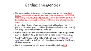
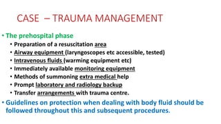
This document provides summaries of various surgical emergencies and their management. It discusses intestinal obstructions, acute appendicitis, acute pancreatitis, gastrointestinal bleeding, perforated peptic ulcers, and other conditions. For each it describes typical symptoms, diagnostic tests, and treatment approaches which may include conservative management, surgery, or other procedures. Conditions covered include hernias, sigmoid volvulus, appendicitis, pancreatitis, ulcers, hemorrhages, burns, fractures, and more.
1. The document discusses various types of surgical bleeding including arterial, venous, and capillary bleeding. It also discusses primary, reactionary, and secondary hemorrhage.
2. Examples of conditions that can cause bleeding in surgical practice are discussed in detail, including peptic ulcer disease, hemorrhoids, ectopic pregnancy, abortions, ruptured spleen, and femur fractures. Signs, symptoms, investigations, and treatments are described for each condition.
3. Types of abortions including threatened, inevitable, complete, incomplete, septic, missed, and habitual abortions are defined and their management is outlined. Complications of abortions including infection and injuries are also mentioned.
This document provides guidelines from a surgical registrar on when and how to contact them regarding surgical emergencies that present to the emergency department. It outlines some key situations that should prompt a call, such as a hole or blockage in something, something in the wrong place, or significant infection or inflammation. It also provides advice on what information to provide when calling, including who you are, where you're calling from, and what you need from the registrar. The document gives some brief overviews of specific conditions like hernias, pancreatitis, ischemic gut and limb, and massive GI bleeds.
General surgery and abdominal emergencies revisionULVAN OZAD
Due to embryonic development, the gastrointestinal tract can be divided into three sections - the foregut, midgut, and hindgut. Pain is localized to different abdominal regions depending on the affected section: epigastric for foregut, umbilical for midgut, and suprapubic for hindgut. Common acute abdominal conditions present with pain in specific locations and include appendicitis in the right lower quadrant, cholecystitis in the right upper quadrant, and diverticulitis in the left lower quadrant. These conditions are diagnosed based on symptoms, physical exam findings, and imaging studies, and treated through medications, surgery, or both.
Lecture on abdominal trauma during Basic Life Support 2018 course in Sibu Hospital. Encompasses blunt and penetrating trauma, principles and tips of management
Acute appendicitis is the most common acute surgical condition of the abdomen. It involves inflammation and infection of the appendix. The appendix arises from the cecum in the right lower quadrant of the abdomen. Clinical diagnosis is based on symptoms of shifting abdominal pain, anorexia, vomiting, and localized tenderness in the right lower quadrant. Treatment involves appendectomy to remove the infected appendix.
Some slides are taken from different textbooks of medicine like Davidson, Kumar and Clark and Oxford, and some from other presentations made by respected tutors. I'm barely responsible for compilation of various resources per my interest. These resources are free for use, and I do not claim any copyright. Hoping knowledge remains free for all, forever.
- Perforation of the gastrointestinal tract can occur due to various causes like perforated ulcers, penetrating injuries, or ischemic bowel. Signs include severe abdominal pain, fever, and tenderness.
- Diagnosis is suggested by imaging showing free air or fluid in the abdomen. Treatment requires emergency surgery to repair any perforations followed by intensive care and broad-spectrum antibiotics to treat peritonitis.
- Surgical management involves thorough irrigation and drainage of the abdominal cavity followed by resection of non-viable bowel and primary anastomosis or stoma formation as needed. Close postoperative monitoring in the ICU is important to support organ function and detect any complications.
The document discusses pancreatic trauma, including relevant anatomy, epidemiology, etiology, presentation, workup, staging, and treatment. Some key points:
- The pancreas is protected by surrounding structures but can be injured by penetrating trauma or direct blunt force.
- Injury is staged based on severity from grade I (minor) to grade V (massive disruption). Treatment depends on grade and location of injury.
- Workup may include labs, CT, MRCP, ERCP. Surgical treatment ranges from observation for minor injuries to distal pancreatectomy or pancreaticoduodenectomy for more severe injuries.
- Complications include pancreatic fistula, abscess, and pseudocyst.
Appendicitis is inflammation of the appendix, which is the most common emergency surgery indication. Early symptoms include pain near the belly button shifting to the lower right abdomen. Untreated appendicitis can lead to complications like perforation and abscess. A clinical exam looking for tenderness in the right lower quadrant combined with blood tests and imaging help make the diagnosis. Treatment is usually an appendectomy, although antibiotics and observation are occasionally used. Complications from untreated or delayed treatment of appendicitis can be serious.
This document discusses the acute abdomen, defined as abdominal pain that requires immediate intervention. It lists common symptoms like pain, vomiting, bleeding and diarrhea. Potential causes can be abdominal or extra-abdominal. Examination may reveal tenderness, guarding, rigidity or rebound tenderness. Common diagnoses include appendicitis, cholecystitis, pancreatitis and diverticulitis. General management involves blood tests, urinalysis, x-rays, IV fluids, pain relief, antibiotics and DVT prophylaxis. Specific management plans are outlined for different conditions.
Laparostomy management - ABThera™ Open Abdomen Negative Pressure Therapy Syst...Dr Edward Fitzgerald
Laparostomy Management with ABThera™
Case Experience: ABThera™ Open Abdomen Negative Pressure Therapy System in a Grade IV Open Abdomen Secondary to Acute Pancreatitis
Acute Abdomen describes sudden, severe abdominal pain of unclear origin lasting less than 24 hours, which may indicate a medical or surgical emergency. A thorough history, physical exam including assessment of the abdomen and vital signs, and diagnostic tests are needed to determine the cause. Relieving pain with analgesics is recommended while diagnosis is pursued. The document provides guidance on evaluating patients with acute abdominal pain.
Dr. Guy Nicastri, Associate Professor of Surgery and Family Medicine at the Warren Alpert School of Medicine at Brown University takes us through some of the pearls of the Acute Abdomen Examination in the Adult
This document discusses the use of CT imaging in evaluating acute abdominal pain. It outlines common causes of acute abdomen including gastrointestinal issues like appendicitis, diverticulitis, bowel obstruction, and perforation. It also mentions genitourinary causes and hepatobiliary and vascular etiologies. The document reviews the CT protocol and imaging appearances of various acute abdominal pathologies like appendicitis, diverticulitis, small bowel obstruction, pancreatitis, aortic aneurysm and dissection. It emphasizes that CT allows for a rapid and cost-effective evaluation of the acute abdomen when performed with the proper technique and protocol.
This document discusses acute abdomen, including causes, symptoms, and examination findings. It notes that acute abdomen involves sudden severe abdominal pain that may be life-threatening. Common intra-abdominal causes include inflammation, perforation, obstruction, and hemorrhage. Examination involves inspection, palpation, percussion, and auscultation of the abdomen, as well as relevant history and lab/imaging tests. Imaging tests like x-ray, ultrasound, and CT scan can help identify conditions like perforation, obstruction, or masses.
This document defines appendicitis and provides details about its historical perspective, epidemiology, etiology, classification, pathophysiology, clinical presentation, differential diagnosis, workup, treatment, and complications. Specifically, it notes that appendicitis is inflammation of the vermiform appendix and was first described in 1886. It occurs most commonly in the second and third decades of life and has a higher incidence in males. Obstruction of the appendix is a major cause and results in increased intraluminal pressure and bacterial overgrowth. Clinical presentation involves shifting pain from the periumbilical region to the right lower quadrant. Treatment is an appendectomy, which can be open or laparoscopic. Complications include appendiceal
This document discusses a case of a 20-year-old man presenting with abdominal pain and diarrhea who was found to have elevated inflammatory markers and free fluid on ultrasound. It then provides a general overview of acute abdomen including common causes, diagnostic approach, important physical exam findings and signs, and appropriate imaging studies.
This document discusses three conditions that affect the esophagus: perforation, Mallory-Weiss syndrome, and corrosive injury. Esophageal perforation can be caused by swallowed foreign bodies, corrosives, endoscopy procedures, or violent vomiting. It presents with chest, neck, and abdominal pain and requires antibiotics and surgical repair if large. Mallory-Weiss syndrome involves a tear in the esophagus from forceful vomiting, usually presenting with blood in the vomit. Corrosive injury results from ingesting substances like drain cleaner or bleach, causing severe esophagitis, pain, and complications like bleeding, perforation, or stricture if not treated promptly with neutralizing agents, antibiotics, and possibly surgery
FISSURE-IN-ANO – Lower GI Hemorrhage
Dear Viewers,
Greetings from “Surgical Educator”
Today in this episode, I have talked about yet another cause for lower GI haemorrhage- “Fissure-in-Ano”. Unlike other causes of lower GI haemorrhage, fissure-in-ano present with painful bleeding per rectum. I am talking on etiology, types, clinical features and treatment of fissure-in-ano. I have also included a mind map, a diagnostic algorithm and a treatment algorithm. You can watch this video in the following links:
surgicaleducator.blogspot.com
youtube.com/c/surgicaleducator
Thank you for watching the video.
Acute appendicitis in adults - Hoang Cuong HMUCường Hoàng
Acute appendicitis is one of the most common causes of abdominal pain requiring emergency surgery. The appendix is located near the cecum but can be in retrocecal or pelvic positions, complicating diagnosis. Obstruction of the appendix is a key factor in pathogenesis as it leads to ischemia, necrosis, and inflammation. Classic symptoms include migrating pain starting around the umbilicus and later localized to the right lower quadrant, along with anorexia, fever, nausea, and vomiting. Physical exam may reveal tenderness at McBurney's point or other signs depending on appendix position. Laboratory tests typically show leukocytosis. Imaging like CT, ultrasound, or MRI can help in diagnosis by identifying an enlarged or fluid-
The patient suffered multiple injuries including rib fractures, a ruptured spleen and pelvic fracture from a traffic accident. He developed abdominal compartment syndrome due to increased abdominal pressure after surgery. His condition worsened with rising ventilator pressures. Measurement of intra-abdominal pressure via bladder catheter confirmed abdominal compartment syndrome. He underwent decompressive laparotomy to reduce pressure, which improved his condition, though he required temporary abdominal closure and developed complications before final closure.
1) Blunt abdominal trauma is injury to the abdomen from non-penetrating forces and is a common cause of injury from motor vehicle accidents. Symptoms can range from minimal signs to cardiovascular collapse.
2) The spleen, liver, and kidneys are most commonly injured in blunt trauma due to their solid nature. Injuries to hollow organs like the stomach and intestines also occur from shearing forces.
3) Initial assessment focuses on the ABCDEs - Airway, Breathing, Circulation, Disability, and Exposure. Patients are fully evaluated and stabilized, with two IV lines placed and fluid resuscitation started if indicated. Ongoing monitoring of vitals and input/output is important
The document provides information about acute abdomen including potential causes, relevant history questions, physical exam findings, and management strategies. It discusses signs and symptoms of several specific conditions that can cause acute abdomen such as appendicitis, duodenal ulcer, kidney stones, abdominal aortic aneurysm, pancreatitis, cholecystitis, and bowel obstruction. The physical exam involves assessing vital signs, palpating the abdomen, and listening to bowel sounds while the management focuses on airway, nothing by mouth, and considering other potential causes like cardiac or gynecological issues.
1) A 66-year-old male presented to the emergency department with worsening abdominal pain and was found to have diffuse abdominal tenderness and guarding. Abdominal imaging would most likely reveal colonic dilation with loss of haustral markings, indicative of diverticulitis.
2) A 65-year-old female presented with abdominal pain and CT showed sigmoid diverticula and perisigmoid stranding. She returned days later with worsening symptoms and a new CT showed a 5 cm rim-enhancing perisigmoid fluid collection, requiring laparotomy for drainage and debridement.
3) Surgical management of diverticulitis may include laparoscopic or open sigmoid
An acute abdomen is severe abdominal pain lasting less than 24 hours with signs of tenderness. It requires rapid diagnosis and treatment as some causes like perforation require urgent surgery. Common causes are hemorrhage, infection, perforation, blockage, and ischemia. Physical exam, lab tests, imaging like CT scans, and diagnostic tools help determine the cause. Indications for urgent surgery include signs of peritonitis, shock, deterioration on conservative care, and radiologic findings suggestive of a condition like perforation requiring operation. Preoperative preparation focuses on IV access, fluid resuscitation, antibiotics, and correcting electrolyte abnormalities.
Handling the emergencies in radiology and first aid in the x ray departmentAnupam Niraula
1) Emergency departments are designed to treat acute medical issues without appointments and are staffed by trauma physicians. They classify patients into non-urgent, urgent, and acute categories to prioritize care.
2) For trauma patients, MDCT is often the preferred imaging method and should be located near the emergency room along with radiography. Interventional radiology may perform procedures like embolization to stop hemorrhaging.
3) In reaction emergencies, treatments vary based on symptoms but may include oxygen, antihistamines, epinephrine, saline, and moving the patient to stabilize their condition. Staff are trained to recognize and respond to different types and severities of reactions.
This document outlines emergency procedures for a dialysis unit. It provides guidelines for responding to various medical emergencies that may occur during dialysis treatment, including cardiac arrest, air embolism, anaphylactic reaction, seizure, shortness of breath, cardiac arrhythmias, and chest pain. For each emergency, it describes signs and symptoms to look for and steps staff should take to assess and stabilize the patient, such as notifying the charge nurse, administering oxygen, treating hypotension, and contacting the patient's physician.
This document presents an evaluation of trauma by Dr. Amr Shaddad. It discusses the objectives of understanding types of trauma, the ATLS protocol, and signs of urological injury. The ATLS protocol is described in detail, outlining the primary and secondary surveys with their respective components of cABCDE and a head-to-toe evaluation. Signs of potential urological injuries from trauma to the kidneys, ureters, bladder, and urethra are also summarized. The presentation aims to educate on proper trauma evaluation and management according to established guidelines.
The document provides guidance on evaluating and treating polytrauma patients. It outlines the goals of trauma resuscitation which include identifying life-threatening injuries. The Advanced Trauma Life Support (ATLS) approach is recommended as a safe standardized method, beginning with the primary survey of ABCDE (airway, breathing, circulation, disability, exposure). Key assessments include mechanism of injury, vital signs, neurological status, and bleeding control. A thorough secondary survey then involves a full head-to-toe examination. Guidance is given on managing specific injuries such as abdominal trauma, with operative intervention prioritized for unstable patients or those with signs of internal bleeding.
Anesthesia carries risks that can lead to patient death or injury. Several factors contribute to risks in the operating room including equipment issues, patient health factors, human performance errors, and system failures. Some key errors that can cause severe harm are airway issues, medication errors, and procedure mistakes. Maintaining vigilance, checklists, standards, training, and learning from adverse events can help improve safety. Thorough documentation and review of incidents is important for quality assurance.
This document provides guidance on the acute management of trauma patients. It outlines the following key points:
1) Trauma patients require a systematic approach involving preparation, a primary survey to address life threats (airway, breathing, circulation, disability, exposure), and management of specific injuries.
2) The primary survey follows an ABCDE approach and includes airway management, breathing and ventilation assessment, circulation assessment and intravenous access, disability assessment, and full body exposure/examination.
3) Specific guidance is given for managing potential airway issues, tension pneumothorax, chest trauma, hemorrhage, cardiac injuries, and other concerns uncovered during the primary survey. Checklists, guidelines, and multidisciplinary
This document discusses primary trauma care and outlines the steps for assessing and managing trauma patients. It covers triaging patients, performing a primary and secondary survey, identifying life-threatening injuries, providing initial resuscitation and stabilization, and determining appropriate disposition. The primary goals are to assess and treat airway, breathing, circulation and disability issues; control hemorrhage; and identify injuries requiring surgical intervention. Proper trauma management in the first hours can significantly impact outcomes.
This document provides an overview of initial assessment and management of trauma patients in remote environments. It discusses the primary survey using CABCDE to rapidly identify life threats, including controlling hemorrhage, maintaining the airway, and assessing breathing and circulation. It also covers the secondary survey, prolonged field care, definitive care, and obtaining a thorough history including mechanism of injury to help predict injuries. The systematic approach outlined aims to stabilize patients and prepare them for evacuation.
The document summarizes the steps of the Advanced Trauma Life Support (ATLS) process. It describes the primary survey with simultaneous resuscitation to identify life-threatening injuries, followed by a secondary survey to identify all other injuries. Finally, a definitive care plan is developed. The primary survey focuses on the ABCDE approach - Airway, Breathing, Circulation, Disability, and Exposure. Specific assessments and interventions are outlined for each step, including cervical spine precautions, tube thoracostomy, and full-body examination during the secondary survey. Maintaining spinal immobilization is emphasized throughout the trauma evaluation process.
1) Trauma deaths follow a trimodal distribution, with peaks occurring within seconds-minutes of injury due to severe brain or spinal cord injury, within minutes-hours due to hemorrhage, and days-weeks later due to sepsis or multiple organ failure.
2) The initial assessment and management of polytrauma patients follows the ABCDE approach, with simultaneous attention to the airway, breathing, circulation, disability, and exposure while preparing for further care and monitoring.
3) Secondary surveys involve a full physical exam, history taking, and diagnostic tests to identify all injuries and guide definitive care, which may involve patient transfer to a higher level trauma center if needed.
name of person assisting with transfer
Background: brief history of present illness and injury
Assessment: vital signs, GCS, injuries identified, procedures performed
Recommendation: level of care required, pending procedures or tests,
anticipated problems during transport
This document discusses the management of various types of medicolegal emergencies from an intensivist's perspective. It outlines procedures for handling cases involving polytrauma, poisoning, drug overdoses, burns, assaults, gunshot wounds, drowning, hanging, and snake/animal bites. Priority is given to stabilizing the patient and addressing life-threatening injuries before legal formalities. Proper consent, confidentiality, evidence collection and medico-legal report preparation are also emphasized.
Based on the information provided, here is how I would triage the patients:
1. Patient C (older male in pool of blood) - He has obvious severe hemorrhage and would be treated first. Direct pressure and a tourniquet would be applied to control bleeding while preparing for transport.
2. Patient B (cyanotic and noisy breathing) - She shows signs of a life-threatening respiratory problem like a tension pneumothorax and would be treated second. Needle decompression would be performed.
3. Patient F (crying holding abdomen) - She likely has an intra-abdominal injury and would be treated third. Bleeding would be addressed and she would be prepared for transport.
1) ATLS was developed in the 1970s by Dr. James Styner after a tragic plane crash injured his children and he found the emergency care provided was inadequate.
2) The goals of ATLS are to save lives through identifying and treating life threats in trauma situations within the "golden hour" window.
3) The ATLS approach involves a primary survey to address airway, breathing, circulation, disability and exposure, followed by secondary and tertiary surveys.
1. The primary survey in trauma management focuses on the ABCDE approach to identify and treat life-threatening injuries. This includes assessing the airway, breathing, circulation, disability, and exposure.
2. Maintaining an open airway is critical and may require techniques like head tilt/chin lift or jaw thrust. Oropharyngeal or nasopharyngeal airways can be used if needed. Tension pneumothorax requires urgent needle decompression and chest drain placement.
3. In addition to airway management, the primary survey involves evaluating breathing/ventilation and looking for signs of respiratory distress or reduced breath sounds. Circulation is also assessed to control hemorrhage and ensure adequate end-organ
Trauma Management in Primary Care Settingssnsharifa
1. The primary survey involves rapidly assessing and treating life-threatening injuries by evaluating the patient's airway, breathing, circulation, disability, and exposure (ABCDE). This includes establishing an open airway, assessing breathing and ventilation, treating hemorrhagic shock, and providing spinal immobilization and intravenous access when needed.
2. The secondary survey is a head-to-toe examination to identify any injuries that may have been missed during the primary survey. It includes detailed examination of specific body regions like the head, neck, chest, abdomen, and extremities.
3. Effective trauma management requires a coordinated team approach with assigned roles. The goals are to rapidly identify and treat life-threatening injuries,
Advanced trauma life support, management of a polytrauma patientLawrenceWanderi
The document outlines the objectives and procedures of the Advanced Trauma Life Support (ATLS) protocol for rapidly assessing and stabilizing patients. It describes the "golden hour" window for treatment, and conducting an initial assessment including primary and secondary surveys, with resuscitation of life-threatening injuries and consideration of patient transfer needs. The primary survey involves assessing the ABCDEs (airway, breathing, circulation, disability, exposure) while the secondary survey is a full physical exam and history. Adjuncts aid diagnosis while continual reassessment is important for trauma patients.
Glaucoma, all what you have to know about.Amr Eldakroury
Glaucoma is a group of progressive disorders characterized by retinal ganglion cell apoptosis and optic neuropathy associated with cupping of the optic disc. There are three main mechanisms that can cause glaucomatous optic neuropathy: high intraocular pressure physically damaging the optic nerve; malfunctioning autoregulatory mechanisms; and genetic factors that induce programmed cell death. Glaucoma is a leading cause of blindness globally and in Qatar it accounts for 39% of bilateral blindness cases and 16% of low vision cases. Risk factors include older age, family history, higher eye pressure, and conditions like diabetes. Treatment involves lowering intraocular pressure through various eye drop medications which can have side effects like superficial punctate keratitis.
1. Identify the difference between vertigo, disequilibrium,, near-syncope, and Undifferentiated dizziness.
2. Identify helpful tests to distinguish peripheral from central vertigo.
3. Understand how to treat different kinds of vertigo
Hair diseases are disorders primarily associated with the follicles of the hair. Many hair diseases can be associated with distinct underlying disorders. Hair disease may refer to excessive shedding or baldness (or both). Balding can be localized or diffuse, scarring or non-scarring.
Communication is the act of conveying meanings from one entity or group to another through the use of mutually understood signs and semiotic rules
The ability to communicate effectively is an essential skill in today's world. Communication is a dynamic process.
This document discusses syphilis, a sexually transmitted disease caused by the spirochete Treponema pallidum. It defines syphilis and describes the causative organism. It discusses the modes of transmission for acquired and congenital syphilis. The clinical stages of syphilis are outlined including primary, secondary, latent, and tertiary syphilis. The diagnosis, treatment, and prognosis of syphilis are summarized. Congenital syphilis is also described. References are provided.
Here are the key steps the emergency department would take after obtaining history and performing an examination:
1. Stabilize the patient. Given the high fever and altered mental status, immediate focus would be on stabilizing vital signs through IV fluids and antipyretics. Oxygen supplementation may also be needed.
2. Obtain imaging. A non-contrast head CT would be done urgently to evaluate for complications like abscess, thrombosis or meningitis. Contrast may be added based on findings.
3. Start empiric antibiotics. Broad spectrum IV antibiotics like vancomycin and piperacillin-tazobactam would be started immediately based on suspicion of orbital or intracranial infection.
This document provides information about bronchiectasis, including its causes, symptoms, diagnosis, and management. It describes bronchiectasis as an abnormal permanent dilation of the bronchi due to destruction of the bronchial wall muscles and elastic tissue. Common causes include infection, aspiration, obstruction, and genetic conditions like cystic fibrosis. Symptoms include chronic cough, sputum production, and recurrent lung infections. Diagnosis is made through clinical history and characteristic findings on CT scan. Management focuses on controlling infections with antibiotics, clearing secretions, and treating underlying causes.
This document discusses onychomycosis (nail fungus). It defines onychomycosis and describes the most common presentation as poor cosmetic appearance of the nail. It then discusses the epidemiology, risk factors, infecting organisms, clinical presentations including distal and lateral subungual onychomycosis, superficial white onychomycosis, and total dystrophic onychomycosis. It also covers investigations, interpretation of results, associated diseases, treatment including medical and surgical options, complications, prognosis, and references.
This document provides an overview and agenda for a presentation on quality management systems and ISO 9001:2008. The presentation covers definitions of quality and the evolution of quality management systems. It discusses the key drivers for quality management systems and the PDCA cycle that ISO 9001 is based on. The eight quality management principles and benefits of ISO 9001 certification are also reviewed. The presentation provides an overview of the clauses and elements of ISO 9001:2008, including requirements for documentation, management responsibility, and product realization. Implementation and certification processes are discussed along with potential pitfalls.
The document provides information on Hepatitis C virus (HCV) including its definition, background prevalence data, transmission routes, screening and testing approaches, natural history, symptoms, treatment options, and standard precautions. It notes that HCV is a blood-borne virus that infects the liver and is transmitted through exposure to infected blood. An estimated 3% of the global population has been infected with HCV.
This document provides information on sarcoidosis, a disease characterized by the growth of granulomas in multiple organs of the body. It begins by discussing the causes of sarcoidosis, which is an immune reaction that continues even after the initial trigger is cleared. Common areas affected include the lungs, eyes, skin, and lymph nodes. The stages of pulmonary sarcoidosis and associated symptoms are described. Treatment options are outlined, ranging from symptomatic treatment for mild cases to corticosteroids and immunosuppressants for more severe symptoms.
One third of the world is infected with tuberculosis, with over 1 million deaths reported annually. Tuberculosis is caused by bacteria in the Mycobacterium tuberculosis complex and is more common in developing countries. Symptoms include cough and abnormal chest x-rays. Diagnosis involves sputum smear, culture, and molecular tests. Treatment for active tuberculosis involves a combination of antibiotics over 6 months, while latent tuberculosis is treated with one or two antibiotics. Granulomas formed during infection help regulate the immune response and minimize tissue damage. Tuberculosis remains a major global health problem despite efforts to diagnose and treat those affected.
This document discusses various lung conditions including bronchiolitis obliterans, organizing pneumonia, and interstitial lung disease. It defines key terms and describes the symptoms, causes, diagnosis, and treatment of these conditions. Bronchiolitis obliterans involves scarring and obstruction of the bronchioles. Organizing pneumonia refers to unresolved pneumonia where exudates persist and cause fibrosis. Interstitial lung disease is a class of diffuse lung diseases involving inflammatory responses with varying degrees of pulmonary fibrosis. Specific forms like BOOP, COP, and IPF are mentioned along with their clinical presentation and pathological features.
The thyroid gland secretes hormones that regulate metabolism. Disorders can cause hyperthyroidism (overactive thyroid) or hypothyroidism (underactive thyroid). Examination of the thyroid involves inspection for size/shape, palpation for consistency/nodules, and auscultation for bruits. Blood tests measure thyroid hormone levels while ultrasound images the gland. Mild cases may only require monitoring but severe or abnormal cases should be referred to an endocrinologist.
Shingles is caused by the reactivation of the varicella zoster virus which causes chickenpox, presenting as a painful rash on one side of the body that is soon followed by blisters. It occurs more often in elderly people and can be prevented with vaccination. Treatment with antiviral drugs within 48 hours can limit post-herpetic neuralgia.
NAVIGATING THE HORIZONS OF TIME LAPSE EMBRYO MONITORING.pdfRahul Sen
Time-lapse embryo monitoring is an advanced imaging technique used in IVF to continuously observe embryo development. It captures high-resolution images at regular intervals, allowing embryologists to select the most viable embryos for transfer based on detailed growth patterns. This technology enhances embryo selection, potentially increasing pregnancy success rates.
Histololgy of Female Reproductive System.pptxAyeshaZaid1
Dive into an in-depth exploration of the histological structure of female reproductive system with this comprehensive lecture. Presented by Dr. Ayesha Irfan, Assistant Professor of Anatomy, this presentation covers the Gross anatomy and functional histology of the female reproductive organs. Ideal for students, educators, and anyone interested in medical science, this lecture provides clear explanations, detailed diagrams, and valuable insights into female reproductive system. Enhance your knowledge and understanding of this essential aspect of human biology.
PGx Analysis in VarSeq: A User’s PerspectiveGolden Helix
Since our release of the PGx capabilities in VarSeq, we’ve had a few months to gather some insights from various use cases. Some users approach PGx workflows by means of array genotyping or what seems to be a growing trend of adding the star allele calling to the existing NGS pipeline for whole genome data. Luckily, both approaches are supported with the VarSeq software platform. The genotyping method being used will also dictate what the scope of the tertiary analysis will be. For example, are your PGx reports a standalone pipeline or would your lab’s goal be to handle a dual-purpose workflow and report on PGx + Diagnostic findings.
The purpose of this webcast is to:
Discuss and demonstrate the approaches with array and NGS genotyping methods for star allele calling to prep for downstream analysis.
Following genotyping, explore alternative tertiary workflow concepts in VarSeq to handle PGx reporting.
Moreover, we will include insights users will need to consider when validating their PGx workflow for all possible star alleles and options you have for automating your PGx analysis for large number of samples. Please join us for a session dedicated to the application of star allele genotyping and subsequent PGx workflows in our VarSeq software.
These lecture slides, by Dr Sidra Arshad, offer a simplified look into the mechanisms involved in the regulation of respiration:
Learning objectives:
1. Describe the organisation of respiratory center
2. Describe the nervous control of inspiration and respiratory rhythm
3. Describe the functions of the dorsal and respiratory groups of neurons
4. Describe the influences of the Pneumotaxic and Apneustic centers
5. Explain the role of Hering-Breur inflation reflex in regulation of inspiration
6. Explain the role of central chemoreceptors in regulation of respiration
7. Explain the role of peripheral chemoreceptors in regulation of respiration
8. Explain the regulation of respiration during exercise
9. Integrate the respiratory regulatory mechanisms
10. Describe the Cheyne-Stokes breathing
Study Resources:
1. Chapter 42, Guyton and Hall Textbook of Medical Physiology, 14th edition
2. Chapter 36, Ganong’s Review of Medical Physiology, 26th edition
3. Chapter 13, Human Physiology by Lauralee Sherwood, 9th edition
Pictorial and detailed description of patellar instability with sign and symptoms and how to diagnose , what investigations you should go with and how to approach with treatment options . I have presented this slide in my 2nd year junior residency in orthopedics at LLRM medical college Meerut and got good reviews for it
After getting it read you will definitely understand the topic.
STUDIES IN SUPPORT OF SPECIAL POPULATIONS: GERIATRICS E7shruti jagirdar
Unit 4: MRA 103T Regulatory affairs
This guideline is directed principally toward new Molecular Entities that are
likely to have significant use in the elderly, either because the disease intended
to be treated is characteristically a disease of aging ( e.g., Alzheimer's disease) or
because the population to be treated is known to include substantial numbers of
geriatric patients (e.g., hypertension).
5-hydroxytryptamine or 5-HT or Serotonin is a neurotransmitter that serves a range of roles in the human body. It is sometimes referred to as the happy chemical since it promotes overall well-being and happiness.
It is mostly found in the brain, intestines, and blood platelets.
5-HT is utilised to transport messages between nerve cells, is known to be involved in smooth muscle contraction, and adds to overall well-being and pleasure, among other benefits. 5-HT regulates the body's sleep-wake cycles and internal clock by acting as a precursor to melatonin.
It is hypothesised to regulate hunger, emotions, motor, cognitive, and autonomic processes.
Summer is a time for fun in the sun, but the heat and humidity can also wreak havoc on your skin. From itchy rashes to unwanted pigmentation, several skin conditions become more prevalent during these warmer months.
2. Cardiac emergencies
• The signs and symptoms of cardiac emergencies include chest
pain ! , shortness of breath, fast and slow heart rates, increased
respiratory rate, low blood pressure ! , poor peripheral perfusion
(indicated by prolonged capillary refill time) and altered mental
state.
• If there is a history of angina the patient will probably carry
glyceryl trinitrate spray or tablets (or isosorbide dinitrate tablets)
and they should be allowed to use them.
• Where symptoms are mild and resolve rapidly with the patient’s
own medication, hospital admission is not normally necessary.
• Sudden alterations in the patient’s heart rate (very fast or very
slow) may lead to a sudden reduction in cardiac output with loss
of consciousness.
• Medical assistance should be summoned by dialling 999.
3. Case. Myocardial infarction
Symptoms and signs of myocardial infarction
• Progressive onset of severe, crushing pain in the centre
and across the front of chest. The pain may radiate to the
shoulders and down the arms (more commonly the left),
into the neck and jaw or through to the back.
• Skin becomes pale and clammy.
• Nausea and vomiting are common.
• Pulse may be weak and blood pressure may fall.
• Shortness of breath.
4.
5. localisation ST elevation Reciprocal ST depression coronary artery
Anterior MI V1-V6 None LAD
Septal MI
V1-V4, disappearance of septum Q in
leads V5,V6
none LAD-septal branches
Lateral MI I, aVL, V5, V6 II,III, aVF LCX or MO
Inferior MI II, III, aVF I, aVL RCA (80%) or RCX (20%)
Posterior MI V7, V8, V9
high R in V1-V3 with ST depression
V1-V3 > 2mm (mirror view)
RCX
Right Ventricle MI V1, V4R I, aVL RCA
Help with the localisation of a myocardial infarct
6. Case. Myocardial infarction
• Call 999 immediately for an ambulance.
• Allow the patient to rest in the position that feels most comfortable;
in the presence of breathlessness this is likely to be the sitting
position. Patients who faint or feel faint should be laid flat; often an
intermediate position (dictated by the patient) will be most
appropriate.
• Give high flow oxygen (10 litres per minute).
• Give sublingual GTN spray if this has not already been given.
• Reassure the patient as far as possible to relieve further anxiety.
• Give aspirin in a single dose of 300 mg orally, crushed or chewed.
Ambulance staff should be made aware that aspirin has already been
given as should the hospital.
• Many ambulance services in the UK will administer thrombolytic
therapy before hospital admission.
• If the patient becomes unresponsive always check for ‘signs of life’
(breathing and circulation) and start CPR in the absence of signs of
life or normal breathing (ignore occasional ‘gasps’).
10. CASE - TRAUMA
Trauma Assessment
The initial assessment and management of seriously
injured patients is a challenging task and requires a rapid
and systematic approach. This systematic approach can
be practised to increase speed and accuracy of the
process but good clinical judgement is also required.
Although described in sequence some of the steps will
be taken simultaneously.
The aim of good trauma care is to prevent early trauma
mortality. Early trauma deaths occur because of failure
of oxygenation of vital organs or central nervous system
injury or both.
11. CASE – TRAUMA MANAGEMENT
First and most important:
Is it safe to approach?
12. CASE – TRAUMA MANAGEMENT
•Aims of the initial evaluation of trauma patients
• Stabilise the patient
• Identify life threatening conditions in order of risk and initiate
supportive treatment
• Organise definitive treatments or organise transfer for definitive
treatments
•Preparation and coordination of care Assessment and
management will begin out of hospital at the scene of injury
and good communication with the receiving hospital is
important. The preparatory measures are outlined below to
'set the scene':
13. CASE – TRAUMA MANAGEMENT
• The prehospital phase
• Preparation of a resuscitation area
• Airway equipment (laryngoscopes etc accessible, tested)
• Intravenous fluids (warming equipment etc)
• Immediately available monitoring equipment
• Methods of summoning extra medical help
• Prompt laboratory and radiology backup
• Transfer arrangements with trauma centre.
• Guidelines on protection when dealing with body fluid should be
followed throughout this and subsequent procedures.
14. CASE – TRAUMAMANAGEMENT
General principles
1. Follow the Airway, Breathing, Circulation, Disability, and Exposure approach
(ABCDE) to assess and treat the patient.
2. Treat life-threatening problems as they are identified before moving to the next
part of the assessment.
3. Continually re-assess starting with Airway if there is further deterioration.
4. Assess the effects of any treatment given.
5. Recognise when you need extra help and call for help early. This may mean
dialling 999 for an ambulance.
6. Use all of your resources – ask members of public for help. This will allow you to
do several things at once, e.g., collect emergency drugs and equipment, dial 999.
15. CASE – TRAUMA MANAGEMENT
Initial assessment
This comprises:
• Primary survey
• Resuscitation
• Secondary survey
• Definitive treatment or transfer for definitive care
16. CASE – TRAUMA MANAGEMENT
• A= Airway maintenance cervical spine protection
• B= Breathing and ventilation
• C= Circulation with haemorrhage control
• D= Disability: Neurological status
• E= Exposure/ environmental control
17. CASE – TRAUMA MANAGEMENT
As part of the secondary survey
•History:
• A=Allergies
• M=Medication currently used
• P=Past illnesses/Pregnancy
• L=Last meal
• E=Events/Environment related to injury
18. CASE – TRAUMA MANAGEMENT
A= Airway maintenance cervical spine protection
• Are there signs of airway obstruction, foreign bodies, facial,
mandibular or laryngeal fractures?
• Establish a clear airway (chin lift or jaw thrust) but protect the cervical
spine at all times. If the patient can talk the airway is likely to be safe
but remain vigilant and recheck. GCS less than 8 requires definitive
airway.
20. CASE – TRAUMAMANAGEMENT
C = Circulation with haemorrhage control
• Blood loss is the main preventable cause of death after trauma. To assess blood loss rapidly
observe:
• Level of consciousness
• Skin colour
• Pulse.
• Bleeding should be assessed and controlled:
• Direct manual pressure should be used (not tourniquets except for traumatic
amputation as these cause distal ischemia).
• Transparent pneumatic splinting devices may control bleeding and allow visual
monitoring.
• Occult bleeding into the abdominal cavity and around long bone or pelvic
fractures is problematic.
21.
22. CASE – TRAUMA MANAGEMENT
D = Disability: Neurological status
• After A,B and C above rapid neurological assessment is made to establish
• Level of consciousness, using Glasgow Coma Scale
• Pupils: size, symmetry and reaction
• Any lateralising signs
• Level of any spinal cord injury (limb movements, spontaneous respiratory effort)
• Note: remember oxygenation, ventilation, perfusion, drugs, alcohol and
hypoglycaemia may all also affect level of consciousness.
23. CASE – TRAUMA MANAGEMENT
E= Exposure/ environmental control
• Undress patient, but prevent hypothermia Clothes may need
to be cut off, but after examination attention to prevention
of heat loss with warming devices, warmed blankets etc is
important, Intravenous fluids should be warmed before
infusion.