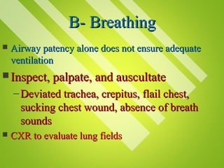
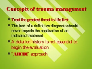
The document discusses the initial assessment and management of trauma patients. It outlines the ABCDE approach to prioritize airway, breathing, circulation, disability, and exposure. Specific interventions are described for addressing life-threatening injuries associated with each category. The goal is to rapidly identify and treat issues that pose the greatest risk to life, such as airway obstruction, tension pneumothorax, and hemorrhagic shock from blood loss.
Patient positioning in operating theatre -gihsgangahealth
This document discusses proper patient positioning during surgery. It outlines common surgical positions like supine, prone, Trendelenburg, and lithotomy. For each position, it describes how to position the patient, nursing precautions to take, and potential complications to avoid. The goal of positioning is to provide optimal surgical access while maintaining patient safety, comfort, and dignity. Careful positioning can prevent injuries, but risks increase for patients with certain medical factors.
This document discusses patient positioning during anesthesia. It covers positioning basics, potential complications, and respiratory and hemodynamic effects of different positions. Key points include positioning being a compromise between surgical exposure and patient comfort. Proper hand positioning and documentation are emphasized to prevent nerve injuries. Respiratory matching is different for awake versus anesthetized patients. The supine position can cause vascular congestion in the lungs.
Patient positioning during surgery Dr Rakesh kaward18rakesh
1. Proper patient positioning is important for surgical access and safety, but can also impact patient physiology.
2. Different positions like supine, prone, lateral, lithotomy and sitting are used for different procedures and come with risks if not done correctly, such as nerve injuries or changes in cardiovascular and respiratory function.
3. Careful positioning and padding of pressure points, along with monitoring of vital signs can help minimize risks, while optimal positions must balance surgical needs with patient well-being.
This document discusses patient positioning for surgery. It notes that positioning usually occurs after anesthesia administration and lists common surgical positions like supine, prone, Trendelenburg, and lithotomy. It also discusses factors that affect positioning sterile team members and equipment, such as the planned procedure, patient position, sizes, and preferences of sterile team members. Finally, it provides images of various patient positioning examples.
The document discusses various surgical patient positioning techniques and their physiological effects. It describes positions such as supine, lithotomy, lateral, prone, Trendelenburg's, and sitting. Positioning must balance exposure for surgery with risks like nerve injury and hypotension. Careful positioning and monitoring are important to prevent complications.
The document provides guidelines for cervical spine immobilization including:
- Proper techniques for applying cervical spine immobilization and the criteria for when to immobilize a patient.
- Spinal immobilization should be provided if there is any reasonable possibility of a spinal or head injury.
- The algorithm outlines the steps for manually stabilizing the cervical spine, logrolling a supine patient onto a backboard, and fully immobilizing standing or seated patients.
The document discusses various patient positioning techniques used in anaesthesia and their goals, risks, and complications. It describes positions like supine, lithotomy, prone, lateral decubitus, and their effects on cardiovascular and respiratory systems. Common risks include nerve injuries, pressure sores, compartment syndrome, and visual complications. Careful patient assessment, padding of pressure points, monitoring for nerve injuries are emphasized. Position changes should be gradual and extremities checked regularly during long procedures to prevent injuries.
1. The document outlines the steps for assessing and treating a trauma patient, including distinguishing those with and without significant mechanisms of injury, performing focused exams, and ongoing reassessment.
2. It describes the eight chapters of trauma assessment - scene size-up, initial assessment, primary survey, history taking, secondary survey, monitoring, transportation considerations, and overall hospital assessment.
3. Guidelines are provided for rapid trauma assessment, detailed physical exam, cervical collar application, and determining whether a detailed exam is needed based on the patient's condition and mechanism of injury.
2018 ATLS PROTOCOL
FOCUSING ON THE PRIMORDIAL MANAGEMENT OF PT IN THE APPROACH OF IMPROVING TRAUMA PT MANAGEMENT AND REDUCING MORTALITY OF TRAUMA PT AT OUR RESPECTIVE HEALTH FACILITIES AS DOCTORS AND CLINICIANS WORKING IN THE EMERGENCY DEPARTMENT.
GIVEN THE NECESSARY EQUIPMENT AND FAVOURABLE AMBIENT WORKING ENVIRONMENT WE SHOULD BE ABLE TO OFFER OUR HUMANITY RACE QUALITY SERVICES BEARING IN MIND THAT LIFE COME FIRST AND ALL THE OTHER ATTRIBUTES IN LIFE FOLLOWS
This document provides an introduction to trauma ultrasound and describes some common diagnostic tools used in trauma patients, including DPL, FAST exam, and abdominal CT scan. It discusses when each tool is most appropriate, limitations, and examples of their use. DPL allows rapid assessment but is invasive, while FAST exam is non-invasive but operator dependent and less sensitive. CT scan is most sensitive but requires more resources. The document advocates initially using FAST exam and then CT scan for trauma patient evaluation.
Patient positioning and anaesthetic considerationIqraa Khanum
This document discusses various surgical body positions and their physiological effects. It describes positions like supine, lithotomy, lateral, and prone. For each position, it details how positioning impacts cardiovascular and pulmonary function, as well as nerves that may be at risk of injury. Complications from prolonged use of each position are also reviewed. The document emphasizes the importance of careful patient positioning to balance surgical access needs with physiological stability and risk of pressure injuries.
This document discusses selective spinal motion restriction (SSMR) and alternatives to traditional full spinal immobilization. It notes the risks of long spine boards and rigid cervical collars, including increased pain, pressure sores, and raised intracranial pressure. Current research supports SSMR, using assessment to determine who needs restriction and allowing removal of extrication devices when possible. For transport, soft restraints like vacuum mattresses are recommended over rigid devices when spinal motion restriction alone is needed. The document outlines indications for SSMR including altered mental status, neck/back pain, and distracting injuries. Innovative solutions discussed include soft foam collars and adjustable collars that reduce risks compared to traditional rigid devices.
Patient positioning is a joint responsibility of the surgeon and anesthesiologist to balance surgical needs and risks to the patient. Key factors to consider include the procedure, patient characteristics, and physiological impacts of different positions. Common positions include supine, lateral, lithotomy and prone, each with benefits and risks requiring precautions like padding pressure points. The team must plan positioning prior to surgery based on these considerations.
The document discusses various surgical positions including supine, prone, Kraske/jackknife, lithotomy, and lateral positions. For each position, it provides the description, common uses in surgery, required equipment, and potential hazards to the patient. The overall document serves to educate medical staff on properly positioning patients for different surgical procedures while avoiding injury and complications.
This document discusses abdominal and pelvic trauma. It covers anatomy, mechanisms of injury including blunt and penetrating trauma, evaluation including physical exam, adjunct studies like FAST and CT scans, classification of pelvic fractures, acute stabilization methods, and outcomes related to different injury patterns. Unrecognized abdominal and pelvic injuries can lead to preventable death due to difficulty evaluating bleeding from solid organs or pelvic structures. Pelvic fractures are associated with high mortality, especially if the patient is hypotensive on admission or has an open injury.
The document discusses patient positioning considerations for anesthesia. It notes that positioning is a joint responsibility of the surgeon and anesthesiologist to balance surgical needs with risks to the patient. Key positions discussed include supine, lateral, prone, lithotomy, and variations like Trendelenburg. Physiological concerns for each position are outlined such as effects on circulation, pulmonary function, and nerve injuries. Proper padding and stabilization are emphasized to prevent pressure injuries.
1) A medic responds to an IED explosion where his convoy was attacked. The person next to him has bilateral mid-thigh amputations with heavy bleeding from one leg. 2) The medic's top priority is to return fire and take cover since they are still under attack. 3) Once there is suppressive fire from the rest of the convoy, the medic applies a tourniquet to the leg with arterial bleeding to control the life-threatening hemorrhage.
Incisions and position in general surgery by dr chandrakant sabaleCHANDRAKANT SABALE
This document discusses surgical incisions and patient positioning in general surgery. It provides details on:
1. Types of incisions like vertical, transverse, oblique and their uses in different abdominal and pelvic surgeries.
2. Principles of incision placement and closure.
3. Langer's lines and their importance in wound healing.
4. Common patient positions used in surgery like supine, lateral, lithotomy and their advantages.
This document discusses the benefits of early mobilization for mechanically ventilated patients in the ICU. Prolonged bed rest can lead to increased morbidity, mortality, costs, and length of stay. Early mobilization, which involves getting patients sitting up and out of bed when minimally able, provides several benefits like improving respiratory function and reducing adverse effects of immobility. Two studies presented found that early mobilization was feasible and safe for respiratory failure patients, with adverse events being rare. Transferring patients to an ICU that prioritizes early activity was also found to substantially improve patient ambulation levels.
Prehospital care in trauma is as important as in hospital care. The presentation addresses simple and basic approach to care a polytrauma victim in platinum 10 minutes based on BTLS.
1. The document discusses the evaluation and management of patients presenting to the emergency department with trauma or acute diseases.
2. It outlines the clinical examination, laboratory tests, radiology imaging and procedures used for trauma and acute care patients.
3. Specific radiological evaluations are described for trauma patients, including cervical spine, chest and pelvis x-rays, and CT scans of the brain, chest, abdomen and pelvis depending on the mechanism and sites of injury.
The document discusses Tactical Combat Casualty Care (TC-3) training for combat medics, which focuses on providing medical care on the battlefield in 3 stages - Care Under Fire, Tactical Field Care, and Combat Casualty Evacuation Care - with an emphasis on controlling hemorrhage, maintaining airways, and fluid resuscitation when possible given limited medical resources and ongoing tactical threats.
The Essentials Of Patient Positioning For Interventional Radiology ProceduresJames_DuCanto_MD
Proper patient positioning is essential for interventional radiology procedures. The anesthesiologist or CRNA guides patient transfer and positioning to maintain airway, breathing, and circulation. When positioning a patient prone, teamwork is needed to safely transfer the patient to the procedure table where all pressure points are padded and no body parts extend beyond the table. Positioning is tailored to each patient's individual needs and ensures the operative site is accessible while avoiding interference with respiration, circulation, peripheral nerves, or undue discomfort.
Tactical Combat Casualty Care Update: 2015Tetiana Botsva
The document provides an update on Tactical Combat Casualty Care (TCCC). It discusses that coalition forces currently have the best trauma care and evacuation system in history. However, 87% of combat fatalities still occur in the pre-hospital phase before reaching the hospital. TCCC aims to ensure casualties survive to reach the hospital where they can benefit from definitive care. The document reviews changes and advances made in TCCC over the past decade, including increased use of tourniquets, hemostatic dressings, and fluid resuscitation with blood products. It emphasizes that further efforts are needed to fully incorporate TCCC advances across medical and operational units.
This document provides information on maxillofacial trauma and its anaesthetic management. It begins with the relevant anatomy of the maxilla and important structures that can be damaged. It then describes Le Fort fracture patterns and their clinical features. Imaging studies like CT scans are the standard for evaluation. Special considerations for anaesthesia include securing the airway, which can be difficult due to the injuries, and managing blood loss. Various airway techniques are discussed like fiberoptic intubation, retrograde intubation, or surgical airways if needed. Intraoperative management focuses on invasive monitoring, induced hypotension to reduce bleeding, and muscle relaxation.
Diabetic ketoacidosis (DKA) is a medical emergency that occurs in type 1 diabetes patients when there is insufficient insulin, causing the body to break down fat and produce ketone bodies. Common precipitating factors include infection, poor blood sugar control, and non-compliance with treatment. Symptoms include nausea, vomiting, abdominal pain, headache, thirst, and breath that smells of ketones. Management involves fluid resuscitation, insulin administration, and treating any underlying causes. Mortality rates are 2-5% in developed countries and higher in developing areas.
This document discusses maxillofacial injuries, focusing on the importance of the "Golden Hour" period for treatment. It describes how maxillofacial injuries can impact functions like breathing, speaking, and eating. The document outlines the steps for examining and diagnosing maxillofacial trauma, including inspecting and palpating the extra-oral and intra-oral areas as well as ordering radiographs. Primary management goals are described as controlling pain, preventing infection, and surgical planning. The classification of fracture types is also detailed. The overarching message is the time sensitivity of treating maxillofacial injuries to address airway issues, bleeding, and prevent long-term complications.
The document discusses the surgical anatomy of the masseter muscle and facial nerve. It notes the relationships of the masseter muscle anteriorly, posteriorly, laterally, and medially. It then describes the surgical approach for locating the facial nerve, which involves making an incision in front of the ear tragus and dissecting through tissue to identify the styloid process as the first landmark. Finally, it provides measurements for distances between branches of the facial nerve and bony landmarks to help surgeons locate the nerve during procedures.
Cr pediatrics residents airway management part 2Danny Castro
This document discusses airway evaluation and management techniques for pediatric patients. It begins by outlining key anatomical features to evaluate the airway, including the Mallampati classification. It then covers positioning, bag mask ventilation, and various airway adjuncts like oral/nasal airways. Endotracheal intubation techniques using different blade types are described. The laryngeal mask airway is also discussed as an alternative to intubation. Finally, the document outlines several pediatric conditions associated with difficult airways.
This document discusses diabetic emergencies, including hyperglycemic hyperosmolar state (HHS). It notes that HHS is characterized by severe hyperglycemia, hyperosmolarity, and dehydration without significant ketoacidosis. It most commonly occurs in older patients with type 2 diabetes due to reduced fluid intake from an illness. Signs include a plasma glucose over 600 mg/dL, effective serum osmolality over 320 mOsm/kg, and dehydration of 8-12 liters with neurologic changes like drowsiness. The pathophysiology involves reduced insulin and elevated counter-regulatory hormones leading to further hyperglycemia and a shift of fluid out of cells causing intracellular
This document provides an overview of the Approach to Trauma- Advanced Trauma Life Support (ATLS) program. It discusses the history and concepts of ATLS, which was created in 1976 to standardize trauma care. The document outlines the ABCDE approach to the primary and secondary trauma surveys, which are designed to rapidly identify and treat life-threatening injuries. It covers steps for airway management, breathing and ventilation support, circulation stabilization, disability assessment, and full patient exposure and monitoring. Adjunct procedures like IV access, imaging, and fluid resuscitation are also reviewed.
This document discusses trauma epidemiology and management. It begins by outlining the topics to be covered, including trauma epidemiology, rapid assessment of patient condition through triage, identifying life-threatening injuries, resuscitation, secondary survey, and planning further care. It then provides statistics on the burden of trauma as a leading cause of death and disability worldwide. Several principles of trauma management are covered, including the concept of the "golden hour" for early treatment. Mortality rates from trauma are shown to have decreased with advances in pre-hospital care and transportation. The document concludes with details on assessing and managing the airway, breathing, circulation and disability during the primary and secondary trauma surveys.
This document discusses the anatomy, classification, clinical presentation, evaluation, and management of zygomaticomaxillary complex (ZMC) fractures. Some key points:
- The ZMC involves 4 bones and plays an important role in facial structure and function. ZMC fractures can be classified based on which bones are involved.
- Clinical findings may include cheek swelling, ecchymosis, step deformities, and diplopia. Evaluation involves physical exam, imaging like CT, and assessing for associated injuries.
- Treatment goals are to restore facial contour and function. Surgical management often requires an open reduction and internal fixation approach using plates and screws via incisions like lateral eyebrow or intraoral. Aut
Panfacial fractures involve multiple facial bones, including the frontal bones, zygomaticomaxillary complex, naso-orbitoethmoid region, maxilla and mandible. Due to the complex nature of these injuries, management requires careful planning and sequencing of treatment to restore facial functions, features and symmetry. Key goals are to reestablish occlusion, stabilize major facial supports to restore three-dimensional contour, and provide a stable scaffold for soft tissue healing. Proper imaging, surgical approaches and attention to anatomical landmarks are important to achieve accurate reduction and fixation.
This document provides an overview of fractures of the midface, including relevant anatomy, classification systems, evaluation, management considerations, operative techniques, and potential complications. It describes the key bones and structures of the midface, including the zygoma, maxilla, and midface buttresses. Classification systems such as the LeFort fractures and Knight and North system for malar fractures are reviewed. Indications, goals, approaches, reduction maneuvers, fixation methods, and postoperative care of midface fractures are discussed in detail. Complications including enophthalmos, nerve dysfunction, diplopia, and ectropion are also reviewed.
Mid facial fractures and their managementRuhi Kashmiri
The document discusses midfacial fractures, including the causes, anatomy, classifications, and management. It describes the LeFort fracture classifications (I, II, III), which involve horizontal fractures of the midface. LeFort I fractures are low-level fractures, while LeFort III fractures are the most severe, involving separation of the midface from the cranial base. Clinical findings, radiographic imaging, and treatment methods such as manual reduction or open reduction are reviewed for managing different types of midfacial fractures.
The document discusses the physiology of thyroid and parathyroid hormones. It describes how iodine is required for the formation of thyroid hormones thyroxine and triiodothyronine within the thyroid gland. The hormones are synthesized from tyrosine residues on thyroglobulin, stored in follicles, and released into blood. They increase metabolism and cellular activity by activating gene transcription in tissues. Effects include increased growth, carbohydrate and fat metabolism, basal metabolic rate, blood flow, and heart rate.
This document provides guidance on the management of multiple trauma patients. It outlines the ABCDE approach and trauma concept, which emphasizes rapidly assessing and treating the most life-threatening injuries first before making definitive diagnoses.
The primary survey involves simultaneously assessing the patient's airway, breathing, circulation, disability, and exposure. Adjuncts like monitoring, catheters and imaging may be used but not delayed transfer. The secondary survey obtains a more detailed history and physical exam.
Special considerations for pediatric, geriatric, and pregnant trauma patients are discussed. Key physiological differences and injury patterns are highlighted. The document also reviews complications like tension pneumothorax, cardiac tamponade and hemorrhagic shock and their
The document discusses maxillary fractures, their classification, and treatment. It notes that René LeFort classified maxillary fractures into 3 types based on the location of fracture lines. LeFort I involves the alveolar process, LeFort II the maxilla and nasal bones, and LeFort III separates the midface from the cranium. Treatment involves reduction using disimpaction forceps followed by fixation methods like wire osteosynthesis, rigid plates, or semi-rigid miniplates depending on the fracture type and location. Complications can include nerve damage, malocclusion, infection, and nonunion if not treated properly.
This document provides an overview of the management of diabetic emergencies, specifically diabetic ketoacidosis (DKA) and hyperglycemic hyperosmolar state (HHS). It describes the clinical presentation, diagnostic criteria, and treatment approach for each condition. The treatment involves aggressive fluid resuscitation and insulin therapy to lower blood glucose levels, along with potassium supplementation and monitoring of electrolytes and fluid balance. Close monitoring of vital signs and laboratory values is required. Management may involve transfer to a higher level of care such as the ICU if certain instability criteria are met.
The thyroid gland is located in the neck below the larynx. It produces three important hormones - thyroxine (T4), triiodothyronine (T3), and calcitonin. T4 and T3 increase the body's metabolic rate, and their production is regulated by TSH from the pituitary gland. Calcitonin regulates blood calcium levels by inhibiting bone resorption and renal calcium reabsorption. The thyroid receives blood supply from the superior and inferior thyroid arteries and is drained by superior, middle, and inferior thyroid veins.
This document discusses various types of maxillofacial fractures seen on radiographs. It describes recent tooth fractures appearing as thin radiolucent lines through teeth. Alveolar fractures appear as sharply defined radiolucent lines in the alveolus. Mandibular condyle fractures involve the condylar head being "sheared off". Le Fort fractures are classified into types I, II, and III based on the anatomical structures involved. CT is the standard for evaluating maxillary fractures while panoramic radiography is best for the mandible.
Maxillofacial trauma can result from injuries like motor vehicle accidents, falls, or animal bites. It includes soft tissue wounds, bone fractures, or a combination. Diagnosis involves clinical examination looking for signs of injury, and radiographic evaluation using techniques like plain X-rays, panoramic views, and advanced imaging to identify fractures. Treatment depends on the specific injuries but the priorities are always managing the airway, breathing, and circulation during the emergency period.
The document summarizes topics discussed at the 2013 Kentucky Trauma Symposium master nurse panel. It includes 6 topics: 1) pretransfer care, 2) sedation protocols, 3) identifying critical errors, 4) performance improvement/quality assurance/training, 5) managing the clinically brain dead, and 6) starting/maintaining a trauma program. It also provides 6 case studies discussing various trauma injuries and management strategies.
Emergency thoracotomy is a procedure performed in the emergency department or operating room to treat penetrating or blunt chest trauma with signs of life. It involves making an incision in the chest wall to access the heart, lungs, and great vessels to control bleeding, release pericardial tamponade, or perform open cardiac massage. Factors associated with increased survival include signs of life on arrival, penetrating rather than blunt trauma, shorter duration of CPR, and certain cardiac rhythms. Proper patient preparation, equipment, and a trained team are required to perform the procedure. Complications can include bleeding, infection, and injury to surrounding structures.
This document provides information on trauma management from the surgical club of Red Sea University. It discusses the phases of mortality from trauma, principles of triage, biomechanics of different injury types, and the ABCDE approach to the primary survey. For airway management, it describes techniques like chin lift, positioning, adjuncts, and definitive airways. It emphasizes controlling circulation through IV access, fluid resuscitation, and identifying/treating sources of hemorrhage in the primary survey. The goal is to recognize and stabilize life-threatening injuries in the first 10 minutes after trauma.
1. The Advanced Trauma Life Support (ATLS) protocol focuses on simultaneously identifying and treating life-threatening injuries within the crucial "Golden Hour" period after trauma.
2. The ATLS protocol involves two surveys - the Primary Survey to address airway, breathing, circulation, disability, and exposure issues, and the Secondary Survey for a full history and physical exam after initial resuscitation is complete.
3. Key components of the Primary Survey include assessing the airway, identifying tension pneumothorax and hemorrhage, and providing spinal immobilization, followed by full exposure to identify all injuries.
Thoracic trauma can cause life-threatening injuries like tension pneumothorax, cardiac tamponade, and uncontrolled hemorrhage. Pre-hospital management focuses on identifying and treating these immediately life-threatening conditions through assessment of airway, breathing, and circulation. Additional assessments identify injuries like rib fractures, lung contusions, and injuries to major blood vessels. Proper positioning, splinting of injuries, analgesia, and rapid transport to the hospital can help manage thoracic trauma in the pre-hospital setting.
This document discusses the management of chest trauma. It describes various types of chest injuries including rib fractures, flail chest, pneumothorax, hemothorax, and tension pneumothorax. For each injury, it outlines the signs and symptoms and recommended treatment approaches. It emphasizes the importance of assessing airway, breathing, and circulation when treating chest trauma patients. It also provides details on procedures for needle chest decompression to treat tension pneumothorax.
The document outlines the steps of the Advanced Trauma Life Support protocol. It includes: 1) preparing equipment and summoning a trauma team, 2) performing triage on multiple casualties, 3) conducting a primary survey to address life threats like airway, breathing, circulation, disability and exposure, 4) providing resuscitation as needed, 5) using adjuncts like monitoring, IVs and diagnostics, 6) performing a full secondary survey and history, 7) using additional adjuncts, 8) continued re-evaluation of the patient, and 9) arranging for their definite care. The protocol aims to quickly identify and treat life threats in a trauma patient.
Assessment of the multiply injured patient o'connorKenan Kasumagić
This document discusses the assessment and management of multiply injured patients. It describes the ATLS protocol, including the primary survey to address the ABCDEs (airway, breathing, circulation, disability, exposure). Special considerations are given to head injuries, chest injuries, and the elderly. The stages of assessment and resuscitation are outlined to structure the initial approach to trauma patients.
The document discusses the surgical experience, including:
1. The different types and levels of urgency of surgical procedures from emergent to elective.
2. The preparation process from pre-admission testing to admission and holding before surgery.
3. General anesthesia involves altering consciousness through agent inhalation, injection, or instillation and has different depth stages from amnesia to overdosage.
4. The phases of general anesthesia including induction, maintenance, emergence, and recovery.
CPCR Basic Life Support by Midland HealthcareAbhishek Singh
1. James Elam -first to experimentally demonstrate CPR
2. Dr. Peter Safar- brought to light effective procedures putting them together into what he called “the ABCs”
3. Claude Beck- Internal defibrillator
4. Paul Zoll- AC External defibrillator
5. Bernard Lown- DC external defibrillator
6. Foundation of successful ACLS is good BLS
For Help Visit: https://midlandhealthcare.org/
Dr. Abhijit Diwate discusses physiotherapy management of ICU patients. Key points include:
1) Physiotherapists assess patients, set goals like pain relief and secretion clearance, and use techniques like positioning, manual hyperinflation, percussion and breathing exercises.
2) Assessment involves examining the chest and determining ventilator settings. Goals are to prevent complications and improve function.
3) Common techniques are positioning, mobilization, manual hyperinflation, percussion, coughing/huffing, and breathing exercises to clear secretions and strengthen respiratory muscles.
The document discusses the initial approach to trauma care, which consists of an initial primary assessment, rapid resuscitation, and a more thorough secondary assessment. The primary assessment focuses on identifying and treating life-threatening conditions by assessing ABCDE (airway, breathing, circulation, disability, exposure). Key interventions include controlling bleeding, treating pneumothorax, and addressing shock. A secondary assessment then provides a full head-to-toe examination to identify all injuries, with resuscitation continuing throughout. An organized team approach is emphasized to properly manage trauma in a timely manner.
This document provides an overview of initial assessment and management of trauma patients in remote environments. It discusses the primary survey using CABCDE to rapidly identify life threats, including controlling hemorrhage, maintaining the airway, and assessing breathing and circulation. It also covers the secondary survey, prolonged field care, definitive care, and obtaining a thorough history including mechanism of injury to help predict injuries. The systematic approach outlined aims to stabilize patients and prepare them for evacuation.
Ninety percent of combat fatalities die before reaching medical treatment. Traditional trauma care like ATLS is not directly applicable to combat settings which involve prolonged evacuations, limited resources, and hostile conditions. Tactical Combat Casualty Care was developed to address the unique prehospital challenges of combat medicine. It focuses on controlling hemorrhage, airway management, and shock treatment at the point of injury through various stages of evacuation. Understanding the operational environment is essential, as standard procedures designed for civilian and hospital settings may not apply to combat prehospital care.
This document discusses the management of polytrauma patients. It defines polytrauma as injuries involving two or more major body systems. The goals of management are to save the patient's life, salvage limbs, and restore function if possible. A team approach is needed involving surgeons, physicians, and other specialists. The initial focus is a thorough primary survey and resuscitation to address life-threatening injuries like airway obstruction, hemorrhage, and spinal cord injury.
This document provides an overview of chest trauma. It begins by defining chest trauma as any injury to the chest, including the ribs, heart and lungs. Chest injuries are categorized as open or closed. Common causes are discussed, including blunt trauma from accidents or penetrating trauma from objects. Signs and symptoms, diagnostic tests, and specific injuries like pneumothorax are described. Treatment focuses on ABCs - airway, breathing and circulation while performing tests to evaluate cardiac and pulmonary function.
Trauma Management PPT for MBBS Students by Dr Anil Kumar,AIIMS-PatnaAnil Kumar
This document discusses trauma management and the burden of trauma in India. It begins with definitions of key terms like trauma, emergency, and triage. It then summarizes trauma statistics from 2005 and 2011 reports, showing over 4 million hospitalizations and 140,000 deaths annually. The document recommends establishing standardized pre-hospital and hospital protocols based on ATLS guidelines to improve outcomes. Key components of trauma management covered include scene safety, triage, primary and secondary surveys, hemorrhage control, spinal immobilization, and hypothermia prevention.
Polytrauma, or multisystem trauma, refers to injuries that affect multiple body systems and require a team-based approach to management. It is a leading cause of death among younger individuals. The document outlines the definition of polytrauma and discusses the priorities and processes for managing polytrauma patients, including establishing airway and breathing, controlling circulation through fluid resuscitation, conducting thorough primary and secondary surveys, obtaining diagnostic imaging, and managing specific life-threatening injuries like those involving the head, spine, pelvis or long bones. A team-based approach is emphasized to efficiently evaluate and treat multiple injuries.
The document discusses World No Tobacco Day which is observed on May 31st each year to highlight the health risks of tobacco use and advocate for policies to reduce consumption. Tobacco use is one of the leading preventable causes of death globally, killing nearly 6 million people annually including over 600,000 from secondhand smoke exposure. If no action is taken, tobacco is projected to kill over 8 million people worldwide by 2030 with 80% of these deaths occurring in low and middle income countries.
The mausoleum of Mughal Emperor Jahangir, who ruled India from 1605 to 1627, is located near Shahdara Bagh in Lahore, Pakistan. Jahangir's son Shah Jahan built the mausoleum 10 years after his father's death in the style of Mughal architecture, featuring a quadrangle design with gardens and four minarets surrounding the tomb. The interior is decorated with beautiful paintings and patterns, and the sepulcher itself is decorated with precious gemstones and inscriptions of Allah's name, augmenting its splendor.
Lake SaifulMuluk is a high-altitude lake located in northern Kaghan Valley, near Naran, Pakistan. At over 3,200 meters above sea level, it is one of the highest lakes in Pakistan. Surrounded by snow-covered mountains, with water that is clear and slightly green in color, Lake SaifulMuluk provides a scenic setting for visitors.
Medical ethics is primarily about applying moral values and judgments to medicine. It originated from the Hippocratic Oath taken by Greek physicians to practice medicine according to their ability and judgment and do no harm. Early Muslim physicians like Ishaq bin Ali Rahawi and Muhammad ibnZakariyaar-Razi contributed significantly to the development of medical ethics. Rahawi established the first medical peer-review process and wrote the first treatise on medical ethics. Razi emphasized the importance of continuing education for physicians. Modern medical ethics focuses on seven principles - autonomy, beneficence, non-maleficence, justice, dignity, truthfulness, and the physician's character. Confidentiality of the patient-physician
The Minaret of Punjab, also known as Minar-E-Pakistan, is a national monument located in Lahore, Pakistan. It was designed free of cost by two architects and took eight years to construct from 1960 to 1968.
Ghanta Ghar is an historic clock tower located in Faisalabad, Pakistan that was built during British rule in the 19th century.
Jahangir's Tomb is the mausoleum of Mughal Emperor Jahangir, located in Lahore. It was commissioned by his son Shah Jahan and built 10 years after Jahangir's death.
World No Tobacco Day is celebrated annually on May 31st to raise awareness about the health risks of tobacco use and advocate for policies to reduce consumption. Tobacco use is the second leading cause of preventable death globally and is projected to kill over 8 million people annually by 2030 if urgent action is not taken. On World No Tobacco Day in 2011, a walk and seminar were held at K E Medical University in Lahore to educate students and doctors about the dangers of tobacco and advocate for smoking bans in medical facilities.
Muktapishti is a traditional Ayurvedic preparation made from Shoditha Mukta (Purified Pearl), is believed to help regulate thyroid function and reduce symptoms of hyperthyroidism due to its cooling and balancing properties. Clinical evidence on its efficacy remains limited, necessitating further research to validate its therapeutic benefits.
share - Lions, tigers, AI and health misinformation, oh my!.pptxTina Purnat
• Pitfalls and pivots needed to use AI effectively in public health
• Evidence-based strategies to address health misinformation effectively
• Building trust with communities online and offline
• Equipping health professionals to address questions, concerns and health misinformation
• Assessing risk and mitigating harm from adverse health narratives in communities, health workforce and health system
Does Over-Masturbation Contribute to Chronic Prostatitis.pptxwalterHu5
In some case, your chronic prostatitis may be related to over-masturbation. Generally, natural medicine Diuretic and Anti-inflammatory Pill can help mee get a cure.
Osteoporosis - Definition , Evaluation and Management .pdfJim Jacob Roy
Osteoporosis is an increasing cause of morbidity among the elderly.
In this document , a brief outline of osteoporosis is given , including the risk factors of osteoporosis fractures , the indications for testing bone mineral density and the management of osteoporosis
Basavarajeeyam is a Sreshta Sangraha grantha (Compiled book ), written by Neelkanta kotturu Basavaraja Virachita. It contains 25 Prakaranas, First 24 Chapters related to Rogas& 25th to Rasadravyas.
1. APPROACH TO TRAUMAAPPROACH TO TRAUMA
Initial Assessment and ManagementInitial Assessment and Management
DR AMER BHUTTA
K E M U
LAHORE
2. ObjectivesObjectives
Demonstrate concepts ofDemonstrate concepts of
primary and secondary patientprimary and secondary patient
assessmentassessment
Establish managementEstablish management
priorities in trauma situationspriorities in trauma situations
Initiate primary and secondaryInitiate primary and secondary
management as necessarymanagement as necessary
Arrange appropriateArrange appropriate
dispositiondisposition
3. TraumaTrauma
180,000 people die each year180,000 people die each year (USA)(USA)
580,000people each yearworld580,000people each yearworld
1 person every 3 minutes.USA1 person every 3 minutes.USA
9 person every minutes..9 person every minutes..
leading cause of death ages 1–44.leading cause of death ages 1–44.
Injury is a majoreconomic burden to societies
cost more than $406-500 billion annuallycost more than $406-500 billion annually
4. Motorvehicle accidents responsible for80% ofMotorvehicle accidents responsible for80% of
(blunt) trauma &50% deaths.(blunt) trauma &50% deaths.
ROAD TRAFFIC INJURIES
An estimated 3,500 people are killed each day, including 1,000
children, around the world in road traffic crashes involving cars,
buses, motorcycles, bicycles, trucks, or pedestrians.
Annually, 1.3 million are killed and at least 50 million are injured
each year from traffic injuries—a number likely to double by 2020.
5. Deathsfollowing TraumaDeathsfollowing Trauma
Trimodal distributionTrimodal distribution
minutes
hour
golden days
weeks
lethal
injuries
Apnea, sever brain injury,
high spinal cord , rupture
heart, major vessel, aorta,
life threatening
injuries
complications
(sepsis, MOF)
6. Concepts of trauma managementConcepts of trauma management
Treat thegreatest threat to lifefirstTreat thegreatest threat to lifefirst
Thelack of adefinitivediagnosisshouldThelack of adefinitivediagnosisshould
never impedetheapplication of annever impedetheapplication of an
indicated treatmentindicated treatment
A detailed history isnot essential to
begin theevaluation
“ABCDE” approach
7. Pre-hospital triage
Triage is the process of grouping injury
victims according to risk of death or other
adverse outcome.
Pre hospital care providers can be trained
to carry out this process according to a
predetermined checklist of criteria or a
system of injury severity scoring.
8. Pre-hospital triage
This triage of trauma patients usually depends on
three simple groups of factors:
Physiology: the vital signs (e.g. pulse >120/min,
systolic blood pressure <90 mmHg, Glasgow
Coma Scale score [GCS] <15)
Anatomy: the immediately evident injuries (e.g.
fractured long bones, spinal cord injury,
penetrating injury)
Mechanism of injury: e.g. fall >5 m, injury to
two or more body regions, vehicle crash with
ejection
9. Primary SurveyPrimary Survey
Patientsareassessed andPatientsareassessed and
treatment prioritiestreatment priorities
established based on theirestablished based on their
injuries, vital signs, andinjuries, vital signs, and
injury mechanismsinjury mechanisms
10. Initial Assessment and ManagementInitial Assessment and Management
ABCDEs of trauma careABCDEs of trauma care
–AA Airway and c-spine protectionAirway and c-spine protection
–BB Breathing and ventilationBreathing and ventilation
–CC Circulation with hemorrhageCirculation with hemorrhage
controlcontrol
–DD Disability/Neurologic statusDisability/Neurologic status
–EE Exposure/Environmental controlExposure/Environmental control
11. AirwayAirway
How do we evaluate the airway?How do we evaluate the airway?
Airway compromise is likelyAirway compromise is likely
Maxillofacial traumaMaxillofacial trauma
Neck traumaNeck trauma
Laryngeal traumaLaryngeal trauma
Airway obstructionAirway obstruction
12. A- AirwayA- Airway
Airway should be assessed forAirway should be assessed for
patencypatency
– Is the patient able to communicate verbally?Is the patient able to communicate verbally?
– AgitationAgitation
– Inspect for any foreign bodiesInspect for any foreign bodies
– Examine for stridor, hoarseness, gurgling,Examine for stridor, hoarseness, gurgling,
pooledpooled secrecretion or bloodsecrecretion or blood
–Pulse oximetryPulse oximetry
13. Assume c-spine injury in patients withAssume c-spine injury in patients with
blunt multisystem traumablunt multisystem trauma
– C-spine clearance is both clinical andC-spine clearance is both clinical and
radiographicradiographic
– C-collar should remain in place until patientC-collar should remain in place until patient
can cooperate with clinical exacan cooperate with clinical examm
– Patient, head and neck should not bePatient, head and neck should not be
– Hyperextended, hyper flexed or rotated;;;;;Hyperextended, hyper flexed or rotated;;;;;
14. Airway InterventionsAirway Interventions
Supplemental oxygenSupplemental oxygen
SuctionSuction
Chin lift/jaw thrustChin lift/jaw thrust
Oral/nasal airwaysOral/nasal airways
Definitive airwaysDefinitive airways
– RSI for agitated patients with c-spine immobilizationRSI for agitated patients with c-spine immobilization
– ETI for comatose patients (GCS<8)ETI for comatose patients (GCS<8)
21. Cervical Spine ProtectionCervical Spine Protection
High index of suspicion depending on theHigh index of suspicion depending on the
history of the accident: (traffic accidents, falls,history of the accident: (traffic accidents, falls,
certain sports).certain sports).
Avoid rough manipulation of the head andAvoid rough manipulation of the head and
neck. Use hard collars to immobilize the neck.neck. Use hard collars to immobilize the neck.
Immobilize the whole body on a long spinalImmobilize the whole body on a long spinal
board.board.
22. BreathingBreathing
What can we look for clinically to assess aWhat can we look for clinically to assess a
patient’s ‘breathing’ status?patient’s ‘breathing’ status?
23. B- BreathingB- Breathing
Airway patency alone does not ensure adequateAirway patency alone does not ensure adequate
ventilationventilation
Inspect, palpate, and auscultateInspect, palpate, and auscultate
– Deviated trachea, crepitus, flail chest,Deviated trachea, crepitus, flail chest,
sucking chest wound, absence of breathsucking chest wound, absence of breath
soundssounds
CXR to evaluate lung fieldsCXR to evaluate lung fields
24. Chest TraumaChest Trauma
The Primary Killers Of Acute TraumaThe Primary Killers Of Acute Trauma
PatientsPatients
1.1.HypoxiaHypoxia
2.2.hypoventilationhypoventilation
26. What would we do for this patient who is havingWhat would we do for this patient who is having
difficulty breathing?difficulty breathing?
X.RAYX.RAY
27.
28. HemothoraxHemothorax
COLLECTION OF BLOOD IN THECOLLECTION OF BLOOD IN THE
PLEURAL SPACEPLEURAL SPACE
CAUSED BY BLUNT ORCAUSED BY BLUNT OR
PENETRATING TRAUMA.PENETRATING TRAUMA.
MOST HAEMOTHORACES AREMOST HAEMOTHORACES ARE
THE RESULT OFTHE RESULT OF
RIB FRACTURES,RIB FRACTURES,
LUNG PARENCHYMAL ANDLUNG PARENCHYMAL AND
MINOR VENOUS INJURIES, ANDMINOR VENOUS INJURIES, AND
AS SUCH ARE SELF-LIMITINGAS SUCH ARE SELF-LIMITING..
30. Flail SegmentFlail Segment
•• 2 or more consecutive ribs broken in 2 or2 or more consecutive ribs broken in 2 or
more placesmore places
•• Paradoxical movement of the flail segmentParadoxical movement of the flail segment
interferes with thoracic volume and createsinterferes with thoracic volume and creates
pain/crepitus, forcing the pt to minimizepain/crepitus, forcing the pt to minimize
volumevolume
•• Stabilize segment and assist ventilations ifStabilize segment and assist ventilations if
neededneeded
33. Breathing InterventionsBreathing Interventions
Ventilatewith 100% oxygenVentilatewith 100% oxygen
Needle decompression if tensionNeedle decompression if tension
pneumothorax suspectedpneumothorax suspected
Chest tubes forpneumothorax /Chest tubes forpneumothorax /
hemothoraxhemothorax
Occlusive dressing to sucking chestOcclusive dressing to sucking chest
woundwound
If intubated, evaluateETT positionIf intubated, evaluateETT position
34. C- CirculationC- Circulation
Hemorrhagic shock should beassumed in anyHemorrhagic shock should beassumed in any
hypotensivetraumapatienthypotensivetraumapatient
Rapid assessment of hemodynamic statusRapid assessment of hemodynamic status
– Level of consciousnessLevel of consciousness
– Skin colorSkin color
– Pulsesin four extremitiesPulsesin four extremities
– Blood pressureand pulsepressureBlood pressureand pulsepressure
35. Hemorrhage -four classesHemorrhage -four classes
Class IClass I
Hemorrhage involves up to 15% ofHemorrhage involves up to 15% of
blood volume.blood volume.
There is typically no change in vitalThere is typically no change in vital
signs and fluid resuscitation is notsigns and fluid resuscitation is not
usually necessary.usually necessary.
36. Class IIClass II
Hemorrhage involves 15-30% of total blood volume.Hemorrhage involves 15-30% of total blood volume.
A patient is often tachycardic (rapid heart beat) withA patient is often tachycardic (rapid heart beat) with
a narrowing of the difference between the systolica narrowing of the difference between the systolic
and diastolic blood pressures.and diastolic blood pressures.
The body attempts to compensate with peripheralThe body attempts to compensate with peripheral
vasoconstriction. Skin may start to look pale and bevasoconstriction. Skin may start to look pale and be
cool to the touch. The patient may exhibit slightcool to the touch. The patient may exhibit slight
changes in behavior.changes in behavior.
Volume resuscitation with crystalloids (SalineVolume resuscitation with crystalloids (Saline
solution or Lactated Ringer's solution) is all that issolution or Lactated Ringer's solution) is all that is
typically required.typically required.
Blood transfusion is not typically required.Blood transfusion is not typically required.
37. Hemorrhage -four classesHemorrhage -four classes
Class IIIClass III
HemorrhageHemorrhage
involves loss of 30-40% of circulating bloodinvolves loss of 30-40% of circulating blood
volume.volume.
blood pressure drops, the heart rate increases,blood pressure drops, the heart rate increases,
peripheral perfusion (shock), such as capillary refillperipheral perfusion (shock), such as capillary refill
worsens, and the mental status worsens.worsens, and the mental status worsens.
Fluid resuscitation with crystalloid and bloodFluid resuscitation with crystalloid and blood
transfusion are usually necessary.transfusion are usually necessary.
Class IVClass IV
Hemorrhage involves loss of >40% of circulatingHemorrhage involves loss of >40% of circulating
blood volume. The limit of the body's compensation isblood volume. The limit of the body's compensation is
reached and aggressive resuscitation is required toreached and aggressive resuscitation is required to
prevent death.prevent death.
38. Circulation InterventionsCirculation Interventions
Cardiac monitorCardiac monitor
Apply pressure to sites of external hemorrhageApply pressure to sites of external hemorrhage
Establish IV accessEstablish IV access
– 2 large bore IVs2 large bore IVs
– Central lines if indicatedCentral lines if indicated
Cardiac tamponade decompression if indicatedCardiac tamponade decompression if indicated
Volume resuscitationVolume resuscitation
– Have blood ready if neededHave blood ready if needed
– Level One infusers availableLevel One infusers available
– Foley catheterto monitorresuscitationFoley catheterto monitorresuscitation
39. Hemorrhagic shockHemorrhagic shock
RAPID HEMOSTASISRAPID HEMOSTASIS
BALANCED RESUSCITATIONBALANCED RESUSCITATION
CRYSTALLOIDSCRYSTALLOIDS
BLOODBLOOD
EARLY IDENTIFICATION ANDEARLY IDENTIFICATION AND
CONTROL OF A SOURCE OFCONTROL OF A SOURCE OF
HEMORRAHAGE IS ESSENTIALHEMORRAHAGE IS ESSENTIAL
40. D- DisabilityD- Disability
Abbreviated neurological examAbbreviated neurological exam
– Level of consciousnessLevel of consciousness
– Pupil size and reactivityPupil size and reactivity
– Motor functionMotor function
– GCSGCS
» Utilized to determine severity of injuryUtilized to determine severity of injury
» Guide for urgency of head CT and ICPGuide for urgency of head CT and ICP
monitoringmonitoring
45. Secondary SurveySecondary Survey
AMPLE historyAMPLE history
– Allergies, medications, PMH, last meal, eventsAllergies, medications, PMH, last meal, events
Physical exam from head to toe, includingPhysical exam from head to toe, including
rectal examrectal exam
Frequent reassessment of vitalsFrequent reassessment of vitals
Diagnostic studies at this timeDiagnostic studies at this time
simultaneouslysimultaneously
– X-rays, lab work, CT orders if indicatedX-rays, lab work, CT orders if indicated
– FAST examFAST exam
47. Diagnostic AidsDiagnostic Aids
Standard trauma labsStandard trauma labs
– CBC, K, Cr, PTT, Utox, EtOH, ABGCBC, K, Cr, PTT, Utox, EtOH, ABG
Standard trauma radiographsStandard trauma radiographs
– CXR, pelvis, lateral C-spine (traditionalCXR, pelvis, lateral C-spine (traditionally)ly)
CT/FAST scansCT/FAST scans
Pt must be monitored in radiologyPt must be monitored in radiology
Pt should only go to radiology if stablePt should only go to radiology if stable
52. Bilateral Pubic Ramus Fractures andBilateral Pubic Ramus Fractures and
Sacroiliac Joint DisruptionSacroiliac Joint Disruption
What should this injury make you worry about?What should this injury make you worry about?
55. FAST ExamFAST Exam
Focused Abdominal Scanning in TraumaFocused Abdominal Scanning in Trauma
4 views: Cardiac, RUQ, LUQ, suprapubic4 views: Cardiac, RUQ, LUQ, suprapubic
Goal: evaluate for free fluidGoal: evaluate for free fluid
See normal
Liver and kidney
Free fluid in Morrison's
Pouch between liver and
kidney
56. The Nature Of Maxillofacial TraumaThe Nature Of Maxillofacial Trauma
There are a number of possible causes of facial traumaThere are a number of possible causes of facial trauma
such as motor vehicle accidents, dog bites, accidentalsuch as motor vehicle accidents, dog bites, accidental
falls, sports injuries, interpersonal violence, and work-falls, sports injuries, interpersonal violence, and work-
related injuries.related injuries.
Types of facial injuries can range from injuries ofTypes of facial injuries can range from injuries of
teeth to extremely severe injuries of the skin and bonesteeth to extremely severe injuries of the skin and bones
of the face.of the face.
Typically, facial injuries are classified as either softTypically, facial injuries are classified as either soft
tissue injuries (skin and gums), bone injuriestissue injuries (skin and gums), bone injuries
(fractures), or injuries to special regions (such as the(fractures), or injuries to special regions (such as the
eyes, facial nerves or the salivary glands).eyes, facial nerves or the salivary glands).
58. MAXILLOFACIAL TRAUMAMAXILLOFACIAL TRAUMA
Facial injuries have the potential to cause facial disfigurement andFacial injuries have the potential to cause facial disfigurement and
loss of function; for example, blindness or difficulty moving the jawloss of function; for example, blindness or difficulty moving the jaw
can result.can result.
Although it is seldom life-threatening, facialAlthough it is seldom life-threatening, facial
trauma has the potential to be fatal as it can causetrauma has the potential to be fatal as it can cause
severe bleeding or interference with the airway;severe bleeding or interference with the airway;
thus a primary concern in treatment is ensuring thatthus a primary concern in treatment is ensuring that
the airway is kept open and not threatened so thatthe airway is kept open and not threatened so that
the patient can breathe.the patient can breathe.
59. SummarySummary
Trauma is best managed by a teamTrauma is best managed by a team
approach (there’s no “I” in trauma)approach (there’s no “I” in trauma)
A thorough primary and secondary surveyA thorough primary and secondary survey
is key to identify life threatening injuriesis key to identify life threatening injuries
Once a life threatening injury is discovered,Once a life threatening injury is discovered,
intervention should not be delayedintervention should not be delayed
Disposition is determined by the patient’sDisposition is determined by the patient’s
condition as well as available resources.condition as well as available resources.
64. Abdominal TraumaAbdominal Trauma
Common source of traumatic injuryCommon source of traumatic injury
Mechanism is importantMechanism is important
– Bike accident over the handlebarsBike accident over the handlebars
– MVC with steering wheel traumaMVC with steering wheel trauma
High suspicion with tachycardia,High suspicion with tachycardia,
hypotension, and abdominal tendernesshypotension, and abdominal tenderness
Can be asymptomatic early onCan be asymptomatic early on
FAST exam can be early screening toolFAST exam can be early screening tool
Hemorrhagic shock V
65.
66.
67.
68. Abdominal TraumaAbdominal Trauma
Look for distension, tenderness, seatbeltLook for distension, tenderness, seatbelt
marks, penetrating trauma, retroperitonealmarks, penetrating trauma, retroperitoneal
ecchymosisecchymosis
Be suspicious of free fluid without evidence ofBe suspicious of free fluid without evidence of
solid organ injurysolid organ injury
69. Splenic InjurySplenic Injury
Most commonly injured organ in blunt traumaMost commonly injured organ in blunt trauma
Often associated with other injuriesOften associated with other injuries
Left lower rib pain may be indicativeLeft lower rib pain may be indicative
Often can be managed non-operativelyOften can be managed non-operatively
Spleen with
surrounding
blood
Blood from
spleen
Tracking around
liver
70. Liver injuryLiver injury
Second most common solid organ injurySecond most common solid organ injury
Can bedifficult to managesurgicallyCan bedifficult to managesurgically
Often associated with other abdominal injuriesOften associated with other abdominal injuries
Liver contusions
71. What’s wrong with this picture?What’s wrong with this picture?
May only see the nasogastric tube appear to be coiledMay only see the nasogastric tube appear to be coiled
in the lung.in the lung.
Left > right due to liver protection of the diaphragm.Left > right due to liver protection of the diaphragm.
Trace the Diaphragm
Outline. Where is the
Diaphragm on the left?
Abdominal contents
Up in the chest on the
left
72. Hollow Viscous InjuryHollow Viscous Injury
Injury can involve stomach, bowel, orInjury can involve stomach, bowel, or
mesenterymesentery
Symptoms are a result from a combination ofSymptoms are a result from a combination of
blood loss and peritoneal contaminationblood loss and peritoneal contamination
Small bowel and colon injuries result mostSmall bowel and colon injuries result most
often from penetrating traumaoften from penetrating trauma
Deceleration injuries can result in bucket-Deceleration injuries can result in bucket-
handle tears of mesenteryhandle tears of mesentery
Free fluid without solid organ injury is aFree fluid without solid organ injury is a
hollow viscus injury until proven otherwisehollow viscus injury until proven otherwise
73. Mesenteric and bowel injury from blunt abdominal
trauma. Notice the bowel and mesenteric disruption.
bowel
mesentery
74. CT Scan in TraumaCT Scan in Trauma
Abdominal CT scan visualizes solid organsAbdominal CT scan visualizes solid organs
and vessels welland vessels well
CT does NOT see hollow viscus,CT does NOT see hollow viscus,
duodenum, diaphram, or omentum wellduodenum, diaphram, or omentum well
Some recent surgery literature advocatesSome recent surgery literature advocates
whole body scans on all traumawhole body scans on all trauma
– Keep in mind that there is an increase inKeep in mind that there is an increase in
mortality related to cancer from CT scansmortality related to cancer from CT scans
76. Non-accidental TraumaNon-accidental Trauma
Key is SUSPICION!!!Key is SUSPICION!!!
Incongruent stories of mechanismIncongruent stories of mechanism
Delay in seeking treatmentDelay in seeking treatment
Multiple stages of injuriesMultiple stages of injuries
Pattern InjuriesPattern Injuries
Multiple hospital visitsMultiple hospital visits
Injury mechanism beyond the scope of the age ofInjury mechanism beyond the scope of the age of
child (6week old rolled over off the bed)child (6week old rolled over off the bed)
Bite marks, submersion injury, cigarette burnsBite marks, submersion injury, cigarette burns
77. Disposition of Trauma PatientsDisposition of Trauma Patients
Dictated by the patient’s condition and availableDictated by the patient’s condition and available
resources i.e. trauma team availableresources i.e. trauma team available
– OR, admit, or transferOR, admit, or transfer
Transfers should be coordinated effortsTransfers should be coordinated efforts
– Stabilization begun prior to transferStabilization begun prior to transfer
– Decompensation should be anticipatedDecompensation should be anticipated
Serial examinationsSerial examinations
– CHI with regain of consciousnessCHI with regain of consciousness
– Abdominal exams for documented blunt traumaAbdominal exams for documented blunt trauma
– Pulmonary contusions with blunt chest traumaPulmonary contusions with blunt chest trauma
78. SourcesSources
ATLS Student Course Manuel, 6ATLS Student Course Manuel, 6thth
edition.edition.
Rosen’s Emergency Medicine Concepts andRosen’s Emergency Medicine Concepts and
Clinical Practice, 5Clinical Practice, 5thth
edition.edition.
Emergency Medicine A ComprehensiveEmergency Medicine A Comprehensive
Study Guide, 5Study Guide, 5thth
edition.edition.