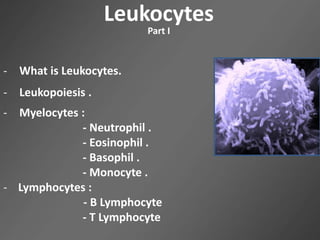
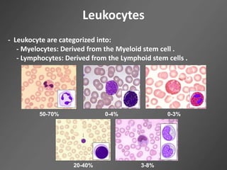
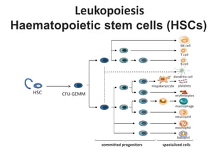
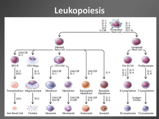
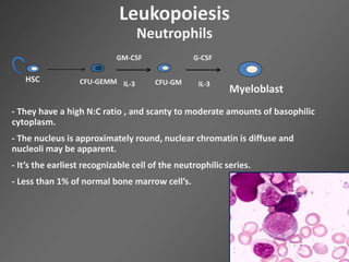
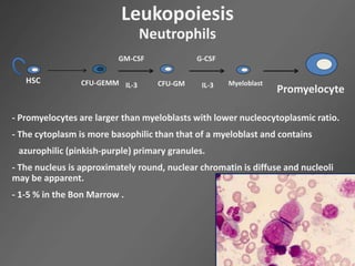
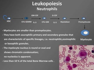
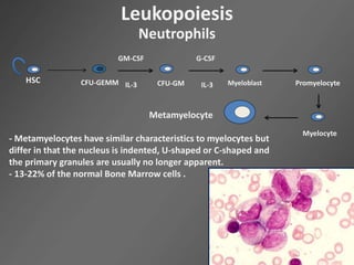
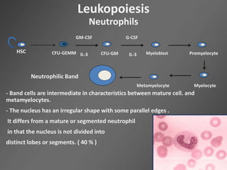
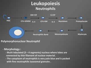
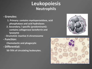
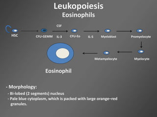
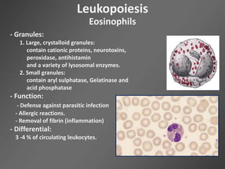
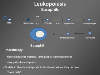
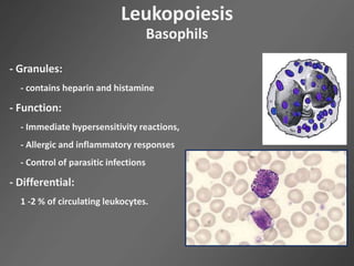
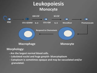
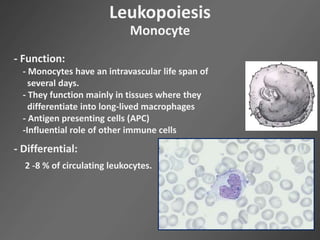
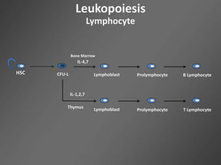
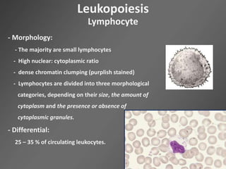
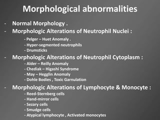
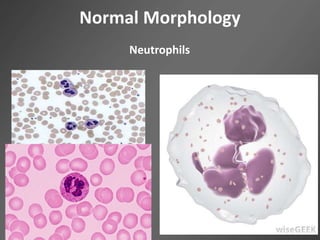
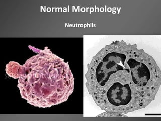
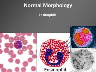
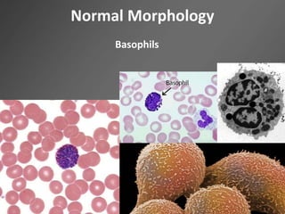
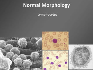
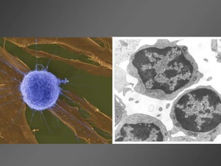
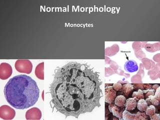
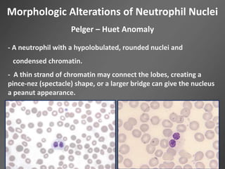
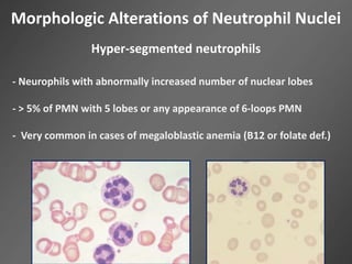
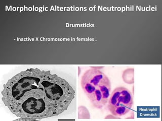
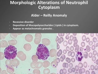
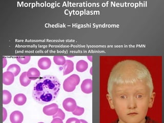
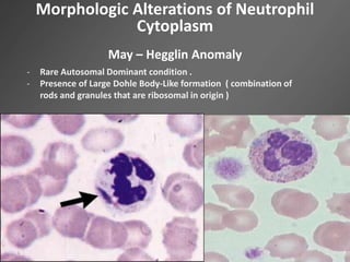
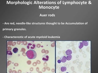
Ahmad A. Al-Qudah
LM751 - ADVANCED HEMATOLOGY
Leukocytes
Leukocytes
Part I
- What is Leukocytes.
- Leukopoiesis .
- Myelocytes :
- Neutrophil .
- Eosinophil .
- Basophil .
- Monocyte .
- Lymphocytes :
- B Lymphocyte
- T Lymphocyte
Leukocytes
- The second category of cellular blood elements
- Leukocytes ≈ white cells (Leuko: white / cyte: cell)
- Got their name from the white buffy coat obtained upon
centrifugation of whole blood.
- Leukocyte are categorized into:
- Myelocytes: Derived from the Myeloid stem cell .
- Lymphocytes: Derived from the Lymphoid stem cells .
Leukocytes
50-70% 0-4% 0-3%
20-40% 3-8%
Leukopoiesis
Haematopoietic stem cells (HSCs)
HSC
committed progenitors
neutrophil
NK cell
erythrocytes
dendritic cell
plateletsmegakaryocyte
macrophage
eosinophil
basophil
B cell
T cell
specialized cells
CFU-GEMM
Leukopoiesis
- Myelocytes are generated and maturate in the BM.
- These cells are divided into:
1. Granulocytes:
- Are granulated cells with poly or bi segmented nuclei
- Include three types of cells
A. Polymorphoneutrophile (PMN)
B. Basophile (Baso)
C. Eosinophile (Eos)
2. Monocytes: Large cells with an asegmented nucleus
Leukopoiesis
Myelocytes
Leukopoiesis
Neutrophils
HSC CFU-GEMM CFU-GM
GM-CSF
IL-3
G-CSF
IL-3
Myeloblast
- They have a high N:C ratio , and scanty to moderate amounts of basophilic
cytoplasm.
- The nucleus is approximately round, nuclear chromatin is diffuse and
nucleoli may be apparent.
- It’s the earliest recognizable cell of the neutrophilic series.
- Less than 1% of normal bone marrow cell’s.
Leukopoiesis
Neutrophils
HSC CFU-GEMM CFU-GM
GM-CSF
IL-3
G-CSF
IL-3 Myeloblast
Promyelocyte
- Promyelocytes are larger than myeloblasts with lower nucleocytoplasmic ratio.
- The cytoplasm is more basophilic than that of a myeloblast and contains
azurophilic (pinkish-purple) primary granules.
- The nucleus is approximately round, nuclear chromatin is diffuse and nucleoli
may be apparent.
- 1-5 % in the Bon Marrow .
Leukopoiesis
Neutrophils
HSC CFU-GEMM CFU-GM
GM-CSF
IL-3
G-CSF
IL-3 Myeloblast Promyelocyte
Myelocyte
- Myelocytes are smaller than promyelocytes.
- They have both azurophilic primary and secondary granules that
are characteristic of specific lineages, i.e. neutrophilic,eosinophilic
or basophilic granules.
- The myelocyte nucleus is round or oval and
shows chromatin condensation;
no nucleolus is apparent.
- Less than 10 % of the total Bone Marrow cells .
Leukopoiesis
Neutrophils
HSC CFU-GEMM CFU-GM
GM-CSF
IL-3
G-CSF
IL-3 Myeloblast Promyelocyte
Myelocyte
Metamyelocyte
- Metamyelocytes have similar characteristics to myelocytes but
differ in that the nucleus is indented, U-shaped or C-shaped and
the primary granules are usually no longer apparent.
- 13-22% of the normal Bone Marrow cells .
Leukopoiesis
Neutrophils
HSC CFU-GEMM CFU-GM
GM-CSF
IL-3
G-CSF
IL-3 Myeloblast Promyelocyte
MyelocyteMetamyelocyte
Neutrophilic Band
- Band cells are intermediate in characteristics between mature cell. and
metamyelocytes.
- The nucleus has an irregular shape with some parallel edges .
It differs from a mature or segmented neutrophil
in that the nucleus is not divided into
distinct lobes or segments. ( 40 % )
Leukopoiesis
Neutrophils
HSC CFU-GEMM CFU-GM
GM-CSF
IL-3
G-CSF
IL-3 Myeloblast Promyelocyte
MyelocyteMetamyelocyteNeutrophilic Band
Polymorphonuclear Neutrophil
- Morphology :
- Multi-lobulated (3 – 4 segments) nucleus where lobes are
connected by thin filament of nuclear material.
- The cytoplasm of neutrophils is very pale blue and is packed
with fine neutrophilic lysosomal granules .
Leukopoiesis
Neutrophils
- Granules:
1. Primary: contains myeloperoxidase, acid
phosphatase and acid hydrolases
2. Secondary / specific (predominant):
contains collagenase lactoferrin and
lysozyme
- Drumstick: inactive X-chromosome
- Function:
Chemotactic and phagocytic
- Differential:
50-70% of circulating leukocytes.
Leukopoiesis
Eosinophils
HSC CFU-GEMM CFU-Eo
CSF
IL-3 IL-5 Myeloblast Promyelocyte
MyelocyteMetamyelocyte
Eosinophil
- Morphology:
- Bi-lobed (2 segments) nucleus
- Pale blue cytoplasm, which is packed with large orange–red
granules.
Leukopoiesis
Eosinophils
- Granules:
1. Large, crystalloid granules:
contain cationic proteins, neurotoxins,
peroxidase, antihistamin
and a variety of lysosomal enzymes.
2. Small granules:
contain aryl sulphatase, Gelatinase and
acid phosphatase
- Function:
- Defense against parasitic infection
- Allergic reactions.
- Removal of fibrin (inflammation)
- Differential:
3 -4 % of circulating leukocytes.
Leukopoiesis
Basophils
HSC CFU-GEMM CFU-Bas
CSF
IL-3 IL-6 Myeloblast Promyelocyte
MyelocyteMetamyelocyte
Basophil
- Morphology:
- have a lobulated nucleus, large purple-staining granules ,
very pale blue cytoplasm.
- Cirulate in blood and migarate to the tissues where they become
“mast cells”.
- Granules:
- contains heparin and histamine
- Function:
- Immediate hypersensitivity reactions,
- Allergic and inflammatory responses
- Control of parasitic infections
- Differential:
1 -2 % of circulating leukocytes.
Leukopoiesis
Basophils
Leukopoiesis
Monocyte
HSC CFU-GEMM CFU-GM
GM-CSF
IL-3
M-CSF
IL-3 MonoBlast Promonocyte
MonocyteMacrophage
Respond to Chemotaxis
Morphology:
- Are the largest normal blood cells.
- Lobulated nuclei and huge greyish- bluecytoplasm
- Cytoplasm is sometimes opaque and may be vacuolated and/or
granulated.
Leukopoiesis
Monocyte
- Function:
- Monocytes have an intravascular life span of
several days.
- They function mainly in tissues where they
differentiate into long-lived macrophages
- Antigen presenting cells (APC)
-Influential role of other immune cells
- Differential:
2 -8 % of circulating leukocytes.
Leukopoiesis
Lymphocyte
HSC CFU-L
IL-4,7
ProlymphocyteLymphoblast B Lymphocyte
ProlymphocyteLymphoblast
IL-1,2,7
T Lymphocyte
Bone Marrow
Thymus
Leukopoiesis
Lymphocyte
- Morphology:
- The majority are small lymphocytes
- High nuclear: cytoplasmic ratio
- dense chromatin clumping (purplish stained)
- Lymphocytes are divided into three morphological
categories, depending on their size, the amount of
cytoplasm and the presence or absence of
cytoplasmic granules.
- Differential:
25 – 35 % of circulating leukocytes.
Leukopoiesis
Lymphocyte
- Function:
1. T lymphocytes:
- Recognition of foreign Ag’s on the context of MHC molecules on surfaces of APC
- Cytotoxic T cells (CD8+) : mediate the destruction of their targets
- T-helper cells (CD4+) : influence the innate and adaptive response
- Natural killer (NK) cells: CD8+ T cells express HLA receptor that are required to mediate
the killing of their targets.
2. B-lymphocytes: maturate in BM
Differentiate into antibody-producing cells (Plasma cells)
Part II
Leukocytes
Morphological abnormalities
- Normal Morphology .
- Morphologic Alterations of Neutrophil Nuclei :
- Pelger – Huet Anomaly .
- Hyper-segmented neutrophils
- Drumsticks
- Morphologic Alterations of Neutrophil Cytoplasm :
- Alder – Reilly Anomaly
- Chediak – Higashi Syndrome
- May – Hegglin Anomaly
- Dohle Bodies , Toxic Garnulation
- Morphologic Alterations of Lymphocyte & Monocyte :
- Reed-Sternberg cells
- Hand-mirror cells
- Sezary cells
- Smudge cells
- Atypical lymphocyte , Activated monocytes
Normal Morphology
Neutrophils
Normal Morphology
Neutrophils
Normal Morphology
Eosinophils
Normal Morphology
Basophils
Normal Morphology
Lymphocytes
Normal Morphology
Monocytes
Morphologic Alterations of Neutrophil Nuclei
Pelger – Huet Anomaly
- A neutrophil with a hypolobulated, rounded nuclei and
condensed chromatin.
- A thin strand of chromatin may connect the lobes, creating a
pince-nez (spectacle) shape, or a larger bridge can give the nucleus
a peanut appearance.
Morphologic Alterations of Neutrophil Nuclei
Hyper-segmented neutrophils
- Neurophils with abnormally increased number of nuclear lobes
- > 5% of PMN with 5 lobes or any appearance of 6-loops PMN
- Very common in cases of megaloblastic anemia (B12 or folate def.)
Morphologic Alterations of Neutrophil Nuclei
Drumsticks
- Inactive X Chromosome in females .
Morphologic Alterations of Neutrophil
Cytoplasm
Alder – Reilly Anomaly
- Recessive disorder
- Deposition of Mucopolysaccharides ( Lipids ) in cytoplasm.
- Appear as metachromatic granules .
Morphologic Alterations of Neutrophil
Cytoplasm
Chediak – Higashi Syndrome
- Rare Autosomal Recessive state .
- Abnormally large Peroxidase-Positive lysosomes are seen in the PMN
(and most cells of the body) results in Albinism.
Morphologic Alterations of Neutrophil
Cytoplasm
May – Hegglin Anomaly
- Rare Autosomal Dominant condition .
- Presence of Large Dohle Body-Like formation ( combination of
rods and granules that are ribosomal in origin )
Morphologic Alterations of Neutrophil
Cytoplasm
Dohle Bodies
- Small blue-gray (single or multiple) inclusions in the cytoplasm of
neutrophils, often at the margins (eccentric).
- Composed of rough endoplasmic reticulum and glycogen granules.
- Associated with inflammatory disorders, burns, MPD and MDS
Morphologic Alterations of Neutrophil
Cytoplasm
Toxic Garnulation
- Neutrophils that are characterized by an increased numbers of granules that
are larger and more basophilic than normal.
- May appear in severe bacterial infections, burns, malignancies,
drug reactions.
Morphologic Alterations of Lymphocyte &
Monocyte
Reed-Sternberg cells
The Mirror Nuclei
Morphologic Alterations of Lymphocyte &
Monocyte
Hand-mirror cells
- Characteristic “hand mirror” shape of T cells in a patient
with T-cell acute lymphocytic leukemia.
Morphologic Alterations of Lymphocyte &
Monocyte
Sezary cells
- Lymphocytes with frequently convoluted nuclei (Sezary cells) in a
patient with advanced mycosis fungoides.
Morphologic Alterations of Lymphocyte &
Monocyte
Smudge cells
- Fragile lymphocytes rupture (during film preparation)
- Nucleus appears spread out with hazy borders and absent
cytoplasm.
Morphologic Alterations of Lymphocyte &
Monocyte
Atypical lymphocyte
- Common in viral infections (e.g. Herpes infection and HIV)
- Large lymph with prominent foamy/vaculated cytoplasm and
irregular nucleus (kidney shaped or lobulated)
- Basophilic cytoplasm and coarse chromatin
Morphologic Alterations of Lymphocyte &
Monocyte
Activated monocytes
- Associated with inflammatory reaction to bacteremia
- Macrophages with increased granulation
Morphologic Alterations of Lymphocyte &
Monocyte
Auer rods
- Are red, needle-like structures thought to be Accumulation of
primary granules.
- Characterestic of acute myeloid leukemia
Part III
Leukocytes
Special stains
Special stains
- Myeloperoxidase ( MPO )
- Sudan Black B (SBB)
- Leukocyte alkaline phosphatase (LAP)
- Specific esterase
- Non-specific esterase
- Acid phosphatase
- Periodic acid schiff (PAS)
Myeloperoxidase ( MPO )
- Myeloperoxidase is present in the primary granules of neutrophils
and the secondary granules of eosinophils
- Principle: Benzidine or diaminobenzidine are converted (oxidized)
inside the granules into brownish precipitate.
- Interpretation:
- PMN’s/Eosinophils and the progenitors (from the
promyelocytic stage on) are positively stained.
- Monocytes lysosomal granulocytes are faintly positive
- Lymphocytes and NRBC’s lack the enzyme
- Purpose: To differentiate a myelogenous or monocytic leukemia
from acute lymphocytic Leukemia.
Myeloperoxidase ( MPO )
Leukemic myeloblasts stained with peroxidase
Myeloperoxidase ( MPO )
Acute lymphocytic leukemia stained with peroxidase, the blast cells are
unreactive (unstained) while the neutrophil is positively stained
Sudan Black B (SBB)
- SBB is a fat soluble stains that stains intracellular lipids as well as
phospholipids.
- Staining pattern is parallel to myeloperoxidase (MPO) staining
- SBB can be used to stain old blood or BM sample and the stain does
not faid with time (MP is sensitive to light; therefore fresh samples are
recommended/ enzymatic activity may diminish on samples older
than 3 weeks)
- Interpretation: Balck/grayish-black staining of the cytoplasm
Sudan Black B (SBB)
Sudan Black B (SBB)
Positive sudan black
B (SBB) stain in a
patient with AML.
Leukocyte alkaline phosphatase (LAP)
- AP activity is found in the cytoplasm of PMN’s, osteoclasts and
some lymphocytes
- Based on the determination of LAP score
- Differential test for CML from leukemoid reactions and other
MPD
- Sodium -naphtyl phsophate (or naphtol-AS-BI phosphate)is
used as a substrate to produce a bright red products
- Interpretation:
- Stain intensity is determined for 100 counted PMN or band
and scored from 0 - 4
- The sum of the scores reflects LAP score (index)
- Normal LAP score is 15 - 130
Leukocyte alkaline phosphatase (LAP)
- Interpretation:
- Low LAP score (<15)
- Chronic myeloid leukemia, PNH, Myelodysplastic
syndrom, rare infections or toxic exposure
- High LAB score (>130)
- Leukomoid reactions in response to infections
and MPD other than CML, inflammatory disorders ,
stress, certain drugs (including lithium, corticosteroids
and estrogen)
Leukocyte alkaline phosphatase (LAP)
Grading:
- (0) No stain
- (+1) Faint stain
- (+2) Moderate stain
- (+3) Strong stain
- (+4) Strong stain without cytoplasmic background
Positive LAP reactionNegative LAP reaction
Specific esterase
- Also called Leder stain
- Is used to identify cells of granulocytic series ONLY.
- Cellular esterase hydrolyze naphtol AS-D chloroacetate substrate
to produce a bright red (red-pink) product at the site of enzymatic
activity
- Neutrophilic granulocyte show a positive reaction from the
promyelocytes stage on
Specific esterase
Non-specific esterase
- This is performed using -naphtyl butyrate or -naphtyl
acetate as a substrate
- Stain positive for monocytic cells but not granulocyteic cells
- Mature T lymphocytes stain positively with a characterestic
focal dot-like pattern.
Acid phosphatase
- Acid phosphatase is found in all hematopoietic cells with the
highest levels in macrophages and osteoclast.
- A localized dot-like pattern is seen in many T-lymphoblasts.
- Tartar-resistance acid phosphatase (TRAP) is an isoenzyme that is
found at high levels in cells of hairy cell leukemia.
- To test for TRAP: In the presence of tartaric acid, Naphtol-AS-BI
phosphate coupled to fast garent GBC salt are used to produce a
bright red homogenous or granular precipitate in the cytoplasm of
cells with enzymatic activity (if a resistance isoenzyme is present)
Acid phosphatase
Periodic acid schiff (PAS)
- Detects intracellular glycogen and neutral mucopolysaccharides
that are found in the majority of hematopoietic cells at variable
quantities.
- Detection is based on the oxidation of -glycols in carbohydrates
and CHO-containing compounds resulting in the formation of
polyaldehyde that can be detected by schiff reagent.
- Products of the staining is a diffuse red stain or pink-to-red
granules or even clumps of varying size.
Periodic acid schiff (PAS)
Marrow film stained with periodic acid Schiff reagent. Intense PAS-positive
staining of leukemic erythroblasts (Acute erythroid leukemia)
Special stains
Blasts Identified
Cellular Element
Stained
Cytochemical Reaction
Myeloblasts strong positive;
monoblasts faint positive
Neutrophil primary
granules
Myeloperoxidase (MPO)
Myeloblasts strong positive;
monoblasts faint positive
PhospholipidsSudan Black B (SBB)
Myeloblasts strong positiveCellular enzymeSpecific esterase
Monoblasts strong positiveCellular enzyme
Nonspecific esterase
(NSE)
Variable, coarse or block-like positivity
often seen in
lymphoblasts and pronormoblasts,
myeloblasts usually
negative although faint diffuse
reaction may
occasionally be seen
Glycogen and
related substances
Periodic acid-Schiff
Leukocytes - Advance Hematology

Leukocytes - Advance Hematology

  • 2.
    Ahmad A. Al-Qudah LM751- ADVANCED HEMATOLOGY Leukocytes
  • 3.
    Leukocytes Part I - Whatis Leukocytes. - Leukopoiesis . - Myelocytes : - Neutrophil . - Eosinophil . - Basophil . - Monocyte . - Lymphocytes : - B Lymphocyte - T Lymphocyte
  • 4.
    Leukocytes - The secondcategory of cellular blood elements - Leukocytes ≈ white cells (Leuko: white / cyte: cell) - Got their name from the white buffy coat obtained upon centrifugation of whole blood.
  • 6.
    - Leukocyte arecategorized into: - Myelocytes: Derived from the Myeloid stem cell . - Lymphocytes: Derived from the Lymphoid stem cells . Leukocytes 50-70% 0-4% 0-3% 20-40% 3-8%
  • 7.
    Leukopoiesis Haematopoietic stem cells(HSCs) HSC committed progenitors neutrophil NK cell erythrocytes dendritic cell plateletsmegakaryocyte macrophage eosinophil basophil B cell T cell specialized cells CFU-GEMM
  • 8.
  • 9.
    - Myelocytes aregenerated and maturate in the BM. - These cells are divided into: 1. Granulocytes: - Are granulated cells with poly or bi segmented nuclei - Include three types of cells A. Polymorphoneutrophile (PMN) B. Basophile (Baso) C. Eosinophile (Eos) 2. Monocytes: Large cells with an asegmented nucleus Leukopoiesis Myelocytes
  • 10.
    Leukopoiesis Neutrophils HSC CFU-GEMM CFU-GM GM-CSF IL-3 G-CSF IL-3 Myeloblast -They have a high N:C ratio , and scanty to moderate amounts of basophilic cytoplasm. - The nucleus is approximately round, nuclear chromatin is diffuse and nucleoli may be apparent. - It’s the earliest recognizable cell of the neutrophilic series. - Less than 1% of normal bone marrow cell’s.
  • 11.
    Leukopoiesis Neutrophils HSC CFU-GEMM CFU-GM GM-CSF IL-3 G-CSF IL-3Myeloblast Promyelocyte - Promyelocytes are larger than myeloblasts with lower nucleocytoplasmic ratio. - The cytoplasm is more basophilic than that of a myeloblast and contains azurophilic (pinkish-purple) primary granules. - The nucleus is approximately round, nuclear chromatin is diffuse and nucleoli may be apparent. - 1-5 % in the Bon Marrow .
  • 12.
    Leukopoiesis Neutrophils HSC CFU-GEMM CFU-GM GM-CSF IL-3 G-CSF IL-3Myeloblast Promyelocyte Myelocyte - Myelocytes are smaller than promyelocytes. - They have both azurophilic primary and secondary granules that are characteristic of specific lineages, i.e. neutrophilic,eosinophilic or basophilic granules. - The myelocyte nucleus is round or oval and shows chromatin condensation; no nucleolus is apparent. - Less than 10 % of the total Bone Marrow cells .
  • 13.
    Leukopoiesis Neutrophils HSC CFU-GEMM CFU-GM GM-CSF IL-3 G-CSF IL-3Myeloblast Promyelocyte Myelocyte Metamyelocyte - Metamyelocytes have similar characteristics to myelocytes but differ in that the nucleus is indented, U-shaped or C-shaped and the primary granules are usually no longer apparent. - 13-22% of the normal Bone Marrow cells .
  • 14.
    Leukopoiesis Neutrophils HSC CFU-GEMM CFU-GM GM-CSF IL-3 G-CSF IL-3Myeloblast Promyelocyte MyelocyteMetamyelocyte Neutrophilic Band - Band cells are intermediate in characteristics between mature cell. and metamyelocytes. - The nucleus has an irregular shape with some parallel edges . It differs from a mature or segmented neutrophil in that the nucleus is not divided into distinct lobes or segments. ( 40 % )
  • 15.
    Leukopoiesis Neutrophils HSC CFU-GEMM CFU-GM GM-CSF IL-3 G-CSF IL-3Myeloblast Promyelocyte MyelocyteMetamyelocyteNeutrophilic Band Polymorphonuclear Neutrophil - Morphology : - Multi-lobulated (3 – 4 segments) nucleus where lobes are connected by thin filament of nuclear material. - The cytoplasm of neutrophils is very pale blue and is packed with fine neutrophilic lysosomal granules .
  • 16.
    Leukopoiesis Neutrophils - Granules: 1. Primary:contains myeloperoxidase, acid phosphatase and acid hydrolases 2. Secondary / specific (predominant): contains collagenase lactoferrin and lysozyme - Drumstick: inactive X-chromosome - Function: Chemotactic and phagocytic - Differential: 50-70% of circulating leukocytes.
  • 17.
    Leukopoiesis Eosinophils HSC CFU-GEMM CFU-Eo CSF IL-3IL-5 Myeloblast Promyelocyte MyelocyteMetamyelocyte Eosinophil - Morphology: - Bi-lobed (2 segments) nucleus - Pale blue cytoplasm, which is packed with large orange–red granules.
  • 18.
    Leukopoiesis Eosinophils - Granules: 1. Large,crystalloid granules: contain cationic proteins, neurotoxins, peroxidase, antihistamin and a variety of lysosomal enzymes. 2. Small granules: contain aryl sulphatase, Gelatinase and acid phosphatase - Function: - Defense against parasitic infection - Allergic reactions. - Removal of fibrin (inflammation) - Differential: 3 -4 % of circulating leukocytes.
  • 19.
    Leukopoiesis Basophils HSC CFU-GEMM CFU-Bas CSF IL-3IL-6 Myeloblast Promyelocyte MyelocyteMetamyelocyte Basophil - Morphology: - have a lobulated nucleus, large purple-staining granules , very pale blue cytoplasm. - Cirulate in blood and migarate to the tissues where they become “mast cells”.
  • 20.
    - Granules: - containsheparin and histamine - Function: - Immediate hypersensitivity reactions, - Allergic and inflammatory responses - Control of parasitic infections - Differential: 1 -2 % of circulating leukocytes. Leukopoiesis Basophils
  • 21.
    Leukopoiesis Monocyte HSC CFU-GEMM CFU-GM GM-CSF IL-3 M-CSF IL-3MonoBlast Promonocyte MonocyteMacrophage Respond to Chemotaxis Morphology: - Are the largest normal blood cells. - Lobulated nuclei and huge greyish- bluecytoplasm - Cytoplasm is sometimes opaque and may be vacuolated and/or granulated.
  • 22.
    Leukopoiesis Monocyte - Function: - Monocyteshave an intravascular life span of several days. - They function mainly in tissues where they differentiate into long-lived macrophages - Antigen presenting cells (APC) -Influential role of other immune cells - Differential: 2 -8 % of circulating leukocytes.
  • 23.
    Leukopoiesis Lymphocyte HSC CFU-L IL-4,7 ProlymphocyteLymphoblast BLymphocyte ProlymphocyteLymphoblast IL-1,2,7 T Lymphocyte Bone Marrow Thymus
  • 24.
    Leukopoiesis Lymphocyte - Morphology: - Themajority are small lymphocytes - High nuclear: cytoplasmic ratio - dense chromatin clumping (purplish stained) - Lymphocytes are divided into three morphological categories, depending on their size, the amount of cytoplasm and the presence or absence of cytoplasmic granules. - Differential: 25 – 35 % of circulating leukocytes.
  • 25.
    Leukopoiesis Lymphocyte - Function: 1. Tlymphocytes: - Recognition of foreign Ag’s on the context of MHC molecules on surfaces of APC - Cytotoxic T cells (CD8+) : mediate the destruction of their targets - T-helper cells (CD4+) : influence the innate and adaptive response - Natural killer (NK) cells: CD8+ T cells express HLA receptor that are required to mediate the killing of their targets. 2. B-lymphocytes: maturate in BM Differentiate into antibody-producing cells (Plasma cells)
  • 29.
  • 30.
    Morphological abnormalities - NormalMorphology . - Morphologic Alterations of Neutrophil Nuclei : - Pelger – Huet Anomaly . - Hyper-segmented neutrophils - Drumsticks - Morphologic Alterations of Neutrophil Cytoplasm : - Alder – Reilly Anomaly - Chediak – Higashi Syndrome - May – Hegglin Anomaly - Dohle Bodies , Toxic Garnulation - Morphologic Alterations of Lymphocyte & Monocyte : - Reed-Sternberg cells - Hand-mirror cells - Sezary cells - Smudge cells - Atypical lymphocyte , Activated monocytes
  • 31.
  • 32.
  • 33.
  • 34.
  • 35.
  • 37.
  • 38.
    Morphologic Alterations ofNeutrophil Nuclei Pelger – Huet Anomaly - A neutrophil with a hypolobulated, rounded nuclei and condensed chromatin. - A thin strand of chromatin may connect the lobes, creating a pince-nez (spectacle) shape, or a larger bridge can give the nucleus a peanut appearance.
  • 39.
    Morphologic Alterations ofNeutrophil Nuclei Hyper-segmented neutrophils - Neurophils with abnormally increased number of nuclear lobes - > 5% of PMN with 5 lobes or any appearance of 6-loops PMN - Very common in cases of megaloblastic anemia (B12 or folate def.)
  • 40.
    Morphologic Alterations ofNeutrophil Nuclei Drumsticks - Inactive X Chromosome in females .
  • 41.
    Morphologic Alterations ofNeutrophil Cytoplasm Alder – Reilly Anomaly - Recessive disorder - Deposition of Mucopolysaccharides ( Lipids ) in cytoplasm. - Appear as metachromatic granules .
  • 42.
    Morphologic Alterations ofNeutrophil Cytoplasm Chediak – Higashi Syndrome - Rare Autosomal Recessive state . - Abnormally large Peroxidase-Positive lysosomes are seen in the PMN (and most cells of the body) results in Albinism.
  • 43.
    Morphologic Alterations ofNeutrophil Cytoplasm May – Hegglin Anomaly - Rare Autosomal Dominant condition . - Presence of Large Dohle Body-Like formation ( combination of rods and granules that are ribosomal in origin )
  • 44.
    Morphologic Alterations ofNeutrophil Cytoplasm Dohle Bodies - Small blue-gray (single or multiple) inclusions in the cytoplasm of neutrophils, often at the margins (eccentric). - Composed of rough endoplasmic reticulum and glycogen granules. - Associated with inflammatory disorders, burns, MPD and MDS
  • 45.
    Morphologic Alterations ofNeutrophil Cytoplasm Toxic Garnulation - Neutrophils that are characterized by an increased numbers of granules that are larger and more basophilic than normal. - May appear in severe bacterial infections, burns, malignancies, drug reactions.
  • 46.
    Morphologic Alterations ofLymphocyte & Monocyte Reed-Sternberg cells The Mirror Nuclei
  • 47.
    Morphologic Alterations ofLymphocyte & Monocyte Hand-mirror cells - Characteristic “hand mirror” shape of T cells in a patient with T-cell acute lymphocytic leukemia.
  • 48.
    Morphologic Alterations ofLymphocyte & Monocyte Sezary cells - Lymphocytes with frequently convoluted nuclei (Sezary cells) in a patient with advanced mycosis fungoides.
  • 49.
    Morphologic Alterations ofLymphocyte & Monocyte Smudge cells - Fragile lymphocytes rupture (during film preparation) - Nucleus appears spread out with hazy borders and absent cytoplasm.
  • 50.
    Morphologic Alterations ofLymphocyte & Monocyte Atypical lymphocyte - Common in viral infections (e.g. Herpes infection and HIV) - Large lymph with prominent foamy/vaculated cytoplasm and irregular nucleus (kidney shaped or lobulated) - Basophilic cytoplasm and coarse chromatin
  • 51.
    Morphologic Alterations ofLymphocyte & Monocyte Activated monocytes - Associated with inflammatory reaction to bacteremia - Macrophages with increased granulation
  • 52.
    Morphologic Alterations ofLymphocyte & Monocyte Auer rods - Are red, needle-like structures thought to be Accumulation of primary granules. - Characterestic of acute myeloid leukemia
  • 54.
  • 55.
    Special stains - Myeloperoxidase( MPO ) - Sudan Black B (SBB) - Leukocyte alkaline phosphatase (LAP) - Specific esterase - Non-specific esterase - Acid phosphatase - Periodic acid schiff (PAS)
  • 56.
    Myeloperoxidase ( MPO) - Myeloperoxidase is present in the primary granules of neutrophils and the secondary granules of eosinophils - Principle: Benzidine or diaminobenzidine are converted (oxidized) inside the granules into brownish precipitate. - Interpretation: - PMN’s/Eosinophils and the progenitors (from the promyelocytic stage on) are positively stained. - Monocytes lysosomal granulocytes are faintly positive - Lymphocytes and NRBC’s lack the enzyme - Purpose: To differentiate a myelogenous or monocytic leukemia from acute lymphocytic Leukemia.
  • 57.
    Myeloperoxidase ( MPO) Leukemic myeloblasts stained with peroxidase
  • 58.
    Myeloperoxidase ( MPO) Acute lymphocytic leukemia stained with peroxidase, the blast cells are unreactive (unstained) while the neutrophil is positively stained
  • 59.
    Sudan Black B(SBB) - SBB is a fat soluble stains that stains intracellular lipids as well as phospholipids. - Staining pattern is parallel to myeloperoxidase (MPO) staining - SBB can be used to stain old blood or BM sample and the stain does not faid with time (MP is sensitive to light; therefore fresh samples are recommended/ enzymatic activity may diminish on samples older than 3 weeks) - Interpretation: Balck/grayish-black staining of the cytoplasm
  • 60.
  • 61.
    Sudan Black B(SBB) Positive sudan black B (SBB) stain in a patient with AML.
  • 62.
    Leukocyte alkaline phosphatase(LAP) - AP activity is found in the cytoplasm of PMN’s, osteoclasts and some lymphocytes - Based on the determination of LAP score - Differential test for CML from leukemoid reactions and other MPD - Sodium -naphtyl phsophate (or naphtol-AS-BI phosphate)is used as a substrate to produce a bright red products - Interpretation: - Stain intensity is determined for 100 counted PMN or band and scored from 0 - 4 - The sum of the scores reflects LAP score (index) - Normal LAP score is 15 - 130
  • 63.
    Leukocyte alkaline phosphatase(LAP) - Interpretation: - Low LAP score (<15) - Chronic myeloid leukemia, PNH, Myelodysplastic syndrom, rare infections or toxic exposure - High LAB score (>130) - Leukomoid reactions in response to infections and MPD other than CML, inflammatory disorders , stress, certain drugs (including lithium, corticosteroids and estrogen)
  • 64.
    Leukocyte alkaline phosphatase(LAP) Grading: - (0) No stain - (+1) Faint stain - (+2) Moderate stain - (+3) Strong stain - (+4) Strong stain without cytoplasmic background Positive LAP reactionNegative LAP reaction
  • 65.
    Specific esterase - Alsocalled Leder stain - Is used to identify cells of granulocytic series ONLY. - Cellular esterase hydrolyze naphtol AS-D chloroacetate substrate to produce a bright red (red-pink) product at the site of enzymatic activity - Neutrophilic granulocyte show a positive reaction from the promyelocytes stage on
  • 66.
  • 67.
    Non-specific esterase - Thisis performed using -naphtyl butyrate or -naphtyl acetate as a substrate - Stain positive for monocytic cells but not granulocyteic cells - Mature T lymphocytes stain positively with a characterestic focal dot-like pattern.
  • 68.
    Acid phosphatase - Acidphosphatase is found in all hematopoietic cells with the highest levels in macrophages and osteoclast. - A localized dot-like pattern is seen in many T-lymphoblasts. - Tartar-resistance acid phosphatase (TRAP) is an isoenzyme that is found at high levels in cells of hairy cell leukemia. - To test for TRAP: In the presence of tartaric acid, Naphtol-AS-BI phosphate coupled to fast garent GBC salt are used to produce a bright red homogenous or granular precipitate in the cytoplasm of cells with enzymatic activity (if a resistance isoenzyme is present)
  • 69.
  • 70.
    Periodic acid schiff(PAS) - Detects intracellular glycogen and neutral mucopolysaccharides that are found in the majority of hematopoietic cells at variable quantities. - Detection is based on the oxidation of -glycols in carbohydrates and CHO-containing compounds resulting in the formation of polyaldehyde that can be detected by schiff reagent. - Products of the staining is a diffuse red stain or pink-to-red granules or even clumps of varying size.
  • 71.
  • 72.
    Periodic acid schiff(PAS) Marrow film stained with periodic acid Schiff reagent. Intense PAS-positive staining of leukemic erythroblasts (Acute erythroid leukemia)
  • 73.
    Special stains Blasts Identified CellularElement Stained Cytochemical Reaction Myeloblasts strong positive; monoblasts faint positive Neutrophil primary granules Myeloperoxidase (MPO) Myeloblasts strong positive; monoblasts faint positive PhospholipidsSudan Black B (SBB) Myeloblasts strong positiveCellular enzymeSpecific esterase Monoblasts strong positiveCellular enzyme Nonspecific esterase (NSE) Variable, coarse or block-like positivity often seen in lymphoblasts and pronormoblasts, myeloblasts usually negative although faint diffuse reaction may occasionally be seen Glycogen and related substances Periodic acid-Schiff