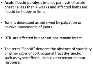
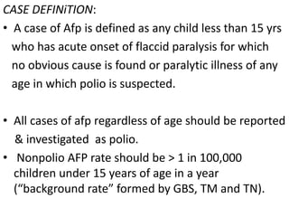
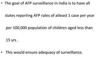
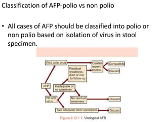
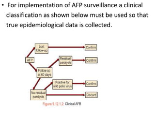
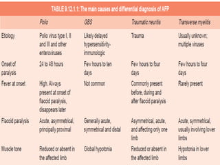
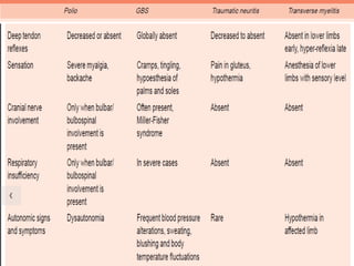
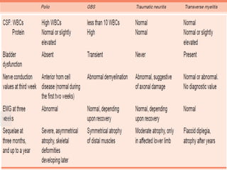
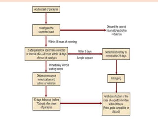
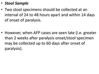
ACUTE FLACCID PARALYSIS
By
Dr subash arun. B
• Acute flaccid paralysis implies paralysis of acute
onset i.e less than 4 weeks and affected limbs are
flaccid i.e floppy or limp.
• Tone is decreased as observed by palpation or
passive movements of joints.
• DTR are affected but sensations remain intact.
• The term “flaccid” denotes the absence of spasticity
or other signs of corticospinal tract dysfunction
such as hyperreflexia, clonus or extensor plantar
response.
CASE DEFINiTION:
• A case of Afp is defined as any child less than 15 yrs
who has acute onset of flaccid paralysis for which
no obvious cause is found or paralytic illness of any
age in which polio is suspected.
• All cases of afp regardless of age should be reported
& investigated as polio.
• Nonpolio AFP rate should be > 1 in 100,000
children under 15 years of age in a year
(“background rate” formed by GBS, TM and TN).
• The goal of AFP surveillance in india is to have all
states reporting AFP rates of atleast 1 case per year
per 100,000 population of children aged less than
15 yrs .
• This would ensure adequacy of surveillance.
Classification of AFP-polio vs non polio
• All cases of AFP should be classified into polio or
non polio based on isolation of virus in stool
specimen.
• For implementation of AFP surveillance a clinical
classification as shown below must be used so that
true epidemiological data is collected.
The global polio eradication initiative was launched
in1988 using opv as the eradication tool and
employing 4 pronged strategy comprising:
• 1,maintaining high routine immunisation coverage.
• 2,supplementary immunisation activities/ppi
• 3,acute flaccid paralysis surveillance
• 4,outbreak response immunisation
• main causes of AFP include:
poliomyelitis, Guillain-Barré syndrome, transverse
myelitis, and traumatic neuritis
Conditions to be considered in a patient with AFP
• 1. Motor neuron
• A. Poliovirus
• B. Other neurotropic viruses
• 2. Peripheral nerves
• A. Acute Guillain-Barre syndrome (GBS)
• B. Porphyria
• C. Toxic neuropathies
Arsenic
Nitrofurantoin
Heavy metals
Thallium
• D. Diphtheria
• E. Collagen disorders
3. Neuromuscular junction (NMJ) disorders
A. Myasthenia gravis
B. Drug-induced neuromuscular blockade
C. Organophosphorus poisoning
D. Botulism
E. Animal venoms and toxins (snake bite)
F. Hypermagnesemia
• 4. Muscle
• A. Periodic paralysis
• B. Hypokalemia
• C. Rhabdomyolysis
• D. Inflammatory myopathies
• 5. Critical illness neuropathy and myopathy
• 6. Acute myelopathy
A. Spinal cord infection
Viruses and retroviruses
Rabies
Herpes simplex virus-2
HIV
Bacteria
Tuberculosis
Brucellosis
Mycoplasma
Borellia
Parasitic
Neurocysticercosis
Hydatid disease
Fungi
• B. Compressive myelopathy
Trauma
Epidural abscess
Hematomyelia
• C. Spinal cord infarction
• D. Tumors
• E. Idiopathic transverse myelitis (post or para-
infectious
• 7. Others
Cerebral venous thrombosis
Acute stroke
Hypoxic-ischemic events
Brainstem/posterior fossa tumors
Mitochondrial disorders
• Other Differential Diagnosis
Non-Polio Enteroviruses
Non-polio enteroviruses are known to cause AFP.
They are coxsackie A virus, coxsackie B virus and
Echo virus, enteroviruses types 70 and 71, and
mumps.
Complete recovery occurs in most of the cases.
In some cases, sequelae may resemble paralysis
caused by wild poliovirus.
Hypokalemia
History of diarrhea and vomiting for few days prior
to onset of paralysis.
Toxic, irritable.
Weakness affects the limbs first, followed by trunk
and respiratory muscles.
Neckdrop is a usual feature.
Death occurs due to respiratory muscle failure and
cardiac arrest
Treatment—Intravenous potassium
Pseudoparalysis
Nonspecific toxic synovitis
Low-grade fever for several days
Unilateral limp
Hip or knee joints are commonly affected
Swelling of the joint, accompanied with painful
movements.
• Scurvy
Age group: 6 months to 2 years
Gradual onset
History of irritability, generalized tenderness
Frog like position of legs, due to pain Spongy swelling
of gums
X-ray knee is characteristic (white line, pencil line
cortex, zone of degeneration
Congenital Syphilitic Osteochondritis:
Found in early infancy.
Osteochondritis is painful, causing pseudoparalysis.
Diagnosis is by X-ray wrist, elbow, knee, ankle,
showing features of osteochondritis
• SURVEILLANCE OF AFP
Surveillance means data collection for action.
“Surveillance is carried out for all cases of acute
flaccid paralysis, not just for poliomyelitis
The surveillance data collected and documented must
support the principle that, “if polio cases had
occurred, they would have been detected, reported
and investigated in an expeditious manner.”
The surveillance is critical for achieving the goal of
polio eradication, which is defined as the absenceof
clinical cases of poliomyelitis for at least 3 years and
the absence of wild type polio virus in the community.
• Stool Sample
• Two stool specimens should be collected at an
interval of 24 to 48 hours apart and within 14 days
of onset of paralysis.
• However, when AFP cases are seen late (i.e. greater
than 2 weeks after paralysis onset/stool specimen
may be collected up to 60 days after onset of
paralysis).
The specimen should be placed in a clean container
such as wide mouth plastic or glass bottle with
screw on cap.
It need not be autoclaved, but should be cleaned.
At least ‘one thumb sized’ 8 gm of stool is required.
Stool sample should be adequate and in good
condition accompanied by all details as required by
laboratories
• The Ministry of Health has sponsored the formation of a
network of laboratories some of which are located at:
Sanjay Gandhi Postgraduate Institute, Lucknow
National Institute of Communicable Diseases, New Delhi
ERC, Mumbai
NIV, Bengaluru
BJMC, Ahmedabad
Enterovirus Research Institute, Kasauli
Pasteur Institute of India, Coonoor
• Stool samples should be stored in the refrigerator or
any container that can maintain a temperature
below 8ºC, till they are reached to the laboratory.
• A vaccine carrier with frozen ice packs is used for
this purpose.
• This vaccine carrier should never be used to
transport vaccine.
• Throughout transport, specimen must be packed to
maintain temperature below 8ºC.
• Indicators of Quality:
1. Nonpolio AFP rate > 1 in 100,000 children under
15 years (annual).
2. At least 80% of AFP cases whose two adequate
stool samples have been examined.
Outbreak Response Efforts:
• These should be initiated promptly without waiting
for the stool culture reports which may take 4 to 8
weeks.
• All cases labeled as “discarded, not polio” require
thorough justification and should be reported with
final diagnosis
Reverse Cold Chain:
• Stool samples (two, at least 24 hrs apart, within 14
days of onset of paralysis) from each AFP case are
obtained and transported to the laboratory within
72 hours of collection (at 4-8oC or frozen at minus
20°C)for virological identification and, if found to be
poliovirus, for finding whether it is a natural wild
virus or vaccine-related virus.
Action
Once a case of AFP has been reported, the following
measures are to be taken:
1, All reported cases should be investigated by
District Immunization Officer (DIO) as per the case
investigation form, within 48 hours after notification
by a reporting unit .
• A reporting unit means a center where paralytic
cases are brought for diagnosis, treatment
&rehabilitation.
• The DIO should send the copy to the state
headquarter.
2, The District Immunization Officer should not wait
for laboratory result to conduct immunization.
• Thedecision should be made on the spot, regardless
of whether the laboratory results are available or
not.
• The DIO should initiate appropriate action for
outbreak response immunization as and when
indicated
• The DIO must revisit every case of AFP 60 days after
the onset of paralysis, to confirm the presence or
absence of residual paralaysis.
• The minimum level of residual weakness can
usually be detected by the measurements of arm
or midthigh circumference as well as by skinfolds
on medial aspect of the thighs
4,The DIO should take part in active surveillance by
regularly visiting the reporting units and private
practitioners.
• All health workers, Aanganwadi workers, traditional
birth attendants must be encouraged to report AFP
cases immediately to the nearest primary health
center.
• The medical officer must in turn immediately
inform the DIO.
• 5. To strengthen the surveillance system, a
Surveillance Medical Officer (SMO) has been
appointed under the National Polio Surveillance
Project whose main task is to supervise the work of
polio related activities in that district and to act in
accordance with the directions of project from time to
time.
• Medical college hospital, specialized pediatric
hospital, district hospital have been named as
reporting unit (RU).
• Very popular pediatrician and quack are made
informers.
• All health service providers (to whom any of the
AFP case have visited in the past 2 years) are part of
network.
• Mopping up: denotes the final strategy when door
to door polio immunization is provided in high risk
districts i.e. where polio cases are detected in the
preceding 3 years.
AFP surveillance is a prime strategy for monitoring the
progress of polio eradication and is a sensitive
instrument for detecting potential poliomyelitis cases
and poliovirus infection.
Role of AFP surveillance:
• To identify high-risk areas or groups where polio
virus transmission is occurring or is likely to occur;
• To monitor progress so as to determine whether
strategies are implemented effectively.
• To certify a country poliofree when no reports of
new cases of polio are received for 3 consecutive years.
• CLINICAL APPROACH
• Most cases of AFP with an intact sensorium result
from disorders that affect the lower motor neuron,
namely, the anterior horn cells, the axons, the
neuromuscular junction, or the muscle.
• If the acute quadriparesis is associated with
alteration of sensorium or cranial nerve
involvement, the evaluation should include an
urgent contrast MRI scan of the brain.
• If the child is alert and upper motor neuron signs or
bowel-bladder involvements exist, an MRI scan of
the cervical and thoracic spine is the investigation
of choice
• The presence of a sensory level, spinal tenderness,
bowel-bladder involvement, even though the tone
and reflexes are reduced or absent, makes a spinal
etiology likely (transverse myelitis, epidural abscess,
metastasis, tuberculosis, spinal cord ischemia from a
dural arteriovenous fistula or other vascular
anomaly).
• A gadolinium enhanced MRI of the cervical and
thoracic spine should be performed.
• It is important to remember that it may take up to
10-14 hours for ischemia to manifest as changes on
the MRI.
• In adults where diagnosis of site of pathology of the
lower motor neuron may be relatively easy,
diagnosis in the child is challenging.
• Clinical observation of the child including his
posture, head control, breathing, speech, difficulty
in swallowing, inability to turn in bed, reach for his
favorite toys or bear weight on his legs afford clues
to the state and stage of illness.
• familial potassium-associated periodic paralysis,
present in a “hyperacute” fashion with weakness
developing over minutes or hours..
• Clues to the Diagnosis
The following are important diagnostic clues:
Pace: Weakness from motor neuron or peripheral
nerve disorders develops over days while the
evolution of symptoms from neuromuscular junction
disorders and periodic paralysis may be marked in
minutes or days.
2. Past or family history: Previous episodes of
weakness or a positive family history should suggest
periodic paralysis, porphyria, rhabdomyolysis or a
metabolic myopathy.
History of vaccination or immunization may precede
Guillian-Barré syndrome (GBS), transverse myelitis
or acute demyelinating encephalomyelitis (ADEM).
A history of drug ingestion including drugs used in
malignancies should be enquired into, to detect
toxic neuropathies
3, Gastrointestinal symptoms: These accompany
porphyria, botulism, sea food poisoning,
organophosphorus toxicity, and arsenic or thallium
ingestion.
• Acute hypokalemic weakness may follow chronic
vomiting or diarrhea
4. Pain: The onset of weakness from GBS, polio,
porphyria or dermatomyositis may be preceded by
neck pain, back pain or myalgias.
• Spinal pain may result from a caries spine,
metastasis or compressive myelopathy.
• Distal paresthesias or dysesthesias may occur with
GBS or toxic neuropathies.
5. Pattern of limb weakness: Polio and occasionally
porphyria are distinguished by asymmetric limb
weakness.
• Distal weakness is a feature of peripheral
neuropathies.
6. Bulbar dysfunction: This is prominent in
neuromuscular junction disorders and may be seen
in GBS, porphyria, polio and diphtheria
7. Ptosis or ophthalmoplegia: Should suggest a
neuromuscular junction disorder, though it may be
seen in GBS, Miller Fisher variant or brainstem
pathologies like infarction or central pontine
myelinolysis, etc.
8. Pupillary changes: Pupillary paralysis is a classic
feature of botulism but is not invariably present.
Paralysis of accommodation occurs with diphtheria
and miosis with organophosphorus poisoning.
Patterns of Weakness
1. Flaccid symmetric weakness with areflexia (+/-
bulbar and respiratory involvement),
• with sensory symptoms and minimal sensory loss:
GBS.
• without sensory symptoms or signs: periodic
paralysis.
• Symmetric proximal weakness with preserved
reflexes:
Acute myopathy (PM-DM);
osteomalacic myopathy
• Flaccid paraplegia or quadriplegia with sensory
level, and bowel-bladder dysfunction: Spinal cord
pathology
• Opthalmoplegia with motor weakness:
GBS; Miller-Fisher variant;
Locked-in-state;
Myasthenia gravis;
Tick paralysis
• Fatigable muscle weakness with bulbar signs/
opthalmoplegia: Myasthenia gravis
• Management:
• The two most important goals in the management
of
a child with AFP are:
1. Stabilization of the patient and attending to
ventilatory needs.
2. History and physical examination to diagnose the
disease, order appropriate tests, and institute specific
therapy.
Whether the child requires ICU admission will
depend on the presence of:
(i) vasomotor and
respiratory insufficiency;
(ii) rapid progression of
disease;
(iii) bulbar symptoms;
(iv) suspected poisoning,
(v) acute severe systemic illness
• Laboratory Investigations
• The following investigations can prove useful for
establishing a diagnosis:
• Complete blood counts: Anemia and leukocytosis as
possible markers of systemic illness.
• Eosinophil count: May be elevated in vasculitic
neuropathy, trichinosis and cysticercosis.
• ESR: Often elevated in infections and autoimmune
disorders.
.
• CPK, aldolase: Elevated in primary muscle diseases.
• BUN: Elevated in myoglobinuria, rhabdomyolysis
and renal insufficiency
• Electrolytes, calcium, phosphorus, and magnesium:
For myopathic disorders and periodic paralysis.
• Thyroid hormone assay: For thyroid related
myopathy.
• Collagen markers: Vasculitic neuropathy,
polymyositis, myasthenia gravis.
• Nerve conduction studies, EMG, repetitive
stimulation:To diagnose GBS, myopathic disorders,
myasthenia gravis.
• MRI/CT scans: Brain, spinal cord.
• Lumbar puncture: Transverse myelitis, TB
arachnoiditis, GBS.
• BIBILOGRAPHY:
1,The short textbook of pediatrics-11th edition.
2,IAP textbook of pediatrics.
3,OP Ghai -8th edition.

Acute flaccid paralysis

  • 1.
  • 2.
    • Acute flaccidparalysis implies paralysis of acute onset i.e less than 4 weeks and affected limbs are flaccid i.e floppy or limp. • Tone is decreased as observed by palpation or passive movements of joints. • DTR are affected but sensations remain intact. • The term “flaccid” denotes the absence of spasticity or other signs of corticospinal tract dysfunction such as hyperreflexia, clonus or extensor plantar response.
  • 3.
    CASE DEFINiTION: • Acase of Afp is defined as any child less than 15 yrs who has acute onset of flaccid paralysis for which no obvious cause is found or paralytic illness of any age in which polio is suspected. • All cases of afp regardless of age should be reported & investigated as polio. • Nonpolio AFP rate should be > 1 in 100,000 children under 15 years of age in a year (“background rate” formed by GBS, TM and TN).
  • 4.
    • The goalof AFP surveillance in india is to have all states reporting AFP rates of atleast 1 case per year per 100,000 population of children aged less than 15 yrs . • This would ensure adequacy of surveillance.
  • 5.
    Classification of AFP-poliovs non polio • All cases of AFP should be classified into polio or non polio based on isolation of virus in stool specimen.
  • 6.
    • For implementationof AFP surveillance a clinical classification as shown below must be used so that true epidemiological data is collected.
  • 7.
    The global polioeradication initiative was launched in1988 using opv as the eradication tool and employing 4 pronged strategy comprising: • 1,maintaining high routine immunisation coverage. • 2,supplementary immunisation activities/ppi • 3,acute flaccid paralysis surveillance • 4,outbreak response immunisation
  • 8.
    • main causesof AFP include: poliomyelitis, Guillain-Barré syndrome, transverse myelitis, and traumatic neuritis
  • 9.
    Conditions to beconsidered in a patient with AFP • 1. Motor neuron • A. Poliovirus • B. Other neurotropic viruses • 2. Peripheral nerves • A. Acute Guillain-Barre syndrome (GBS) • B. Porphyria • C. Toxic neuropathies Arsenic Nitrofurantoin Heavy metals Thallium • D. Diphtheria • E. Collagen disorders
  • 10.
    3. Neuromuscular junction(NMJ) disorders A. Myasthenia gravis B. Drug-induced neuromuscular blockade C. Organophosphorus poisoning D. Botulism E. Animal venoms and toxins (snake bite) F. Hypermagnesemia
  • 11.
    • 4. Muscle •A. Periodic paralysis • B. Hypokalemia • C. Rhabdomyolysis • D. Inflammatory myopathies • 5. Critical illness neuropathy and myopathy
  • 12.
    • 6. Acutemyelopathy A. Spinal cord infection Viruses and retroviruses Rabies Herpes simplex virus-2 HIV Bacteria Tuberculosis Brucellosis Mycoplasma Borellia Parasitic Neurocysticercosis Hydatid disease Fungi
  • 13.
    • B. Compressivemyelopathy Trauma Epidural abscess Hematomyelia • C. Spinal cord infarction • D. Tumors • E. Idiopathic transverse myelitis (post or para- infectious
  • 14.
    • 7. Others Cerebralvenous thrombosis Acute stroke Hypoxic-ischemic events Brainstem/posterior fossa tumors Mitochondrial disorders
  • 18.
    • Other DifferentialDiagnosis Non-Polio Enteroviruses Non-polio enteroviruses are known to cause AFP. They are coxsackie A virus, coxsackie B virus and Echo virus, enteroviruses types 70 and 71, and mumps. Complete recovery occurs in most of the cases. In some cases, sequelae may resemble paralysis caused by wild poliovirus.
  • 19.
    Hypokalemia History of diarrheaand vomiting for few days prior to onset of paralysis. Toxic, irritable. Weakness affects the limbs first, followed by trunk and respiratory muscles. Neckdrop is a usual feature. Death occurs due to respiratory muscle failure and cardiac arrest Treatment—Intravenous potassium
  • 20.
    Pseudoparalysis Nonspecific toxic synovitis Low-gradefever for several days Unilateral limp Hip or knee joints are commonly affected Swelling of the joint, accompanied with painful movements.
  • 21.
    • Scurvy Age group:6 months to 2 years Gradual onset History of irritability, generalized tenderness Frog like position of legs, due to pain Spongy swelling of gums X-ray knee is characteristic (white line, pencil line cortex, zone of degeneration
  • 22.
    Congenital Syphilitic Osteochondritis: Foundin early infancy. Osteochondritis is painful, causing pseudoparalysis. Diagnosis is by X-ray wrist, elbow, knee, ankle, showing features of osteochondritis
  • 23.
    • SURVEILLANCE OFAFP Surveillance means data collection for action. “Surveillance is carried out for all cases of acute flaccid paralysis, not just for poliomyelitis The surveillance data collected and documented must support the principle that, “if polio cases had occurred, they would have been detected, reported and investigated in an expeditious manner.”
  • 24.
    The surveillance iscritical for achieving the goal of polio eradication, which is defined as the absenceof clinical cases of poliomyelitis for at least 3 years and the absence of wild type polio virus in the community.
  • 26.
    • Stool Sample •Two stool specimens should be collected at an interval of 24 to 48 hours apart and within 14 days of onset of paralysis. • However, when AFP cases are seen late (i.e. greater than 2 weeks after paralysis onset/stool specimen may be collected up to 60 days after onset of paralysis).
  • 27.
    The specimen shouldbe placed in a clean container such as wide mouth plastic or glass bottle with screw on cap. It need not be autoclaved, but should be cleaned. At least ‘one thumb sized’ 8 gm of stool is required. Stool sample should be adequate and in good condition accompanied by all details as required by laboratories
  • 28.
    • The Ministryof Health has sponsored the formation of a network of laboratories some of which are located at: Sanjay Gandhi Postgraduate Institute, Lucknow National Institute of Communicable Diseases, New Delhi ERC, Mumbai NIV, Bengaluru BJMC, Ahmedabad Enterovirus Research Institute, Kasauli Pasteur Institute of India, Coonoor
  • 29.
    • Stool samplesshould be stored in the refrigerator or any container that can maintain a temperature below 8ºC, till they are reached to the laboratory. • A vaccine carrier with frozen ice packs is used for this purpose. • This vaccine carrier should never be used to transport vaccine. • Throughout transport, specimen must be packed to maintain temperature below 8ºC.
  • 30.
    • Indicators ofQuality: 1. Nonpolio AFP rate > 1 in 100,000 children under 15 years (annual). 2. At least 80% of AFP cases whose two adequate stool samples have been examined.
  • 31.
    Outbreak Response Efforts: •These should be initiated promptly without waiting for the stool culture reports which may take 4 to 8 weeks. • All cases labeled as “discarded, not polio” require thorough justification and should be reported with final diagnosis
  • 32.
    Reverse Cold Chain: •Stool samples (two, at least 24 hrs apart, within 14 days of onset of paralysis) from each AFP case are obtained and transported to the laboratory within 72 hours of collection (at 4-8oC or frozen at minus 20°C)for virological identification and, if found to be poliovirus, for finding whether it is a natural wild virus or vaccine-related virus.
  • 33.
    Action Once a caseof AFP has been reported, the following measures are to be taken: 1, All reported cases should be investigated by District Immunization Officer (DIO) as per the case investigation form, within 48 hours after notification by a reporting unit . • A reporting unit means a center where paralytic cases are brought for diagnosis, treatment &rehabilitation. • The DIO should send the copy to the state headquarter.
  • 34.
    2, The DistrictImmunization Officer should not wait for laboratory result to conduct immunization. • Thedecision should be made on the spot, regardless of whether the laboratory results are available or not. • The DIO should initiate appropriate action for outbreak response immunization as and when indicated
  • 35.
    • The DIOmust revisit every case of AFP 60 days after the onset of paralysis, to confirm the presence or absence of residual paralaysis. • The minimum level of residual weakness can usually be detected by the measurements of arm or midthigh circumference as well as by skinfolds on medial aspect of the thighs
  • 36.
    4,The DIO shouldtake part in active surveillance by regularly visiting the reporting units and private practitioners. • All health workers, Aanganwadi workers, traditional birth attendants must be encouraged to report AFP cases immediately to the nearest primary health center. • The medical officer must in turn immediately inform the DIO.
  • 37.
    • 5. Tostrengthen the surveillance system, a Surveillance Medical Officer (SMO) has been appointed under the National Polio Surveillance Project whose main task is to supervise the work of polio related activities in that district and to act in accordance with the directions of project from time to time.
  • 38.
    • Medical collegehospital, specialized pediatric hospital, district hospital have been named as reporting unit (RU). • Very popular pediatrician and quack are made informers. • All health service providers (to whom any of the AFP case have visited in the past 2 years) are part of network.
  • 39.
    • Mopping up:denotes the final strategy when door to door polio immunization is provided in high risk districts i.e. where polio cases are detected in the preceding 3 years.
  • 40.
    AFP surveillance isa prime strategy for monitoring the progress of polio eradication and is a sensitive instrument for detecting potential poliomyelitis cases and poliovirus infection. Role of AFP surveillance: • To identify high-risk areas or groups where polio virus transmission is occurring or is likely to occur; • To monitor progress so as to determine whether strategies are implemented effectively. • To certify a country poliofree when no reports of new cases of polio are received for 3 consecutive years.
  • 41.
    • CLINICAL APPROACH •Most cases of AFP with an intact sensorium result from disorders that affect the lower motor neuron, namely, the anterior horn cells, the axons, the neuromuscular junction, or the muscle. • If the acute quadriparesis is associated with alteration of sensorium or cranial nerve involvement, the evaluation should include an urgent contrast MRI scan of the brain.
  • 42.
    • If thechild is alert and upper motor neuron signs or bowel-bladder involvements exist, an MRI scan of the cervical and thoracic spine is the investigation of choice
  • 43.
    • The presenceof a sensory level, spinal tenderness, bowel-bladder involvement, even though the tone and reflexes are reduced or absent, makes a spinal etiology likely (transverse myelitis, epidural abscess, metastasis, tuberculosis, spinal cord ischemia from a dural arteriovenous fistula or other vascular anomaly).
  • 44.
    • A gadoliniumenhanced MRI of the cervical and thoracic spine should be performed. • It is important to remember that it may take up to 10-14 hours for ischemia to manifest as changes on the MRI.
  • 45.
    • In adultswhere diagnosis of site of pathology of the lower motor neuron may be relatively easy, diagnosis in the child is challenging. • Clinical observation of the child including his posture, head control, breathing, speech, difficulty in swallowing, inability to turn in bed, reach for his favorite toys or bear weight on his legs afford clues to the state and stage of illness.
  • 46.
    • familial potassium-associatedperiodic paralysis, present in a “hyperacute” fashion with weakness developing over minutes or hours..
  • 47.
    • Clues tothe Diagnosis The following are important diagnostic clues: Pace: Weakness from motor neuron or peripheral nerve disorders develops over days while the evolution of symptoms from neuromuscular junction disorders and periodic paralysis may be marked in minutes or days.
  • 48.
    2. Past orfamily history: Previous episodes of weakness or a positive family history should suggest periodic paralysis, porphyria, rhabdomyolysis or a metabolic myopathy. History of vaccination or immunization may precede Guillian-Barré syndrome (GBS), transverse myelitis or acute demyelinating encephalomyelitis (ADEM). A history of drug ingestion including drugs used in malignancies should be enquired into, to detect toxic neuropathies
  • 49.
    3, Gastrointestinal symptoms:These accompany porphyria, botulism, sea food poisoning, organophosphorus toxicity, and arsenic or thallium ingestion. • Acute hypokalemic weakness may follow chronic vomiting or diarrhea
  • 50.
    4. Pain: Theonset of weakness from GBS, polio, porphyria or dermatomyositis may be preceded by neck pain, back pain or myalgias. • Spinal pain may result from a caries spine, metastasis or compressive myelopathy. • Distal paresthesias or dysesthesias may occur with GBS or toxic neuropathies.
  • 51.
    5. Pattern oflimb weakness: Polio and occasionally porphyria are distinguished by asymmetric limb weakness. • Distal weakness is a feature of peripheral neuropathies. 6. Bulbar dysfunction: This is prominent in neuromuscular junction disorders and may be seen in GBS, porphyria, polio and diphtheria
  • 52.
    7. Ptosis orophthalmoplegia: Should suggest a neuromuscular junction disorder, though it may be seen in GBS, Miller Fisher variant or brainstem pathologies like infarction or central pontine myelinolysis, etc. 8. Pupillary changes: Pupillary paralysis is a classic feature of botulism but is not invariably present. Paralysis of accommodation occurs with diphtheria and miosis with organophosphorus poisoning.
  • 53.
    Patterns of Weakness 1.Flaccid symmetric weakness with areflexia (+/- bulbar and respiratory involvement), • with sensory symptoms and minimal sensory loss: GBS. • without sensory symptoms or signs: periodic paralysis.
  • 54.
    • Symmetric proximalweakness with preserved reflexes: Acute myopathy (PM-DM); osteomalacic myopathy • Flaccid paraplegia or quadriplegia with sensory level, and bowel-bladder dysfunction: Spinal cord pathology
  • 55.
    • Opthalmoplegia withmotor weakness: GBS; Miller-Fisher variant; Locked-in-state; Myasthenia gravis; Tick paralysis • Fatigable muscle weakness with bulbar signs/ opthalmoplegia: Myasthenia gravis
  • 56.
    • Management: • Thetwo most important goals in the management of a child with AFP are: 1. Stabilization of the patient and attending to ventilatory needs. 2. History and physical examination to diagnose the disease, order appropriate tests, and institute specific therapy.
  • 57.
    Whether the childrequires ICU admission will depend on the presence of: (i) vasomotor and respiratory insufficiency; (ii) rapid progression of disease; (iii) bulbar symptoms; (iv) suspected poisoning, (v) acute severe systemic illness
  • 58.
    • Laboratory Investigations •The following investigations can prove useful for establishing a diagnosis: • Complete blood counts: Anemia and leukocytosis as possible markers of systemic illness. • Eosinophil count: May be elevated in vasculitic neuropathy, trichinosis and cysticercosis. • ESR: Often elevated in infections and autoimmune disorders. .
  • 59.
    • CPK, aldolase:Elevated in primary muscle diseases. • BUN: Elevated in myoglobinuria, rhabdomyolysis and renal insufficiency • Electrolytes, calcium, phosphorus, and magnesium: For myopathic disorders and periodic paralysis. • Thyroid hormone assay: For thyroid related myopathy. • Collagen markers: Vasculitic neuropathy, polymyositis, myasthenia gravis.
  • 60.
    • Nerve conductionstudies, EMG, repetitive stimulation:To diagnose GBS, myopathic disorders, myasthenia gravis. • MRI/CT scans: Brain, spinal cord. • Lumbar puncture: Transverse myelitis, TB arachnoiditis, GBS.
  • 61.
    • BIBILOGRAPHY: 1,The shorttextbook of pediatrics-11th edition. 2,IAP textbook of pediatrics. 3,OP Ghai -8th edition.