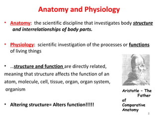
The document defines anatomy and its main subdivisions:
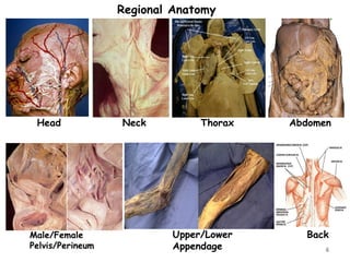
1) Gross anatomy includes regional, systemic, and surface anatomy.
2) Microscopic anatomy includes cytology and histology.
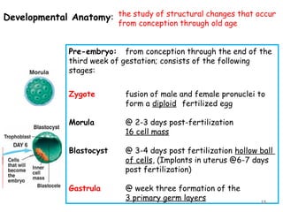
3) Developmental anatomy includes embryology and fetology.
4) Pathological anatomy studies structural changes during disease.
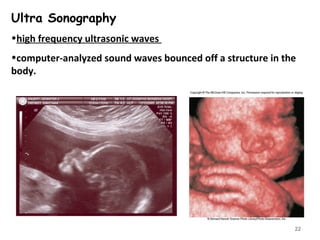
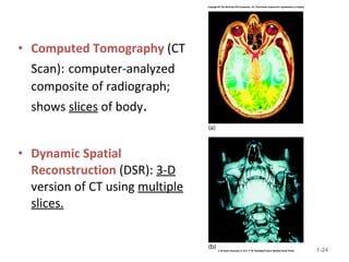
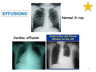
5) Radiographic anatomy uses imaging techniques like X-rays and MRI to study body structures.


![Anatomy introduction[1]](https://cdn.slidesharecdn.com/ss_thumbnails/9rhvq4jzrwacyv5bjs6b-signature-460517c25b85fc4e63c8080c3e27df73c8dfae9e0c6544cc7ea6d9e8b5e79cc7-poli-180213064029-thumbnail.jpg?width=640&height=640&fit=bounds)


