This document describes the key concepts of blood vessels, including:
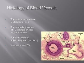
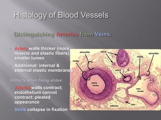
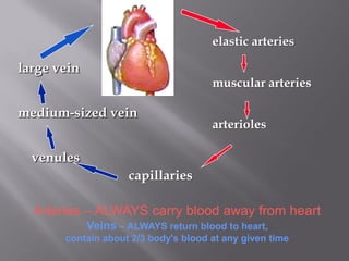
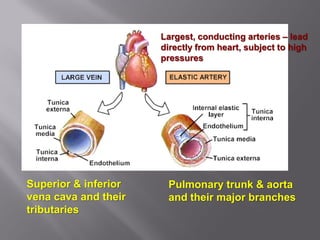
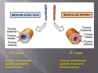
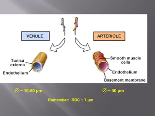
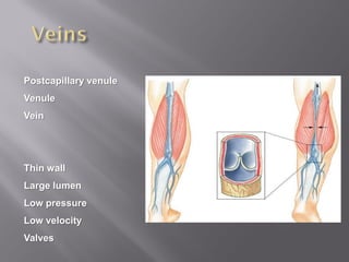
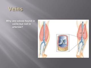
1) The histological similarities and differences between arteries and veins.
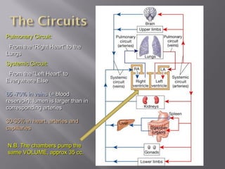
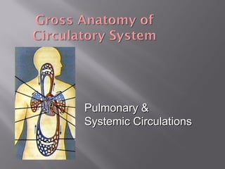
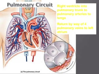
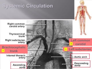
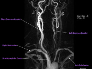
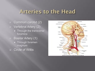
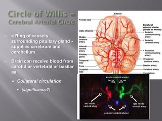
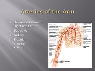
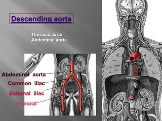
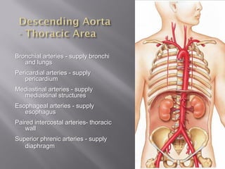
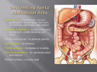
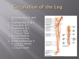
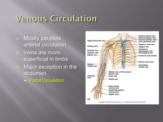
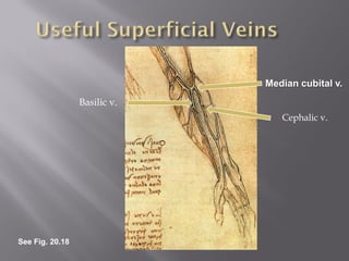
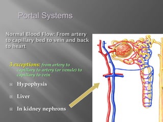
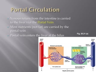
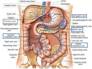
2) The major arteries and veins of the pulmonary and systemic circulations, including their patterns and names.
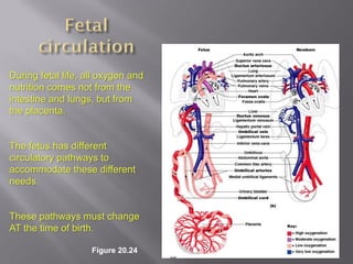
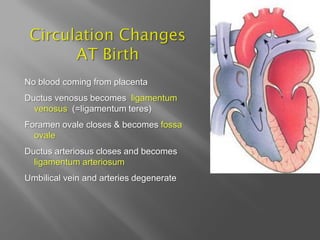
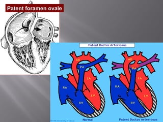
3) The circulatory changes that occur at birth as the fetus transitions from receiving oxygen and nutrition from the placenta to receiving it from the lungs and intestines.