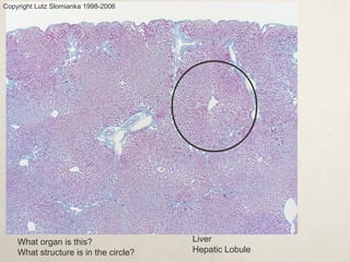
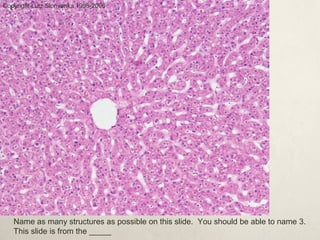
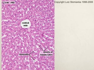
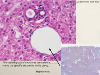
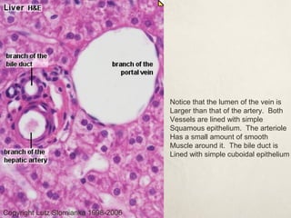
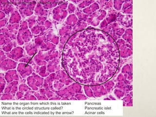
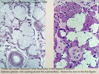
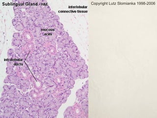
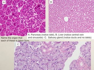
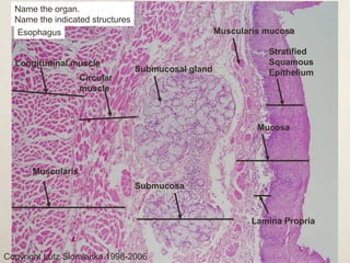
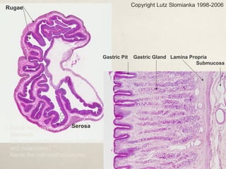
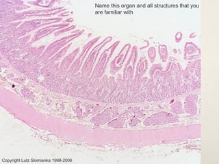
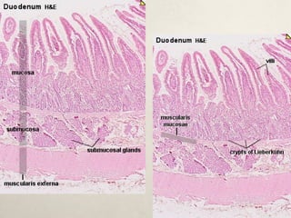
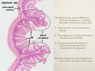
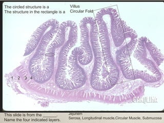
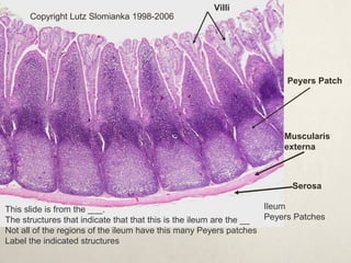
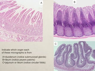
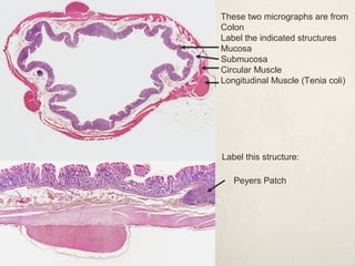
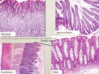
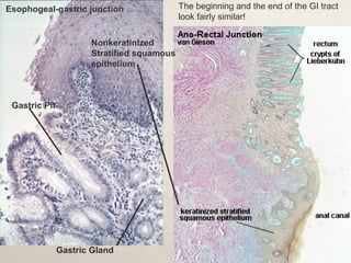
Histology of the Digestive System 
 This presentation will prepare you to identify all of the 
structures on the anatomy lab list. 
 Micrographs in this presentation were obtained from 
various sources which are indicated on each slide. 
 This presentation was created by Arleen Sawitzke
What organ is this? 
What structure is in the circle? 
Liver 
Hepatic Lobule 
Copyright Lutz Slomianka 1998-2006
Copyright Lutz Slomianka 1998-2006 
Name as many structures as possible on this slide. You should be able to name 3. 
This slide is from the _____
Copyright Lutz Slomianka 1998-2006
Copyright Lutz Slomianka 1998-2006 
The circled group of structures are called a_____ 
Name the specific structures in this group. 
Hepatic triad
Notice that the lumen of the vein is 
Larger than that of the artery. Both 
Vessels are lined with simple 
Squamous epithelium. The arteriole 
Has a small amount of smooth 
Muscle around it. The bile duct is 
Lined with simple cuboidal epithelium 
Copyright Lutz Slomianka 1998-2006
Copyright Lutz Slomianka 1998-2006 Pan20he 
Name the organ from which this is taken 
What is the circled structure called? 
What are the cells indicated by the arrow? 
Pancreas 
Pancreatic islet 
Acinar cells
Copyright Addison Wesley Longman 1998 
These two slides represent what structures in the digestive system? 
Salivary glands—the sublingual and the submaxillary. Notice the duct in the first figure
Copyright Lutz Slomianka 1998-2006
A B 
A. Pancreas (notice islet), B. Liver (notice central vein 
and sinusoids) C. Salivary gland (notice ducts and no islets) 
Name the organ that 
each of these is taken from 
C
Name the organ. 
Name the indicated structures 
Esophagus 
Longitudinal muscle 
Copyright Lutz Slomianka 1998-2006 
Muscularis mucosa 
Stratified 
Squamous 
Epithelium 
Mucosa 
Submucosal gland 
Circular 
muscle 
Submucosa 
Lamina Propria 
Muscularis
Copyright Lutz Slomianka 1998-2006 
Rugae 
Serosa 
Gastric Pit Gastric Gland Lamina Propria 
Name this organ 
Stomach 
Can you see the mucosa, submucosa, 
and muscularis? 
Name the indicated structures 
Submucosa
Copyright Lutz Slomianka 1998-2006 
Name this organ and all structures that you 
are familiar with
The jejunum has several differences 
from the duodenum. Compare 
this slide with the previous slide. 
1. There are few or no submucosal 
glands 
2. The submucosa is folded into plicae 
circulari (circular folds) 
3. The mucosal glands (crypts of 
Lieberkuhn) are much less 
prominent in the jejunum. 
Both the duodenum and the jejunum 
have villi to increase surface area.
1 2 3 4 
This slide is from the ______. 
Name the four indicated layers. 
Jejunem 
Serosa, Longitudinal muscle,Circular Muscle, Submucosa 
The circled structure is a 
The structure in the rectangle is a 
Villus 
Circular Fold
Copyright Lutz Slomianka 1998-2006 
This slide is from the ___. 
The structures that indicate that that this is the ileum are the __ 
Not all of the regions of the ileum have this many Peyers patches 
Label the indicated structures 
Ileum 
Peyers Patches 
Villi 
Peyers Patch 
Muscularis 
externa 
Serosa
Indicate which organ each 
of these micrographs is from 
A B 
C 
A=duodenum (notice submucosal glands) 
B=ileum (notice peyers patchs) 
C=jejunum or ileum (notice circular folds)
These two micrographs are from 
Colon 
Label the indicated structures 
Mucosa 
Submucosa 
Circular Muscle 
Longitudinal Muscle (Tenia coli) 
Label this structure: 
Peyers Patch
Stomach Jejunum/ileum 
Name the organs that each of these are taken from 
Goblet cells 
Name the indicated cells 
Duodonem Colon
Esophogeal-gastric junction 
Gastric Pit 
Nonkeratinized 
Stratified squamous 
epithelium 
Gastric Gland 
The beginning and the end of the GI tract 
look fairly similar!

Digestive System Histology

  • 1.
    Histology of theDigestive System  This presentation will prepare you to identify all of the structures on the anatomy lab list.  Micrographs in this presentation were obtained from various sources which are indicated on each slide.  This presentation was created by Arleen Sawitzke
  • 2.
    What organ isthis? What structure is in the circle? Liver Hepatic Lobule Copyright Lutz Slomianka 1998-2006
  • 3.
    Copyright Lutz Slomianka1998-2006 Name as many structures as possible on this slide. You should be able to name 3. This slide is from the _____
  • 4.
  • 5.
    Copyright Lutz Slomianka1998-2006 The circled group of structures are called a_____ Name the specific structures in this group. Hepatic triad
  • 6.
    Notice that thelumen of the vein is Larger than that of the artery. Both Vessels are lined with simple Squamous epithelium. The arteriole Has a small amount of smooth Muscle around it. The bile duct is Lined with simple cuboidal epithelium Copyright Lutz Slomianka 1998-2006
  • 7.
    Copyright Lutz Slomianka1998-2006 Pan20he Name the organ from which this is taken What is the circled structure called? What are the cells indicated by the arrow? Pancreas Pancreatic islet Acinar cells
  • 8.
    Copyright Addison WesleyLongman 1998 These two slides represent what structures in the digestive system? Salivary glands—the sublingual and the submaxillary. Notice the duct in the first figure
  • 9.
  • 10.
    A B A.Pancreas (notice islet), B. Liver (notice central vein and sinusoids) C. Salivary gland (notice ducts and no islets) Name the organ that each of these is taken from C
  • 11.
    Name the organ. Name the indicated structures Esophagus Longitudinal muscle Copyright Lutz Slomianka 1998-2006 Muscularis mucosa Stratified Squamous Epithelium Mucosa Submucosal gland Circular muscle Submucosa Lamina Propria Muscularis
  • 12.
    Copyright Lutz Slomianka1998-2006 Rugae Serosa Gastric Pit Gastric Gland Lamina Propria Name this organ Stomach Can you see the mucosa, submucosa, and muscularis? Name the indicated structures Submucosa
  • 13.
    Copyright Lutz Slomianka1998-2006 Name this organ and all structures that you are familiar with
  • 15.
    The jejunum hasseveral differences from the duodenum. Compare this slide with the previous slide. 1. There are few or no submucosal glands 2. The submucosa is folded into plicae circulari (circular folds) 3. The mucosal glands (crypts of Lieberkuhn) are much less prominent in the jejunum. Both the duodenum and the jejunum have villi to increase surface area.
  • 16.
    1 2 34 This slide is from the ______. Name the four indicated layers. Jejunem Serosa, Longitudinal muscle,Circular Muscle, Submucosa The circled structure is a The structure in the rectangle is a Villus Circular Fold
  • 17.
    Copyright Lutz Slomianka1998-2006 This slide is from the ___. The structures that indicate that that this is the ileum are the __ Not all of the regions of the ileum have this many Peyers patches Label the indicated structures Ileum Peyers Patches Villi Peyers Patch Muscularis externa Serosa
  • 18.
    Indicate which organeach of these micrographs is from A B C A=duodenum (notice submucosal glands) B=ileum (notice peyers patchs) C=jejunum or ileum (notice circular folds)
  • 19.
    These two micrographsare from Colon Label the indicated structures Mucosa Submucosa Circular Muscle Longitudinal Muscle (Tenia coli) Label this structure: Peyers Patch
  • 20.
    Stomach Jejunum/ileum Namethe organs that each of these are taken from Goblet cells Name the indicated cells Duodonem Colon
  • 21.
    Esophogeal-gastric junction GastricPit Nonkeratinized Stratified squamous epithelium Gastric Gland The beginning and the end of the GI tract look fairly similar!

Editor's Notes