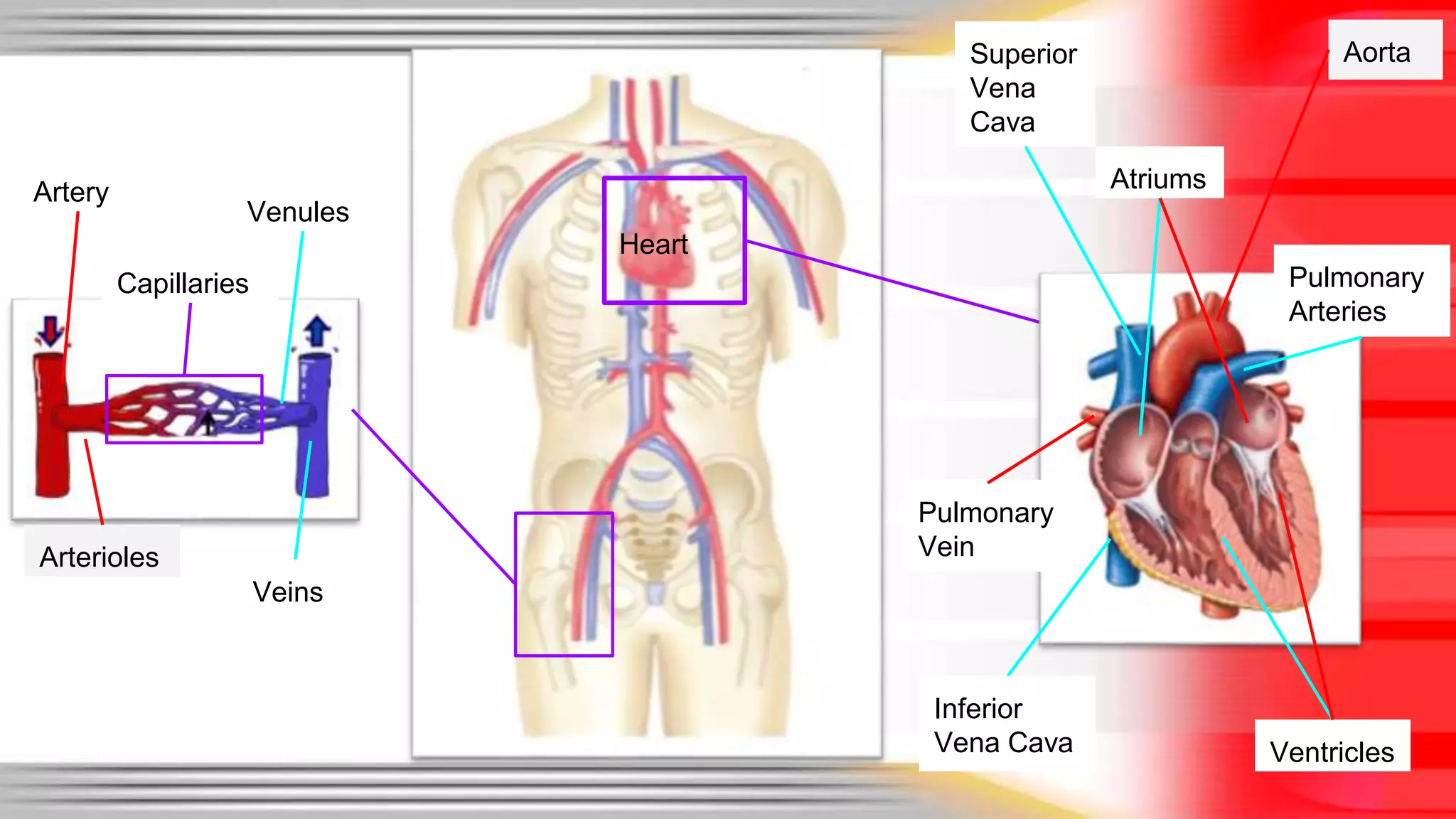
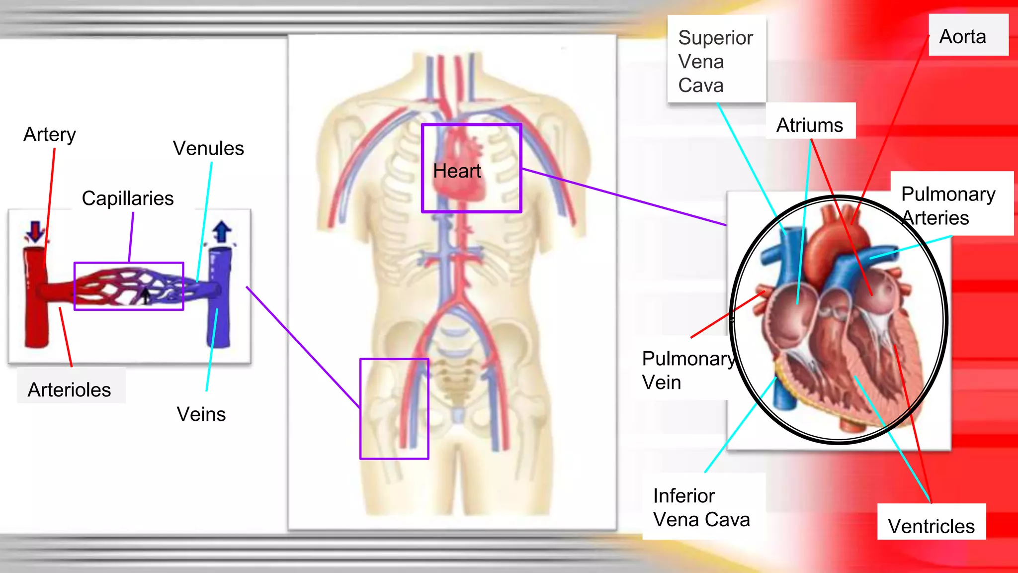
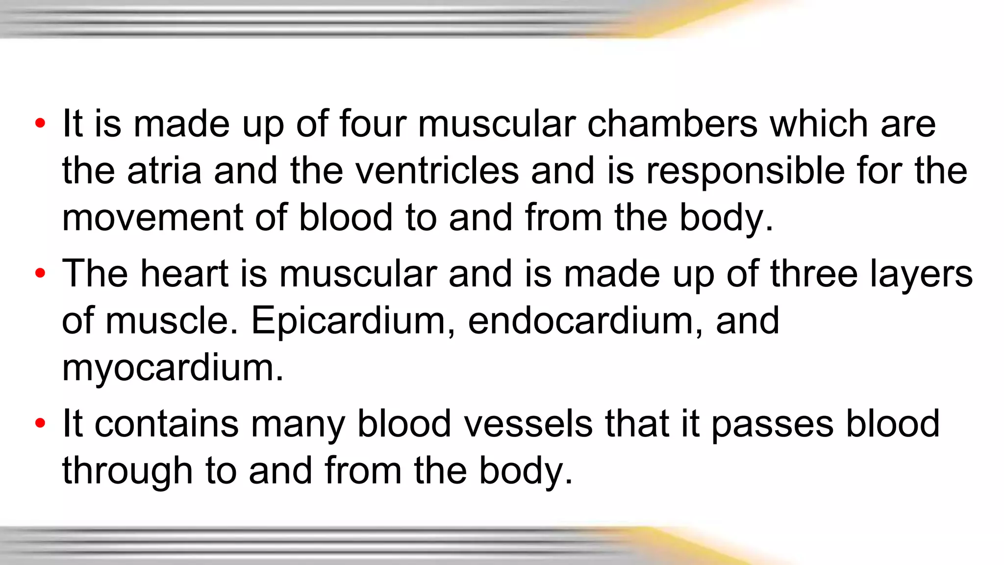
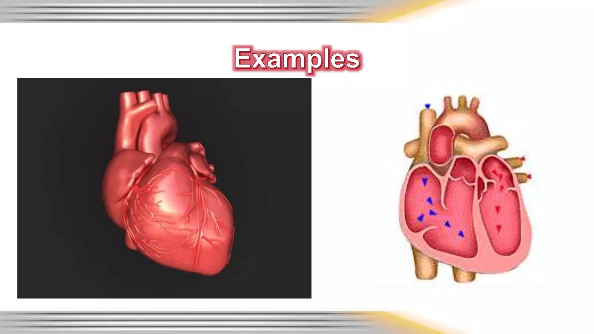
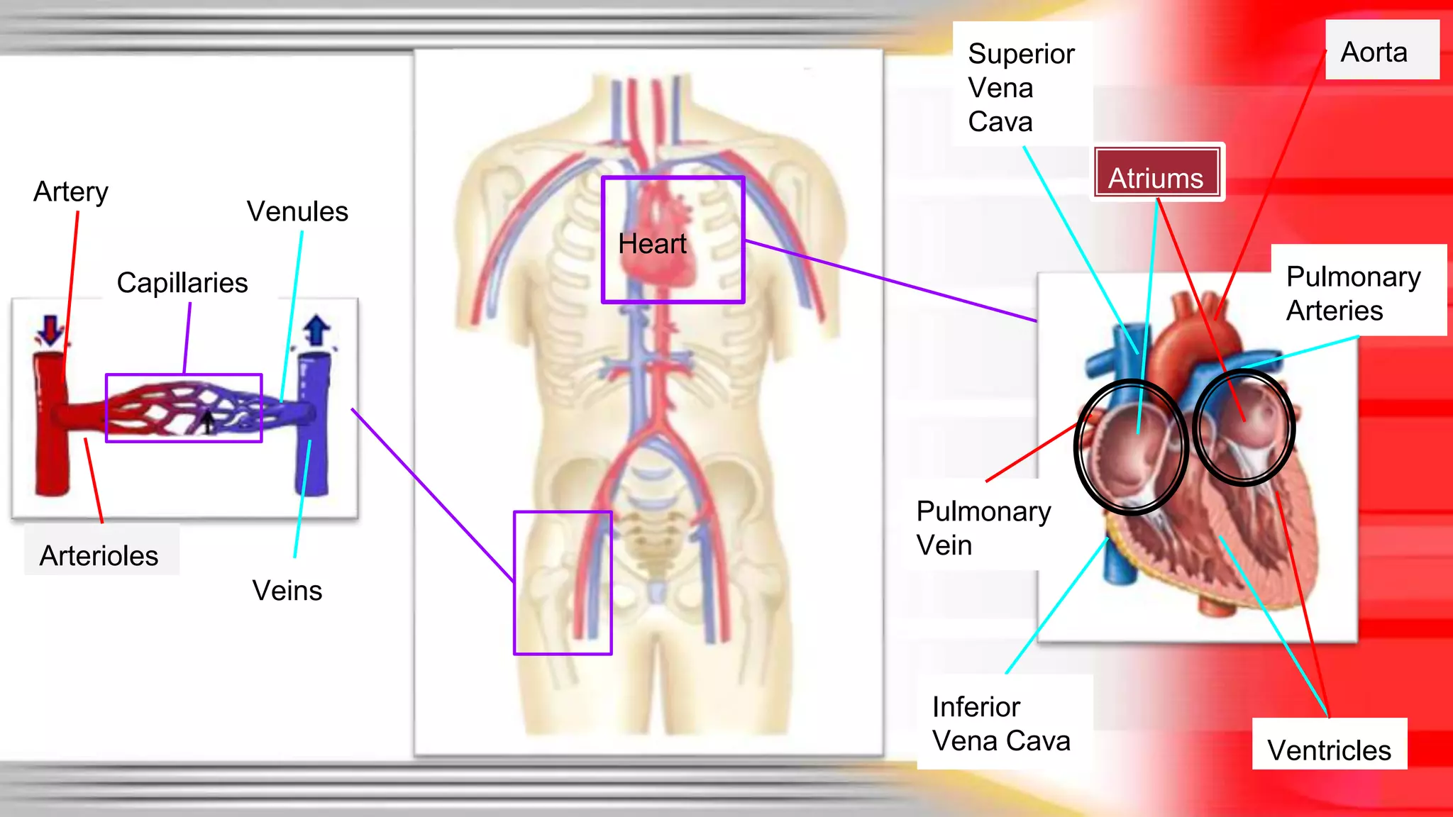
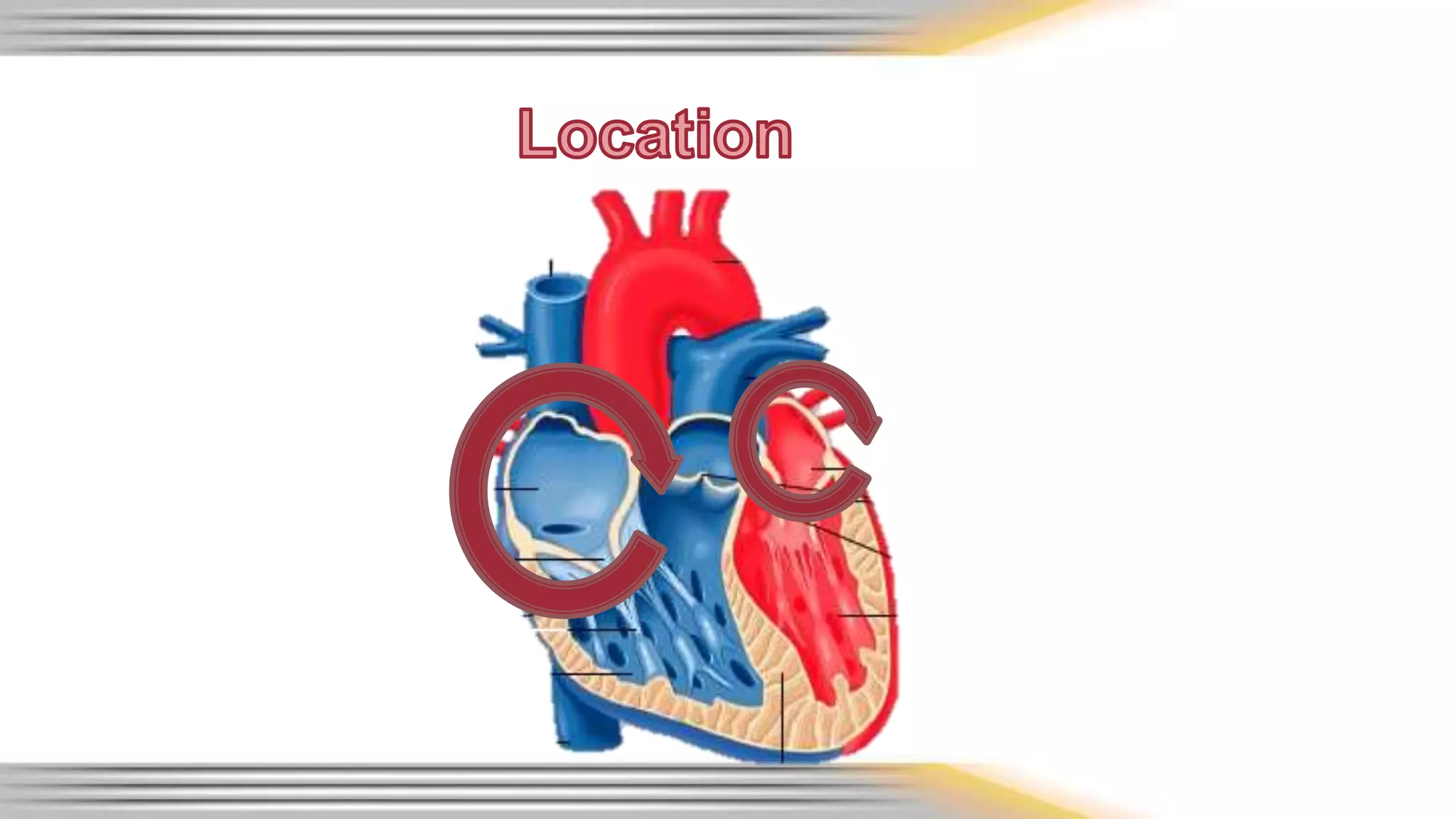
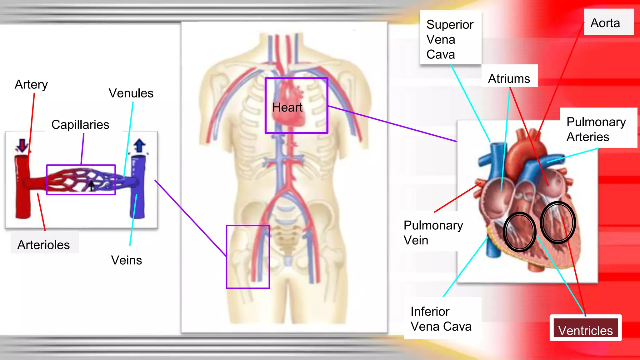
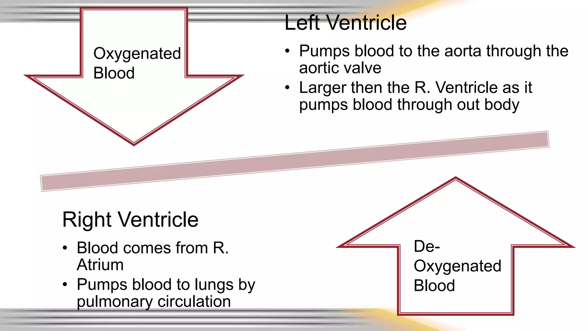
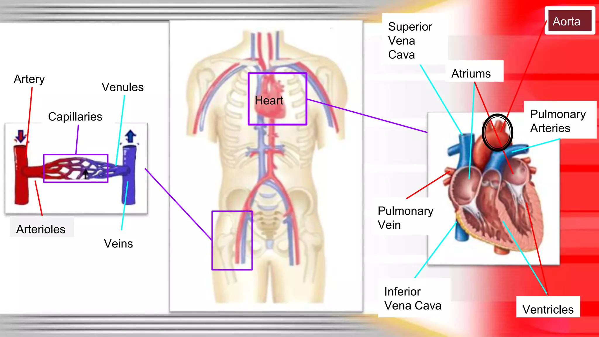
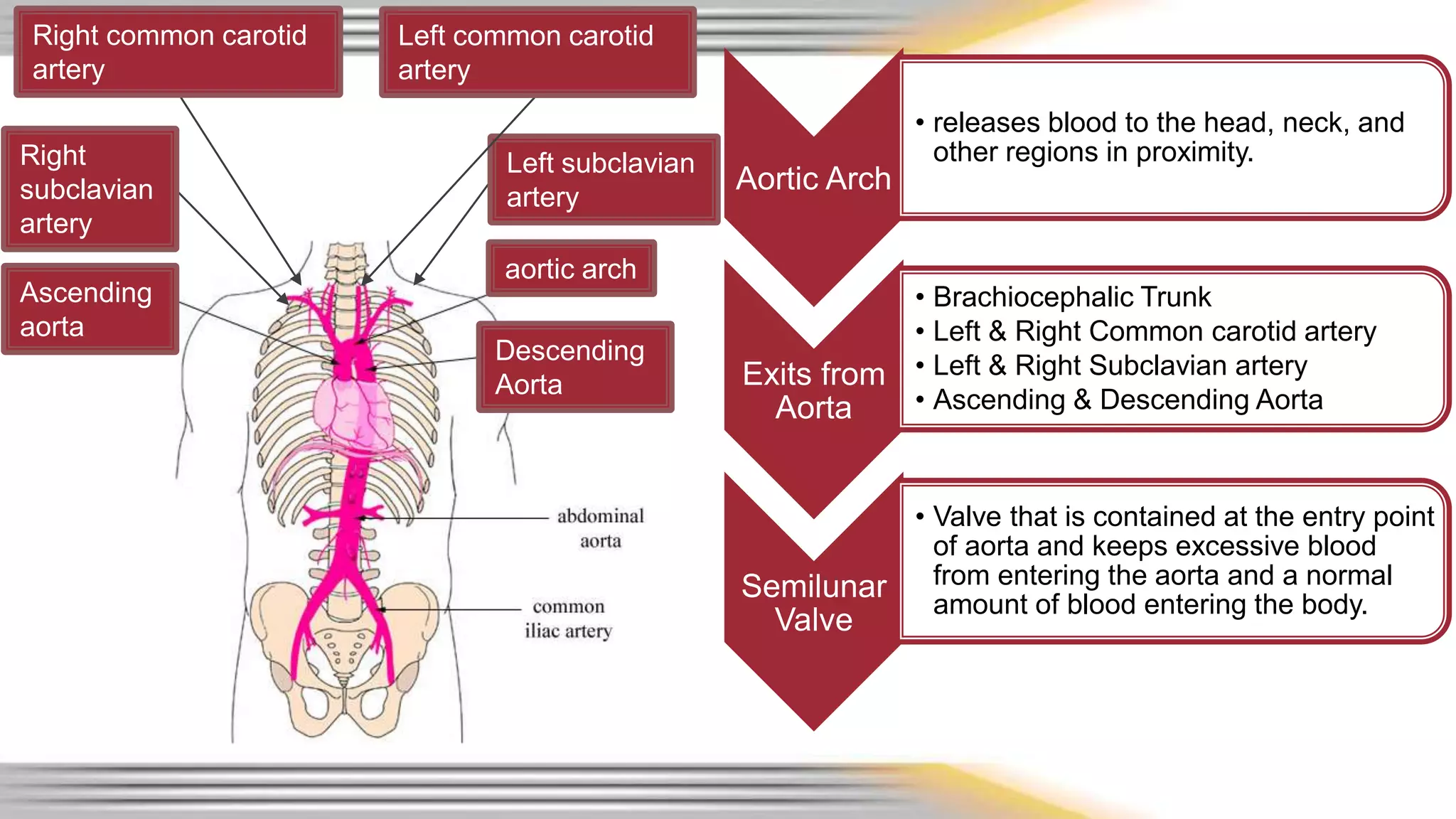
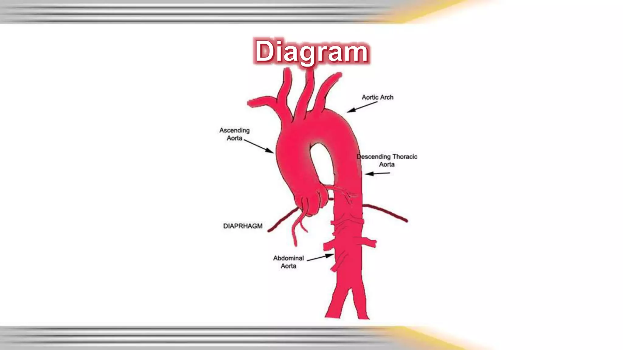
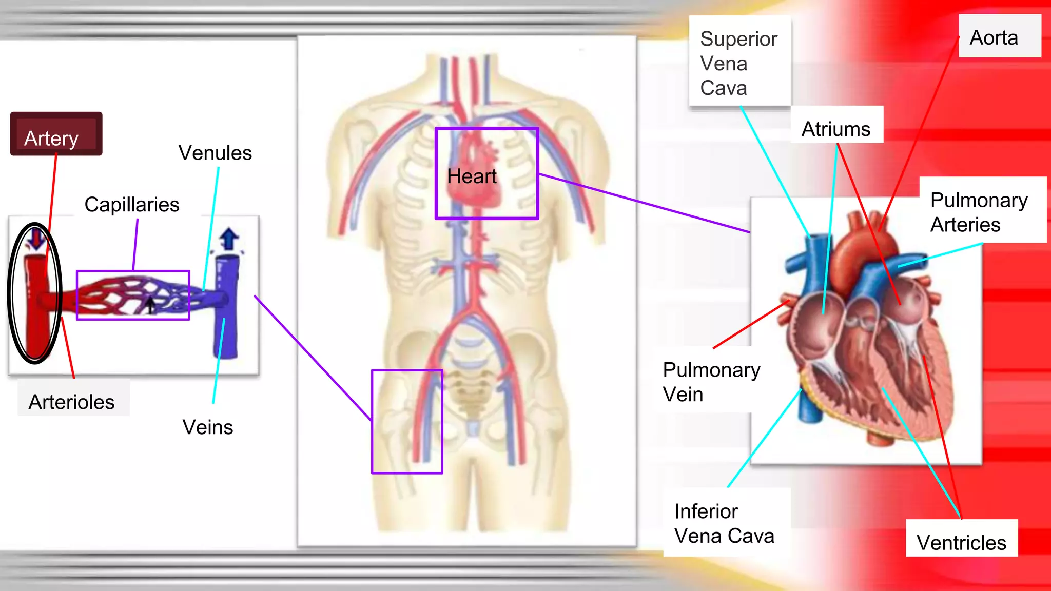
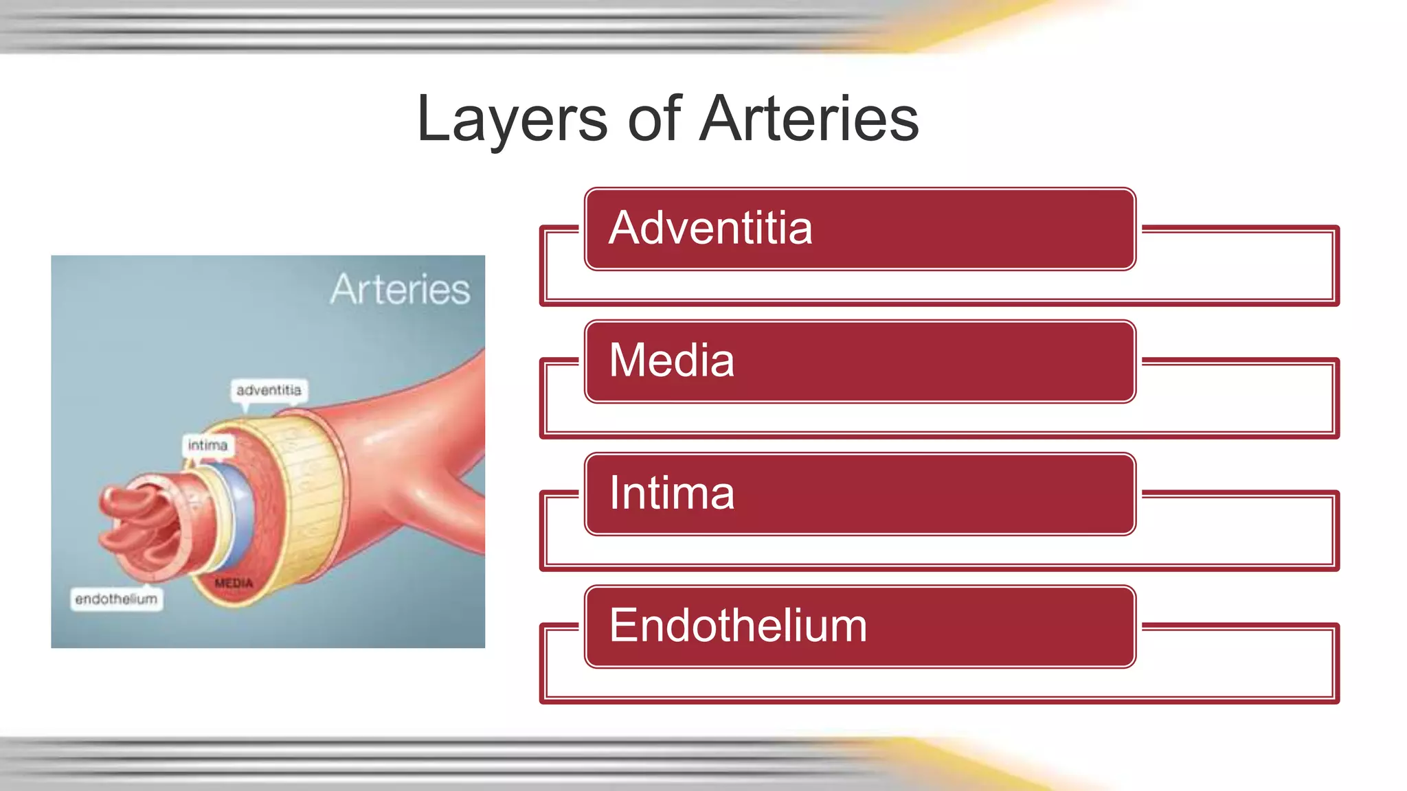
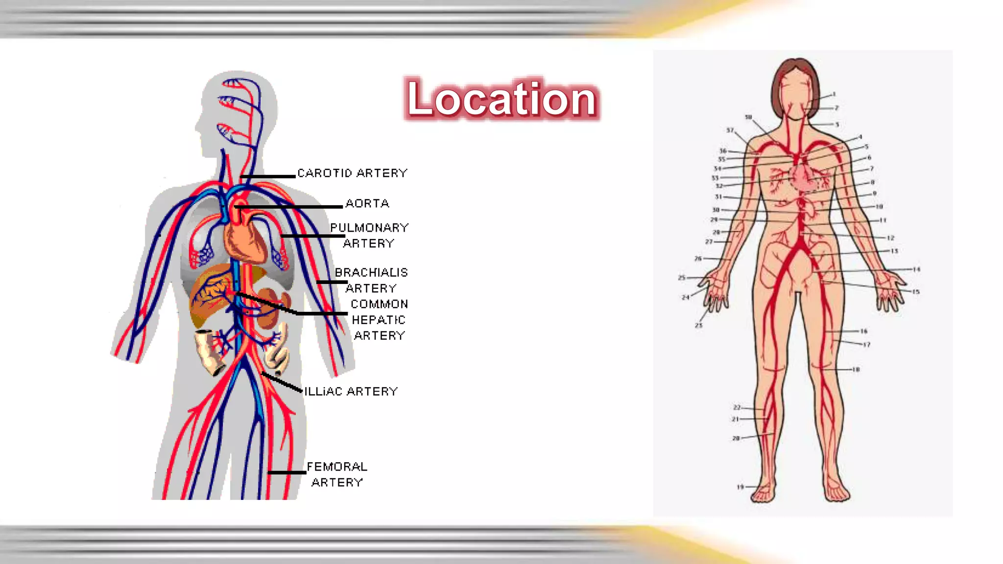
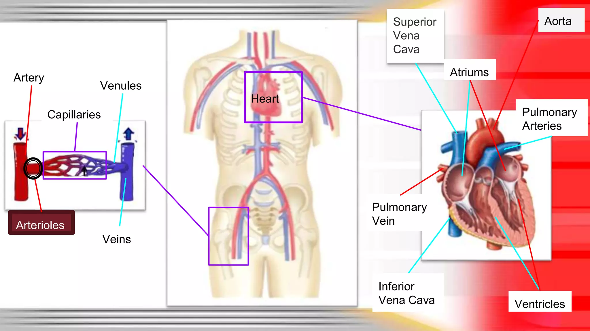
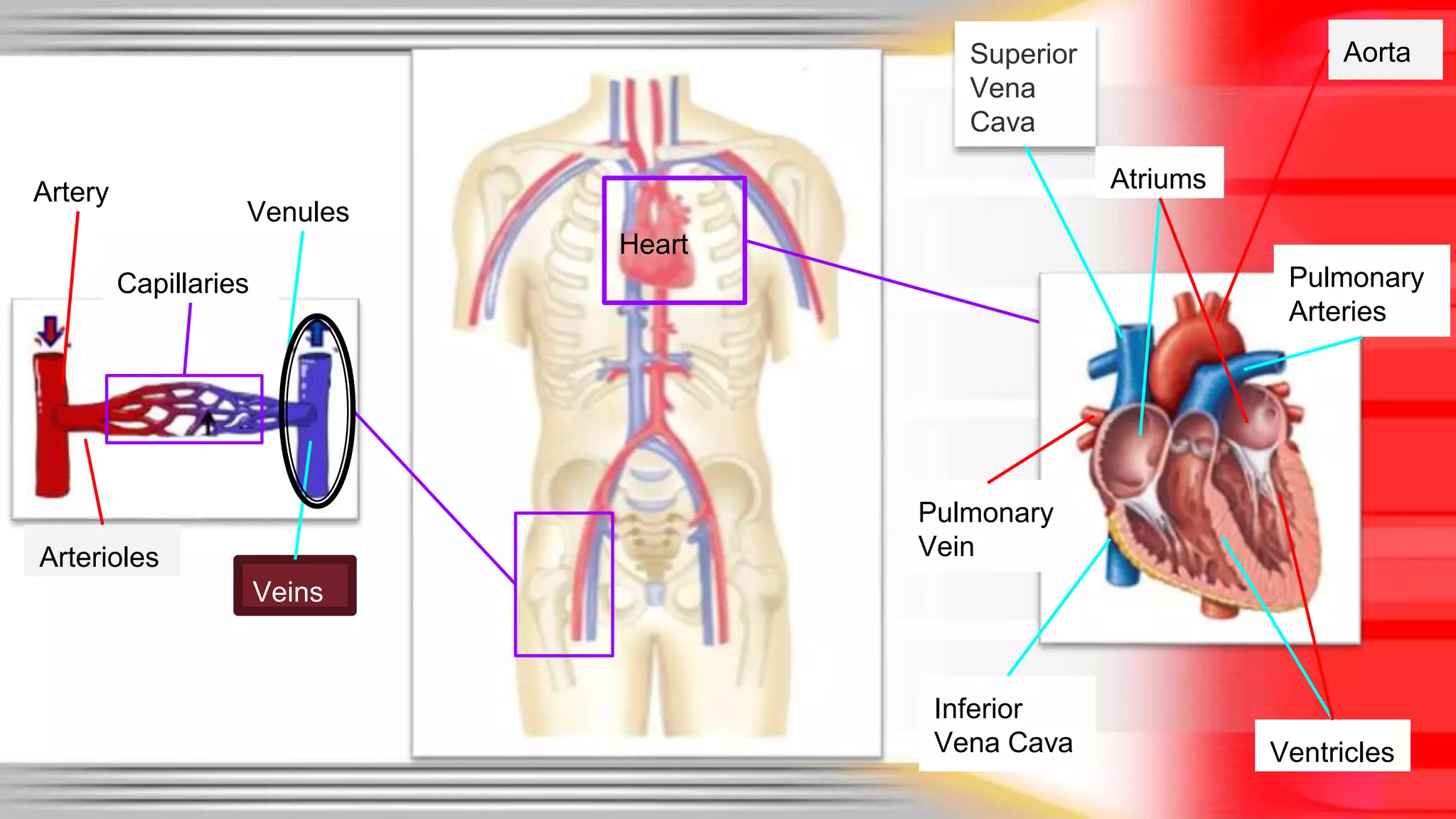
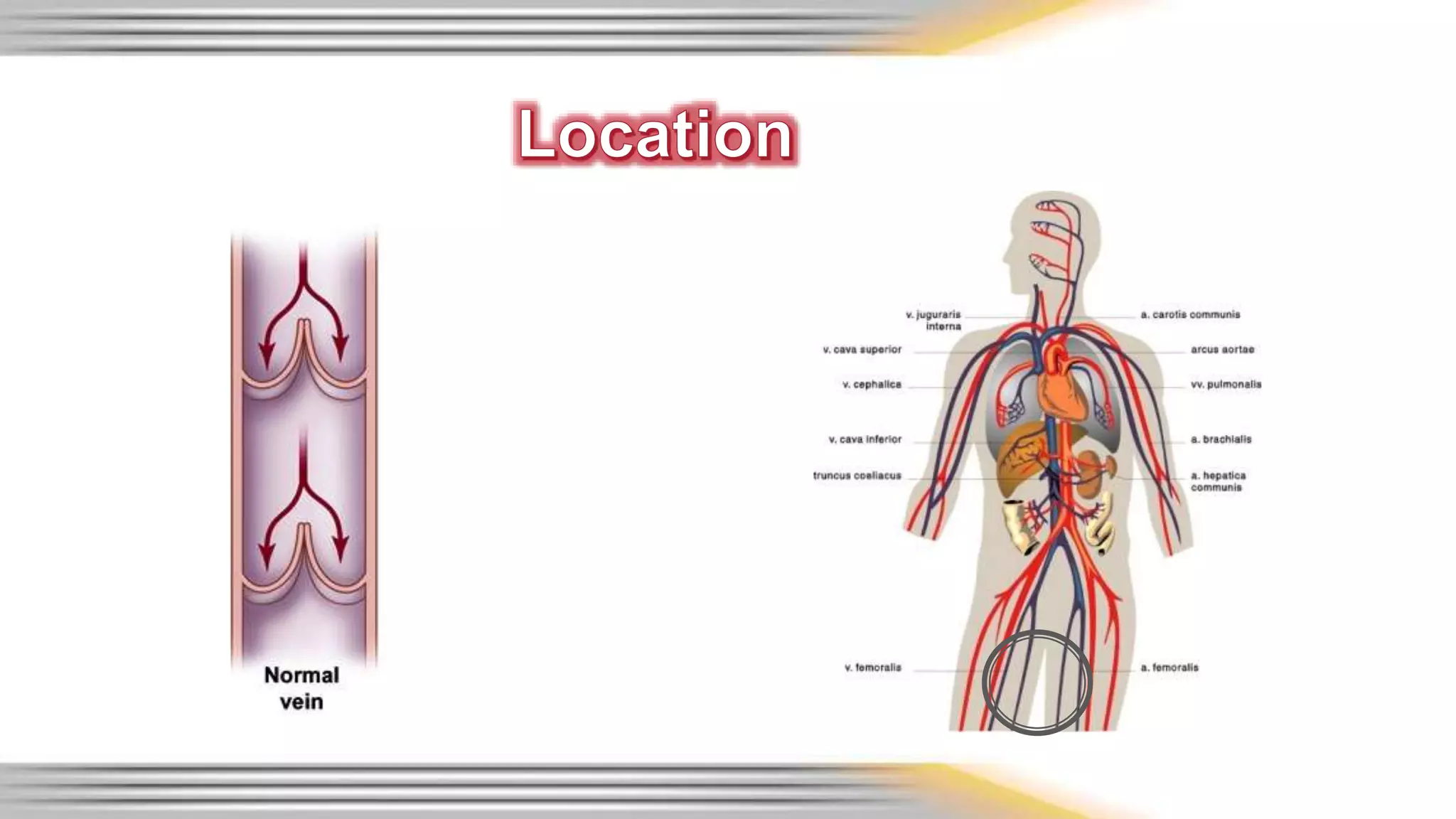
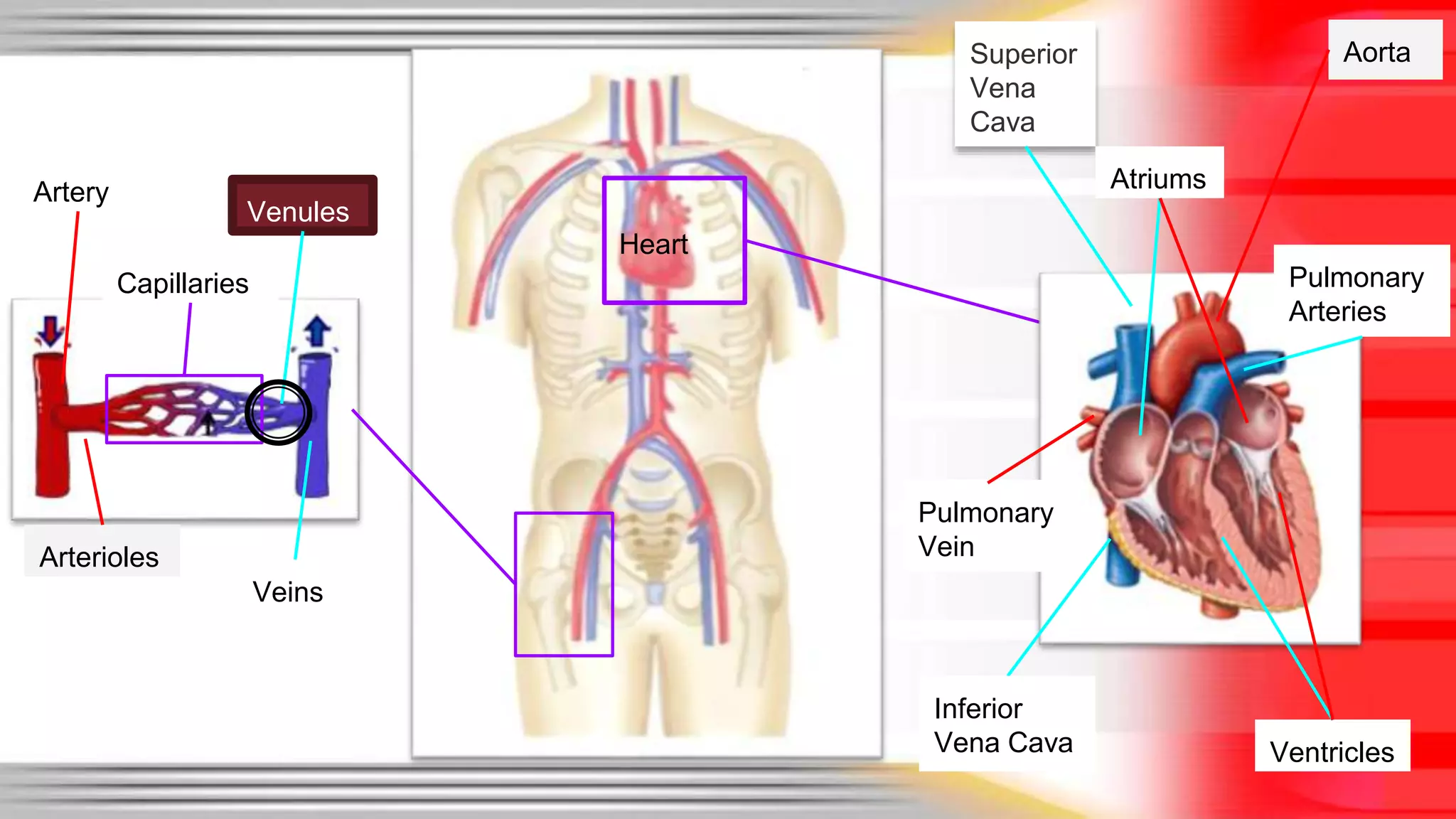
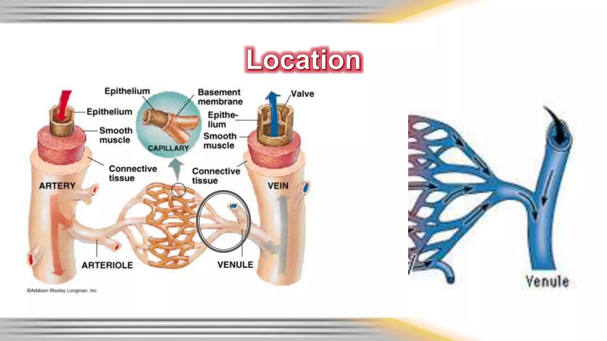
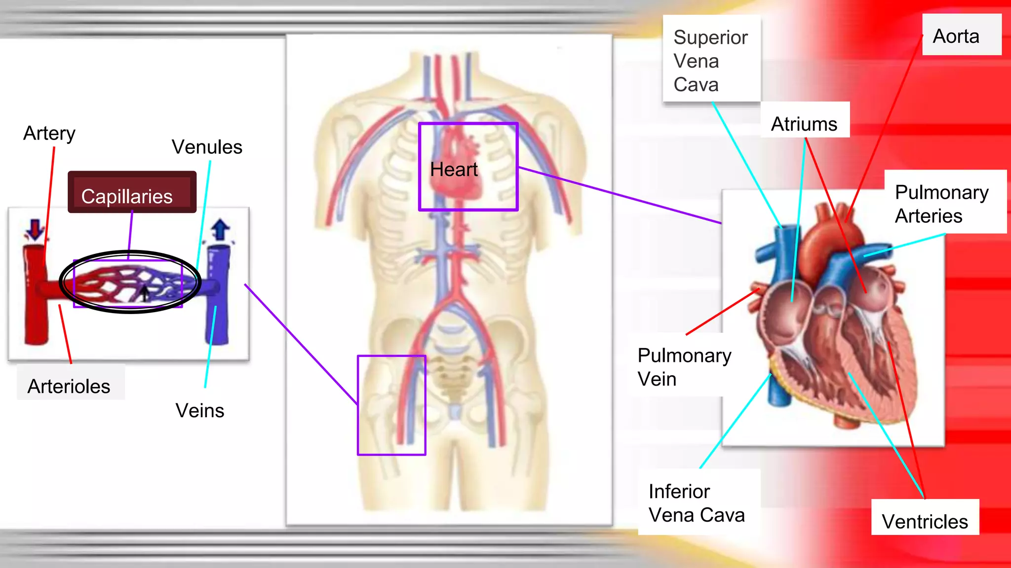
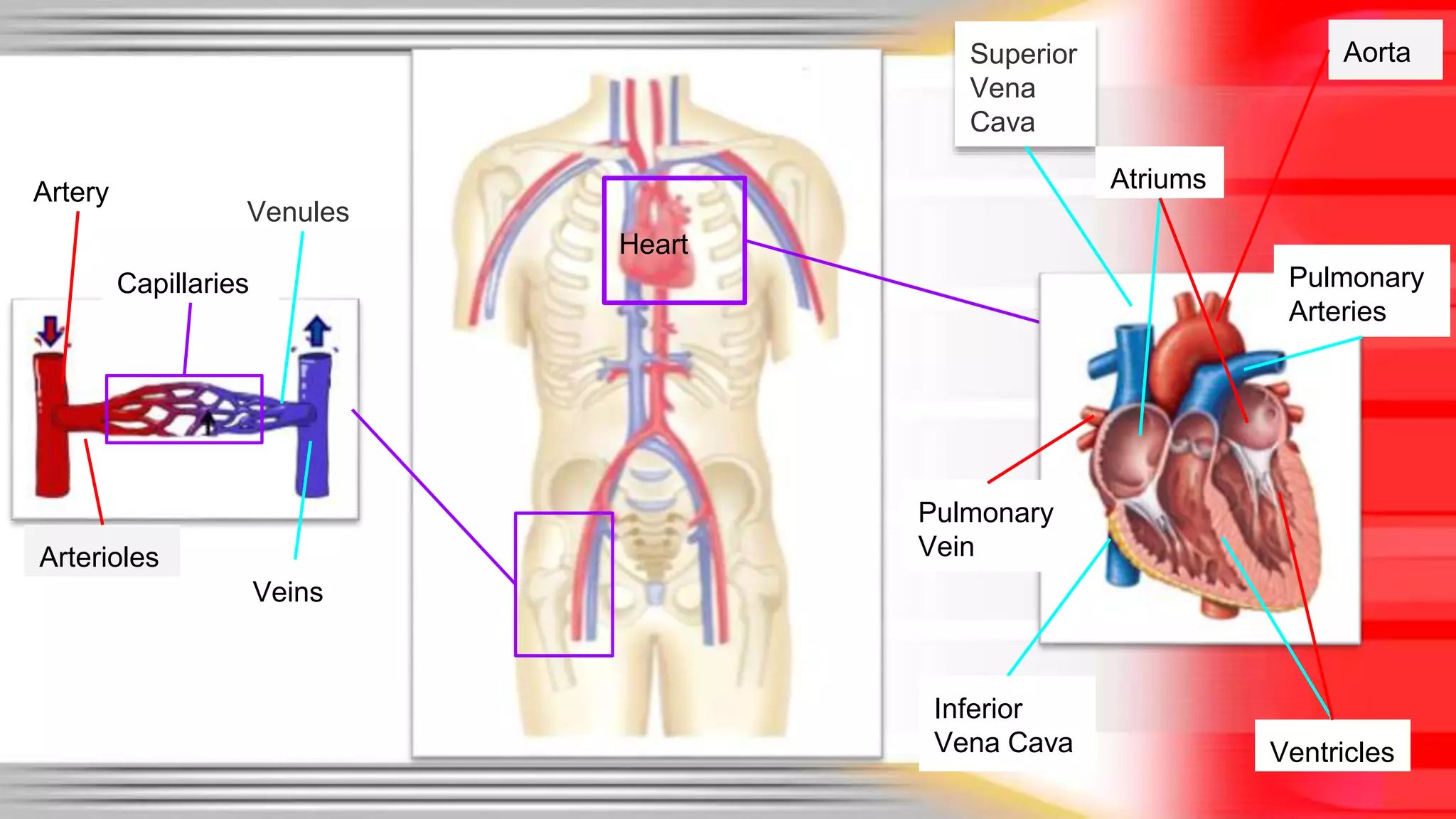
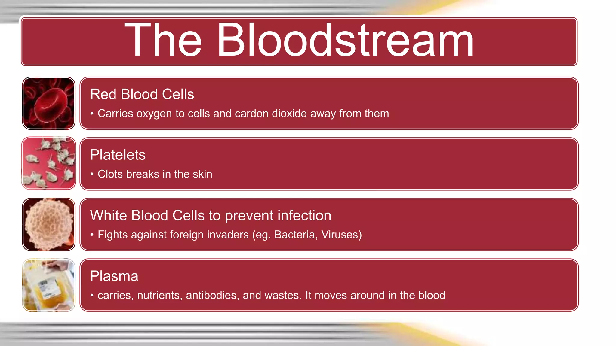
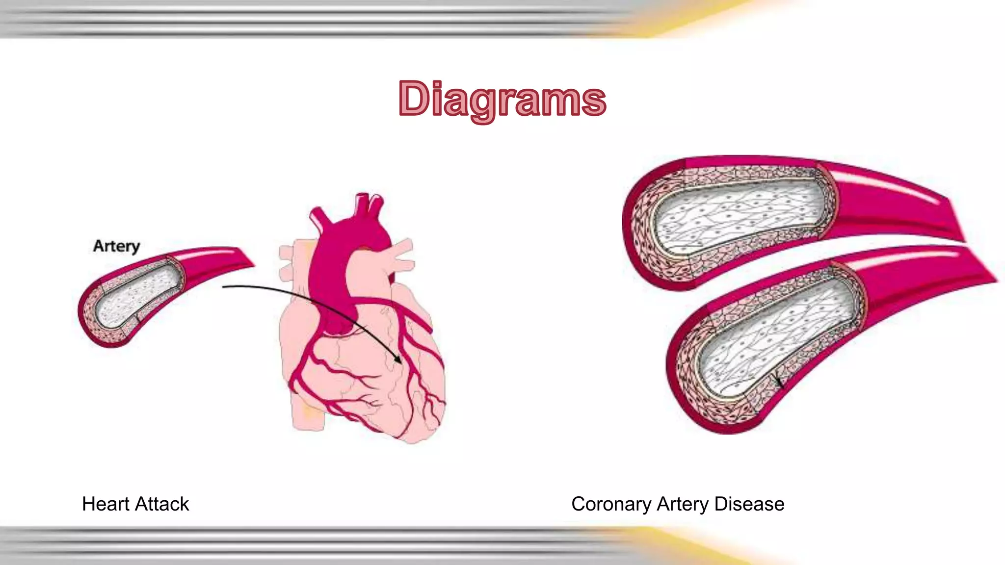
The cardiovascular system consists of the heart, blood vessels, and blood. The heart pumps blood through a closed system of arteries, veins, and capillaries. It is made up of four chambers that receive and pump blood throughout the body and lungs. Blood carries oxygen, nutrients, waste products, hormones, and more as it circulates through the body. Heart disease occurs when the arteries become blocked or narrowed, reducing blood flow and oxygen delivery.
FunctionsTransport oxygen and nutrients to the lungs and tissuesForm blood clots to prevent excess blood lossCarry cells and antibodies that fight infectionBring waste products to the kidneys and liver to filter bloodRegulate body temperature
A closed system of the heart and blood vessels
The heart pumps blood
Blood vessels allow blood to circulate to all parts of the body
The function of the cardiovascular system is to deliver oxygen and nutrients and to remove carbon dioxide and other waste products
A closed system of the heart and blood vessels
The heart pumps blood
Blood vessels allow blood to circulate to all parts of the body
The function of the cardiovascular system is to deliver oxygen and nutrients and to remove carbon dioxide and other waste products
The cardiovascular system can be thought of as the transport system of the body.
This system has three main components: the heart, the blood vessel and the blood itself.
The heart is the system’s pump and the blood vessels are like the delivery routes.
This system has three main components: the heart, the blood vessel and the blood itself. The heart is the system's pump and the blood vessels are like the delivery routes. Blood can be thought of as a fluid which contains the oxygen and nutrients the body needs and carries the wastes which need to be removed.
General Introduction of Cardiovascular System and Anatomy of Cardiovascular System.
In this slide, you will be able to find the general anatomy of the heart and Basic introduction of Cardiovascular Sstem
If you or anyone you know has heart disease, hypertension, disorders of the heart valves, or cholesterol issues then you need this information. In this presentation you will learn about the circulatory system, risk factors, and what supplements you can take to help it achieve optimal health.
Be sure to order any supplements mentioned in this presesntation from NaturesSunshine.com (use sponsor number 2849323 / or search for MCM Products) or from BrilliantNaturalHealth.com.
Human heart anatomy and physiology Part -1Ritu Sharma
The heart is the pump responsible for maintaining adequate circulation of oxygenated blood around the vascular network of the body. It is a four-chamber pump, with the right side receiving deoxygenated blood from the body at low presure and pumping it to the lungs (the pulmonary circulation) and the left side receiving oxygenated blood from the lungs and pumping it at high pressure around the body (the systemic circulation).
1 GNM - Anatomy unit - 4 - CVS by thirumurugan.pptxthiru murugan
By:M. Thiru murugan
Unit – IV:
Heart : Structure, functions including conduction system & cardiac cycle
Blood vessels : Types, Structure and position
Circulation of blood
Blood pressure and pulse
Heart
The circulatory system:
It consisting of blood, blood vessels, and heart.
This supplies oxygen and other nutrients,
Transports hormones
Removes unnecessary waste products.
Heart and its Structure
The heart is a muscular organ about the size of a fist,
located in mediastinum just behind and slightly left of the breastbone (sternum).
The heart pumps blood through the blood vessels (arteries and veins called the cardiovascular system).
Structure of heart:
Layers of the heart (3)
Chambers of the heart (4)
Valves of the heart (4)
Blood vessels of the heart (5)
3 layers of the heart:
Epicardium/pericardium: outer protective layer of the heart. Visceral and parietal (pericardial fluid). Protection for the heart and big vessels and prevent collapse of heart,
Myocardium: muscular middle layer wall of the heart. Responsible for keeping the heart pumping blood around the body.
Endocardium: the inner layer of the heart. Regulate blood flow through the chambers of the heart and pass the electrical impulses
Chambers of the heart:
The atria: These are the 2 upper chambers, which receive blood. RA / LA
The ventricles: These are the 2 lower chambers, which discharge blood. RV/ LV
A wall of tissue called the septum separates the left and right atria called atrial septum and the left and right ventricle called ventricular septum.
Valves in the heart:
There are four valves
Two-atrio ventricular valves: The 2 types: bicuspid (mitral) - LA & LV, and tricuspid valves - RA & RV.
Two-semilunar valves: The aortic valves and the pulmonary valve.
Major blood vessels of the heart
There are 5 major blood vessels
Pulmonary artery
Pulmonary veins
Aorta[artery]
Inferior vena cava [IVC] veins
Superior vena cava [SVC] veins
Functions of heart:
Pumping oxygenated blood to the body parts.
Pumping nutrients and other vital substances
Receiving deoxygenated blood and carrying metabolic waste products from the body
Pumping deoxygenated blood to the lungs for oxygenation.
Maintaining blood pressure.
Conduction system
The electrical conduction system that controls the heart rate.
This system generates electrical impulses and conducts them throughout the muscle of the heart, stimulating the heart to contract and pump blood.
The electrical pulses determine the order in which the chambers contract & the heart rate
Conductive system consist of:
SA Node
AV Node
Bundle of his or His Bundles – bundle of branches
( right and left)
4. Purkinje fibres
Sinoatrial node (SA) : also known as the pace maker of the heart and Located in the upper wall of the right atrium
Made up of both muscle and nervous tissue
Here the electrical impulse begins
Atrioventricular (AV) node:
located between the atria and ventricles of the heart
The electrical impulse is carried fr
This ppt is made to make people understand about the things present in a human body and also to explain the process whicyh happen in the human body . This ppt is basicly for the students of class 7th because it made for there level only .
The presentation that will unable to create a clear cut concept regarding the Vessels the vascular system of the human body. It will let you know about the arteries, veins, capillaries how the exchange of nutrients and other substance takes place..and many more things related to the vessels of the body.
CARDIO VASCULAR SYSTEM.pdf for bsc nursing studentsshanmukhadevi
The cardiovascular system refers to the heart, blood vessels and the blood.
Blood contains oxygen and other nutrients, which your body needs to survive. The body takes these essential nutrients from the blood.
At the same time, the body dumps waste products like carbon dioxide, back into the blood, so they can be removed.
The main function of the cardiovascular system is therefore to maintain blood flow to all parts of the body, to allow it to survive.
Veins deliver used blood from the body back to the heart. Blood in the veins is low in oxygen (as it has been taken out by the body) and high in carbon dioxide (as the body has unloaded it back into the blood).
All the veins drain into the superior and inferior vena cava, which then drain into the right atrium.
The right atrium pumps blood into the right ventricle. Then the right ventricle pumps blood to the pulmonary trunk, through the pulmonary arteries and into the lungs.
In the lungs the blood picks up oxygen that we breathe in and gets rid of carbon dioxide, which we breathe out. The blood is becomes rich in oxygen, which the body can use.
From the lungs, blood drains into the left atrium and is then pumped into the left ventricle. The left ventricle then pumps this oxygen-rich blood out into the aorta, which then distributes it to the rest of the body through other arteries.
This blood will again return back to the heart through the veins and the cycle continues.
The cardiovascular system can be thought of as the transport system of the body.
This system has three main components: the heart, the blood vessel and the blood itself.
The heart is the system's pump and the blood vessels are like the delivery routes.
Blood can be thought of as a fluid, which contains the oxygen and nutrients the body needs and carries the wastes, which need to be removed.
A closed system of the heart and blood vessels
The heart pumps blood
Blood vessels allow blood to circulate to all parts of the body
The function of the cardiovascular system is to deliver oxygen and nutrients and to remove carbon dioxide and other waste products
The heart contributes to homeostasis by pumping blood through blood vessels to the tissues of the body to deliver oxygen and nutrients and remove wastes.
Blood to reach body cells and exchange materials with them, it must be pumped continuously by the heart through the body’s blood vessels.
The heart beats about 100,000 times every day, which adds up to about 35 million beats in a year, and approximately 2.5 billion times in an average lifetime.
The left side of the heart pumps blood through an estimated 100,000 km (60,000 mi) of blood vessels, which is equivalent to traveling around the earth’s equator about three times.
The right side of the heart pumps blood through the lungs, enabling blood to pick up oxygen and unload carbon dioxide.
Prix Galien International 2024 Forum ProgramLevi Shapiro
June 20, 2024, Prix Galien International and Jerusalem Ethics Forum in ROME. Detailed agenda including panels:
- ADVANCES IN CARDIOLOGY: A NEW PARADIGM IS COMING
- WOMEN’S HEALTH: FERTILITY PRESERVATION
- WHAT’S NEW IN THE TREATMENT OF INFECTIOUS,
ONCOLOGICAL AND INFLAMMATORY SKIN DISEASES?
- ARTIFICIAL INTELLIGENCE AND ETHICS
- GENE THERAPY
- BEYOND BORDERS: GLOBAL INITIATIVES FOR DEMOCRATIZING LIFE SCIENCE TECHNOLOGIES AND PROMOTING ACCESS TO HEALTHCARE
- ETHICAL CHALLENGES IN LIFE SCIENCES
- Prix Galien International Awards Ceremony
Flu Vaccine Alert in Bangalore Karnatakaaddon Scans
As flu season approaches, health officials in Bangalore, Karnataka, are urging residents to get their flu vaccinations. The seasonal flu, while common, can lead to severe health complications, particularly for vulnerable populations such as young children, the elderly, and those with underlying health conditions.
Dr. Vidisha Kumari, a leading epidemiologist in Bangalore, emphasizes the importance of getting vaccinated. "The flu vaccine is our best defense against the influenza virus. It not only protects individuals but also helps prevent the spread of the virus in our communities," he says.
This year, the flu season is expected to coincide with a potential increase in other respiratory illnesses. The Karnataka Health Department has launched an awareness campaign highlighting the significance of flu vaccinations. They have set up multiple vaccination centers across Bangalore, making it convenient for residents to receive their shots.
To encourage widespread vaccination, the government is also collaborating with local schools, workplaces, and community centers to facilitate vaccination drives. Special attention is being given to ensuring that the vaccine is accessible to all, including marginalized communities who may have limited access to healthcare.
Residents are reminded that the flu vaccine is safe and effective. Common side effects are mild and may include soreness at the injection site, mild fever, or muscle aches. These side effects are generally short-lived and far less severe than the flu itself.
Healthcare providers are also stressing the importance of continuing COVID-19 precautions. Wearing masks, practicing good hand hygiene, and maintaining social distancing are still crucial, especially in crowded places.
Protect yourself and your loved ones by getting vaccinated. Together, we can help keep Bangalore healthy and safe this flu season. For more information on vaccination centers and schedules, residents can visit the Karnataka Health Department’s official website or follow their social media pages.
Stay informed, stay safe, and get your flu shot today!
Pulmonary Thromboembolism - etilogy, types, medical- Surgical and nursing man...VarunMahajani
Disruption of blood supply to lung alveoli due to blockage of one or more pulmonary blood vessels is called as Pulmonary thromboembolism. In this presentation we will discuss its causes, types and its management in depth.
Report Back from SGO 2024: What’s the Latest in Cervical Cancer?bkling
Are you curious about what’s new in cervical cancer research or unsure what the findings mean? Join Dr. Emily Ko, a gynecologic oncologist at Penn Medicine, to learn about the latest updates from the Society of Gynecologic Oncology (SGO) 2024 Annual Meeting on Women’s Cancer. Dr. Ko will discuss what the research presented at the conference means for you and answer your questions about the new developments.
Title: Sense of Smell
Presenter: Dr. Faiza, Assistant Professor of Physiology
Qualifications:
MBBS (Best Graduate, AIMC Lahore)
FCPS Physiology
ICMT, CHPE, DHPE (STMU)
MPH (GC University, Faisalabad)
MBA (Virtual University of Pakistan)
Learning Objectives:
Describe the primary categories of smells and the concept of odor blindness.
Explain the structure and location of the olfactory membrane and mucosa, including the types and roles of cells involved in olfaction.
Describe the pathway and mechanisms of olfactory signal transmission from the olfactory receptors to the brain.
Illustrate the biochemical cascade triggered by odorant binding to olfactory receptors, including the role of G-proteins and second messengers in generating an action potential.
Identify different types of olfactory disorders such as anosmia, hyposmia, hyperosmia, and dysosmia, including their potential causes.
Key Topics:
Olfactory Genes:
3% of the human genome accounts for olfactory genes.
400 genes for odorant receptors.
Olfactory Membrane:
Located in the superior part of the nasal cavity.
Medially: Folds downward along the superior septum.
Laterally: Folds over the superior turbinate and upper surface of the middle turbinate.
Total surface area: 5-10 square centimeters.
Olfactory Mucosa:
Olfactory Cells: Bipolar nerve cells derived from the CNS (100 million), with 4-25 olfactory cilia per cell.
Sustentacular Cells: Produce mucus and maintain ionic and molecular environment.
Basal Cells: Replace worn-out olfactory cells with an average lifespan of 1-2 months.
Bowman’s Gland: Secretes mucus.
Stimulation of Olfactory Cells:
Odorant dissolves in mucus and attaches to receptors on olfactory cilia.
Involves a cascade effect through G-proteins and second messengers, leading to depolarization and action potential generation in the olfactory nerve.
Quality of a Good Odorant:
Small (3-20 Carbon atoms), volatile, water-soluble, and lipid-soluble.
Facilitated by odorant-binding proteins in mucus.
Membrane Potential and Action Potential:
Resting membrane potential: -55mV.
Action potential frequency in the olfactory nerve increases with odorant strength.
Adaptation Towards the Sense of Smell:
Rapid adaptation within the first second, with further slow adaptation.
Psychological adaptation greater than receptor adaptation, involving feedback inhibition from the central nervous system.
Primary Sensations of Smell:
Camphoraceous, Musky, Floral, Pepperminty, Ethereal, Pungent, Putrid.
Odor Detection Threshold:
Examples: Hydrogen sulfide (0.0005 ppm), Methyl-mercaptan (0.002 ppm).
Some toxic substances are odorless at lethal concentrations.
Characteristics of Smell:
Odor blindness for single substances due to lack of appropriate receptor protein.
Behavioral and emotional influences of smell.
Transmission of Olfactory Signals:
From olfactory cells to glomeruli in the olfactory bulb, involving lateral inhibition.
Primitive, less old, and new olfactory systems with different path
Anti ulcer drugs and their Advance pharmacology ||
Anti-ulcer drugs are medications used to prevent and treat ulcers in the stomach and upper part of the small intestine (duodenal ulcers). These ulcers are often caused by an imbalance between stomach acid and the mucosal lining, which protects the stomach lining.
||Scope: Overview of various classes of anti-ulcer drugs, their mechanisms of action, indications, side effects, and clinical considerations.
Title: Sense of Taste
Presenter: Dr. Faiza, Assistant Professor of Physiology
Qualifications:
MBBS (Best Graduate, AIMC Lahore)
FCPS Physiology
ICMT, CHPE, DHPE (STMU)
MPH (GC University, Faisalabad)
MBA (Virtual University of Pakistan)
Learning Objectives:
Describe the structure and function of taste buds.
Describe the relationship between the taste threshold and taste index of common substances.
Explain the chemical basis and signal transduction of taste perception for each type of primary taste sensation.
Recognize different abnormalities of taste perception and their causes.
Key Topics:
Significance of Taste Sensation:
Differentiation between pleasant and harmful food
Influence on behavior
Selection of food based on metabolic needs
Receptors of Taste:
Taste buds on the tongue
Influence of sense of smell, texture of food, and pain stimulation (e.g., by pepper)
Primary and Secondary Taste Sensations:
Primary taste sensations: Sweet, Sour, Salty, Bitter, Umami
Chemical basis and signal transduction mechanisms for each taste
Taste Threshold and Index:
Taste threshold values for Sweet (sucrose), Salty (NaCl), Sour (HCl), and Bitter (Quinine)
Taste index relationship: Inversely proportional to taste threshold
Taste Blindness:
Inability to taste certain substances, particularly thiourea compounds
Example: Phenylthiocarbamide
Structure and Function of Taste Buds:
Composition: Epithelial cells, Sustentacular/Supporting cells, Taste cells, Basal cells
Features: Taste pores, Taste hairs/microvilli, and Taste nerve fibers
Location of Taste Buds:
Found in papillae of the tongue (Fungiform, Circumvallate, Foliate)
Also present on the palate, tonsillar pillars, epiglottis, and proximal esophagus
Mechanism of Taste Stimulation:
Interaction of taste substances with receptors on microvilli
Signal transduction pathways for Umami, Sweet, Bitter, Sour, and Salty tastes
Taste Sensitivity and Adaptation:
Decrease in sensitivity with age
Rapid adaptation of taste sensation
Role of Saliva in Taste:
Dissolution of tastants to reach receptors
Washing away the stimulus
Taste Preferences and Aversions:
Mechanisms behind taste preference and aversion
Influence of receptors and neural pathways
Impact of Sensory Nerve Damage:
Degeneration of taste buds if the sensory nerve fiber is cut
Abnormalities of Taste Detection:
Conditions: Ageusia, Hypogeusia, Dysgeusia (parageusia)
Causes: Nerve damage, neurological disorders, infections, poor oral hygiene, adverse drug effects, deficiencies, aging, tobacco use, altered neurotransmitter levels
Neurotransmitters and Taste Threshold:
Effects of serotonin (5-HT) and norepinephrine (NE) on taste sensitivity
Supertasters:
25% of the population with heightened sensitivity to taste, especially bitterness
Increased number of fungiform papillae
Explore natural remedies for syphilis treatment in Singapore. Discover alternative therapies, herbal remedies, and lifestyle changes that may complement conventional treatments. Learn about holistic approaches to managing syphilis symptoms and supporting overall health.
HOT NEW PRODUCT! BIG SALES FAST SHIPPING NOW FROM CHINA!! EU KU DB BK substit...GL Anaacs
Contact us if you are interested:
Email / Skype : kefaya1771@gmail.com
Threema: PXHY5PDH
New BATCH Ku !!! MUCH IN DEMAND FAST SALE EVERY BATCH HAPPY GOOD EFFECT BIG BATCH !
Contact me on Threema or skype to start big business!!
Hot-sale products:
NEW HOT EUTYLONE WHITE CRYSTAL!!
5cl-adba precursor (semi finished )
5cl-adba raw materials
ADBB precursor (semi finished )
ADBB raw materials
APVP powder
5fadb/4f-adb
Jwh018 / Jwh210
Eutylone crystal
Protonitazene (hydrochloride) CAS: 119276-01-6
Flubrotizolam CAS: 57801-95-3
Metonitazene CAS: 14680-51-4
Payment terms: Western Union,MoneyGram,Bitcoin or USDT.
Deliver Time: Usually 7-15days
Shipping method: FedEx, TNT, DHL,UPS etc.Our deliveries are 100% safe, fast, reliable and discreet.
Samples will be sent for your evaluation!If you are interested in, please contact me, let's talk details.
We specializes in exporting high quality Research chemical, medical intermediate, Pharmaceutical chemicals and so on. Products are exported to USA, Canada, France, Korea, Japan,Russia, Southeast Asia and other countries.
Lung Cancer: Artificial Intelligence, Synergetics, Complex System Analysis, S...Oleg Kshivets
RESULTS: Overall life span (LS) was 2252.1±1742.5 days and cumulative 5-year survival (5YS) reached 73.2%, 10 years – 64.8%, 20 years – 42.5%. 513 LCP lived more than 5 years (LS=3124.6±1525.6 days), 148 LCP – more than 10 years (LS=5054.4±1504.1 days).199 LCP died because of LC (LS=562.7±374.5 days). 5YS of LCP after bi/lobectomies was significantly superior in comparison with LCP after pneumonectomies (78.1% vs.63.7%, P=0.00001 by log-rank test). AT significantly improved 5YS (66.3% vs. 34.8%) (P=0.00000 by log-rank test) only for LCP with N1-2. Cox modeling displayed that 5YS of LCP significantly depended on: phase transition (PT) early-invasive LC in terms of synergetics, PT N0—N12, cell ratio factors (ratio between cancer cells- CC and blood cells subpopulations), G1-3, histology, glucose, AT, blood cell circuit, prothrombin index, heparin tolerance, recalcification time (P=0.000-0.038). Neural networks, genetic algorithm selection and bootstrap simulation revealed relationships between 5YS and PT early-invasive LC (rank=1), PT N0—N12 (rank=2), thrombocytes/CC (3), erythrocytes/CC (4), eosinophils/CC (5), healthy cells/CC (6), lymphocytes/CC (7), segmented neutrophils/CC (8), stick neutrophils/CC (9), monocytes/CC (10); leucocytes/CC (11). Correct prediction of 5YS was 100% by neural networks computing (area under ROC curve=1.0; error=0.0).
CONCLUSIONS: 5YS of LCP after radical procedures significantly depended on: 1) PT early-invasive cancer; 2) PT N0--N12; 3) cell ratio factors; 4) blood cell circuit; 5) biochemical factors; 6) hemostasis system; 7) AT; 8) LC characteristics; 9) LC cell dynamics; 10) surgery type: lobectomy/pneumonectomy; 11) anthropometric data. Optimal diagnosis and treatment strategies for LC are: 1) screening and early detection of LC; 2) availability of experienced thoracic surgeons because of complexity of radical procedures; 3) aggressive en block surgery and adequate lymph node dissection for completeness; 4) precise prediction; 5) adjuvant chemoimmunoradiotherapy for LCP with unfavorable prognosis.
2. • The meaning of the cardiovascular system
• cardio- Greek word for Heart
• vascular- Greek word for vessel
• Responsible for pumping blood throughout the body
–10 pints of blood a day travels throughout the
blood
• The HARDEST working system in the human body.
6. • It is made up of four muscular chambers which are
the atria and the ventricles and is responsible for the
movement of blood to and from the body.
• The heart is muscular and is made up of three layers
of muscle. Epicardium, endocardium, and
myocardium.
• It contains many blood vessels that it passes blood
through to and from the body.
10. • One of the largest veins in the body
–Carries deoxygenated blood from circulatory
circulation into the right atrium
–Specifically carries blood from the upper region
of the body
• Examples include
– head, neck, arm and chest regions
15. • A hollow chamber in which blood flows through
–Composed of 2 out of 4 chambers in the heart
–Located at the top of the heart near the aorta.
• Function
–Receives blood from veins (pulmonary vein,
superior/inferior venae cavae)
–Pumps blood to ventricles
16. Left
Atrium
Oxygenated Blood enters from
the pulmonary vein
• Following pulmonary circulation
Sends Blood to ventricle via
the mitral valve
Right
Atrium
Deoxygenated blood enters
from the superior and inferior
vena cava
• Following systemic circulation
Sends blood to ventricle via the
tricuspid valve
Location of SA & AV node
20. • A thick chamber in which blood is pumped to
sections of the body
• At the bottom of the heart near the apex
• Function
–Pump blood to entire body and to pulmonary
circulation
• The two ventricles
–Left Ventricle
–Right Ventricle
21. Left Ventricle
• Pumps blood to the aorta through the
aortic valve
• Larger then the R. Ventricle as it
pumps blood through out body
Oxygenated
Blood
Right Ventricle
• Blood comes from R.
Atrium
• Pumps blood to lungs by
pulmonary circulation
De-
Oxygenated
Blood
25. • This muscular artery is responsible for moving
oxygenated blood from the left ventricle of the
heart to the rest of the body .
• The aorta is the largest artery in the heart and
across the entire body.
• The aorta moves blood from the left ventricle to
the rest of the body.
26. • Layer intima: Innermost layer of
the aorta. Interacts with blood as
it passes through.
• Layer media:Middle layer of
aorta. This is the thickest layer
• Layer adventita: Outermost layer
of the aorta.
27. Aortic Arch
• releases blood to the head, neck, and
other regions in proximity.
Exits from
Aorta
• Brachiocephalic Trunk
• Left & Right Common carotid artery
• Left & Right Subclavian artery
• Ascending & Descending Aorta
Semilunar
Valve
• Valve that is contained at the entry point
of aorta and keeps excessive blood
from entering the aorta and a normal
amount of blood entering the body.
aortic arch
Descending
Aorta
Right common carotid
artery
Ascending
aorta
Left subclavian
artery
Right
subclavian
artery
Left common carotid
artery
31. • Arteries are blood vessels that bring blood from
the heart to the rest of the body.
• Arteries have a very strong outer layer and are
soft on the inside
• Arteries move blood from the heart to arterioles
to capillaries and then to organs in the body.
• Examples: Pulmonary artery and aorta
36. • They are small versions of arteries
–Arterioles have similar functions to arteries but are
smaller in size.
–Arterioles are one layer thick and serve as a buffer
between arteries and capillaries.
–They are larger than capillaries which take blood to
different parts of the body.
–Provide blood pressure because of their small size
and this controls the amount of blood flowing
around the body.
40. • Veins are blood vessels that move blood towards the
heart.
–They are not as strong as arteries.
–They contain the same three layers as arteries but
are not as strong
–Veins contain valves that block blood flow
backwards into the veins.
–Veins can include the pulmonary veins , and bodily
veins.
44. • Venules are small veins that emerge from
capillaries and move blood into veins and to the
heart.
• They are an intermediate between capillaries and
veins.
• They are thin and small and are similar to
arterioles.
48. • Capillaries are the smallest blood vessels in the
body.
• They can move blood to and from the body
organs and branch off of venules and arterioles.
• Capillaries are extremely fragile.
• Capillaries release heat and move deoxygenated
blood to veins and take oxygenated blood from
arteries.
52. • Function of blood is that it carries oxygen to the
entire body and then returns to the heart to be re-oxygenated.
• Blood is composed of four parts and is essential to
normal function of the body as it provides the
oxygen and nutrients necessary for the body to
operate.
53.
54. The Bloodstream
Red Blood Cells
• Carries oxygen to cells and cardon dioxide away from them
Platelets
• Clots breaks in the skin
White Blood Cells to prevent infection
• Fights against foreign invaders (eg. Bacteria, Viruses)
Plasma
• carries, nutrients, antibodies, and wastes. It moves around in the blood
55.
56. • Heart disease is caused because for many
reasons but one main reason is atherosclerosis.
• Two well known types of heart disease are,
coronary artery disease and heart attack.
• Heart disease can affect various body systems.
57. • Heart Attack (myocardial infarction): Heart attack is a
condition where there is a blockage in artery like the
coronary artery where blood flow to the heart or other
parts of the body become blocked off. It causes heart
cells to die
• Coronary Artery Disease: Coronary artery disease is
when the coronary arteries of an individual become
blocked off very often and when completely blocked
off, stop blood flow to heart muscle.
59. • Heart diseases can affect many different body systems.
Some of these systems include the digestive, immune,
nervous, respiratory, and urinary system.
• Heart disease affects the nervous system because it can
cause a deficiency in the flow of blood to the brain and
cause many issues.
• Heart disease affects the immune system because it
contains essential nutrients such as white blood cells
that may not be present.
60. • Heart disease affects the digestive system because it can
stop the process of cellular respiration in this vital organ
and glucose cannot be broken down.
• Heart disease affects the respiratory system because
blood is essential for the movement of oxygen and carbon
dioxide in the body.
• Heart disease affects the urinary system because the
kidneys need blood to function properly and the urinary
system is dependent on the kidneys.
61. Works Cited
• Blackburn, V., & Harris, B. (2013, December 20).
What Are Venules?. WiseGeek. Retrieved January 18,
2014, from http://www.wisegeek.com/what-are-venules.
htm
• The Flow of Blood through Blood Vessels. (1999,
September 17). Blood Vessels. Retrieved January 18,
2014, from
http://www.biosbcc.net/doohan/sample/htm/vessel
s.htm
62. Works Cited Continued
• Heart Disease Health Center. (2014, January 2).
WebMD. Retrieved January 18,2014 from
www.webmd.com/heart-disease.default.htm
• Heart Disease Center. (2013, September 4). Center
for Disease Control. Retrieved January 18,2014 from
www.cdc.gov/heart+disease.default.page_1.htm
63. Works Cited Continued
• Arteries. (2011, November 29). Discovery. Retrieved
From www.discovery.org/arteries/page?.htm
• The Flow of Blood through Blood Vessels. (1999,
September 17). Blood Vessels. Retrieved January 18,
2014, from
http://www.biosbcc.net/doohan/sample/htm/vessel
s.htm
64. Works Cited Continued
• Bailey, R. (n.d.). Atria of the Heart. About.com
Biology. Retrieved February 2, 2014, from
http://biology.about.com/od/anatomy/ss/Atria-Of-The-
Heart.htm
• Superior vena cava. (n.d.). Healthlines RSS
News. Retrieved February 2, 2014, from
http://www.healthline.com/human-body-maps/
superior-vena-cava#2/13