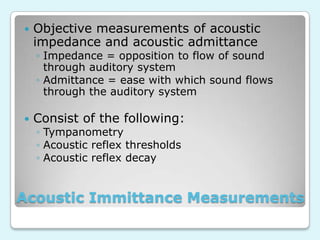
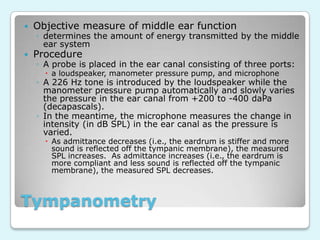
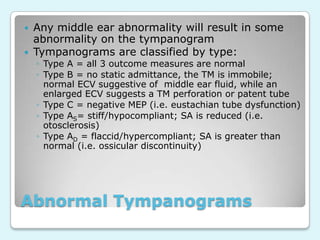
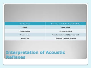
Acoustic Immittance
Measurements
Ozarks Technical Community College
HIS 125


Objective measurements of acoustic
impedance and acoustic admittance

◦ Impedance = opposition to flow of sound
through auditory system
◦ Admittance = ease with which sound flows
through the auditory system



Consist of the following:
◦ Tympanometry
◦ Acoustic reflex thresholds
◦ Acoustic reflex decay

Acoustic Immittance Measurements


Objective measure of middle ear function



Procedure

◦ determines the amount of energy transmitted by the middle
ear system
◦ A probe is placed in the ear canal consisting of three ports:
 a loudspeaker, manometer pressure pump, and microphone

◦ A 226 Hz tone is introduced by the loudspeaker while the
manometer pressure pump automatically and slowly varies
the pressure in the ear canal from +200 to -400 daPa
(decapascals).
◦ In the meantime, the microphone measures the change in
intensity (in dB SPL) in the ear canal as the pressure is
varied.
 As admittance decreases (i.e., the eardrum is stiffer and more
sound is reflected off the tympanic membrane), the measured
SPL increases. As admittance increases (i.e., the eardrum is
more compliant and less sound is reflected off the tympanic
membrane), the measured SPL decreases.

Tympanometry
Admittance decreases,
Reflected SPL increases

Admittance increases,
Reflected SPL
decreases

Normal Tympanogram


Three outcome measures:
static admittance (SA) or maximum
compliance= point along the tympanogram where
the amount of reflected SPL is the least
 ear canal volume (ECV)=physical space between
the end of the probe assembly and the tympanic
membrane
 middle ear pressure (MEP)= pressure along the
x-axis of the tympanogram where the eardrum is
the most compliant or where pressure is equal on
both sides of the tympanic membrane


Tympanometry
Parameter

Normal Range

Ear Canal Volume

0.65-1.75 mL or cm3

Middle Ear Pressure

- 100 to + 50 daPa

Static Admittance

0.3-1.4 mL

Normal Tympanometry Values for
Adults



Any middle ear abnormality will result in some
abnormality on the tympanogram
Tympanograms are classified by type:
◦ Type A = all 3 outcome measures are normal
◦ Type B = no static admittance, the TM is immobile;
normal ECV suggestive of middle ear fluid, while an
enlarged ECV suggests a TM perforation or patent tube
◦ Type C = negative MEP (i.e. eustachian tube dysfunction)
◦ Type AS= stiff/hypocompliant; SA is reduced (i.e.
otosclerosis)
◦ Type AD = flaccid/hypercompliant; SA is greater than
normal (i.e. ossicular discontinuity)

Abnormal Tympanograms
Type B=flat

*Note: based on the
enlarged ECV, this
patient has a patent
tube or a hole in the
TM

Type C=negative
pressure

Abnormal Tympanograms
Type As=stiff,
hypocompliant

Type AD=flaccid,
hypercompliant

Abnormal Tympanograms


If otoscopy reveals significant cerumen
accumulation, a tympanogram will allow
you to determine if an accurate
audiogram can be obtained.
◦ If tympanometry is normal in the presence of
excess cerumen, then there is enough of an
opening in the ear canal for accurate hearing
testing to occur.

Other use of tympanometry


The introduction of a loud sound to the
ear canal of either ear results in the
acoustic reflex, or the contraction of the
stapedius muscle, which causes the
tympanic membrane to stiffen.
◦ This results in a change in middle ear
immittance, which is measured as an increase
in dB SPL by the microphone in the probe
assembly.

The Acoustic Reflex


Acoustic reflex thresholds (ARTs) = the
lowest level in dB HL at which the acoustic
reflex can be elicited (deflection ≥0.02 ml)
◦ Uses the same bracketing technique that is used in
obtaining puretone thresholds
◦ Measured at 500, 1000, 2000, and 4000 Hz
◦ Useful in determining site-of-lesion
 Directly evaluates middle ear status and indirectly
evaluates cochlear and retrocochlear status

◦ ARTs can be measured ipsilaterally (stimulus is in
the probe ear) and contralaterally (stimulus is in
the non-probe ear) via a supra-aural or insert
earphone

Acoustic Reflex Thresholds


Here is a graph of acoustic reflex testing
at 500, with threshold being established
at 85 dBHL
Hearing Status

Expected Acoustic Reflex Threshold (dB HL)

Normal

70-100 dB HL

Conductive Loss

Elevated or Absent

Cochlear Loss

Normal sensation level (SL) or reduced SL

Neural Loss

Normal SL, elevated, or absent

Interpretation of Acoustic
Reflexes
Acoustic Reflex Pathway

From: Martin & Clark, Introduction to Audiology

•As you can see from the
reflex pathway, the
measurement of ARTs not
only provides information
regarding the status of
the middle ear
system, but also of the
inner ear, auditory
nerve, regions of the
lower auditory
brainstem, and the facial
nerve.


Acoustic reflex decay is a measure of the sustainability
of the acoustic reflex, or how long the stapedius muscle
can remain contracted during continuous stimulation.



Procedure:



Outcomes:

◦ Reflex decay is measured with contralateral stimulation at 500 and
1000 Hz at 10 dB SL (re: contralateral acoustic reflex threshold).
◦ The tone is presented continuously for 10 seconds and the patient
is instructed to remain as still as possible.
◦ Negative reflex decay indicates that the magnitude of the
stapedial reflex contraction did not decrease by ≥50% in the first
five seconds of testing
◦ Positive reflex decay indicates that the magnitude of the
stapedial reflex contraction decreased by ≥50% in the first five
seconds of testing
 Positive acoustic reflex decay is highly suggestive of a retrocochlear
pathology and further evaluation by an otologist is strongly
recommended.

Acoustic Reflex Decay


Negative (upper)
and positive
(lower) acoustic
reflex decay at
1000 Hz.



In positive decay,
the stapedius
contracts initially,
but then “lets go”

Acoustic Reflex Decay

Acoustic Immittance Measurements

  • 1.
  • 2.
     Objective measurements ofacoustic impedance and acoustic admittance ◦ Impedance = opposition to flow of sound through auditory system ◦ Admittance = ease with which sound flows through the auditory system  Consist of the following: ◦ Tympanometry ◦ Acoustic reflex thresholds ◦ Acoustic reflex decay Acoustic Immittance Measurements
  • 3.
     Objective measure ofmiddle ear function  Procedure ◦ determines the amount of energy transmitted by the middle ear system ◦ A probe is placed in the ear canal consisting of three ports:  a loudspeaker, manometer pressure pump, and microphone ◦ A 226 Hz tone is introduced by the loudspeaker while the manometer pressure pump automatically and slowly varies the pressure in the ear canal from +200 to -400 daPa (decapascals). ◦ In the meantime, the microphone measures the change in intensity (in dB SPL) in the ear canal as the pressure is varied.  As admittance decreases (i.e., the eardrum is stiffer and more sound is reflected off the tympanic membrane), the measured SPL increases. As admittance increases (i.e., the eardrum is more compliant and less sound is reflected off the tympanic membrane), the measured SPL decreases. Tympanometry
  • 4.
    Admittance decreases, Reflected SPLincreases Admittance increases, Reflected SPL decreases Normal Tympanogram
  • 5.
     Three outcome measures: staticadmittance (SA) or maximum compliance= point along the tympanogram where the amount of reflected SPL is the least  ear canal volume (ECV)=physical space between the end of the probe assembly and the tympanic membrane  middle ear pressure (MEP)= pressure along the x-axis of the tympanogram where the eardrum is the most compliant or where pressure is equal on both sides of the tympanic membrane  Tympanometry
  • 6.
    Parameter Normal Range Ear CanalVolume 0.65-1.75 mL or cm3 Middle Ear Pressure - 100 to + 50 daPa Static Admittance 0.3-1.4 mL Normal Tympanometry Values for Adults
  • 7.
      Any middle earabnormality will result in some abnormality on the tympanogram Tympanograms are classified by type: ◦ Type A = all 3 outcome measures are normal ◦ Type B = no static admittance, the TM is immobile; normal ECV suggestive of middle ear fluid, while an enlarged ECV suggests a TM perforation or patent tube ◦ Type C = negative MEP (i.e. eustachian tube dysfunction) ◦ Type AS= stiff/hypocompliant; SA is reduced (i.e. otosclerosis) ◦ Type AD = flaccid/hypercompliant; SA is greater than normal (i.e. ossicular discontinuity) Abnormal Tympanograms
  • 8.
    Type B=flat *Note: basedon the enlarged ECV, this patient has a patent tube or a hole in the TM Type C=negative pressure Abnormal Tympanograms
  • 9.
  • 10.
     If otoscopy revealssignificant cerumen accumulation, a tympanogram will allow you to determine if an accurate audiogram can be obtained. ◦ If tympanometry is normal in the presence of excess cerumen, then there is enough of an opening in the ear canal for accurate hearing testing to occur. Other use of tympanometry
  • 11.
     The introduction ofa loud sound to the ear canal of either ear results in the acoustic reflex, or the contraction of the stapedius muscle, which causes the tympanic membrane to stiffen. ◦ This results in a change in middle ear immittance, which is measured as an increase in dB SPL by the microphone in the probe assembly. The Acoustic Reflex
  • 12.
     Acoustic reflex thresholds(ARTs) = the lowest level in dB HL at which the acoustic reflex can be elicited (deflection ≥0.02 ml) ◦ Uses the same bracketing technique that is used in obtaining puretone thresholds ◦ Measured at 500, 1000, 2000, and 4000 Hz ◦ Useful in determining site-of-lesion  Directly evaluates middle ear status and indirectly evaluates cochlear and retrocochlear status ◦ ARTs can be measured ipsilaterally (stimulus is in the probe ear) and contralaterally (stimulus is in the non-probe ear) via a supra-aural or insert earphone Acoustic Reflex Thresholds
  • 13.
     Here is agraph of acoustic reflex testing at 500, with threshold being established at 85 dBHL
  • 14.
    Hearing Status Expected AcousticReflex Threshold (dB HL) Normal 70-100 dB HL Conductive Loss Elevated or Absent Cochlear Loss Normal sensation level (SL) or reduced SL Neural Loss Normal SL, elevated, or absent Interpretation of Acoustic Reflexes
  • 15.
    Acoustic Reflex Pathway From:Martin & Clark, Introduction to Audiology •As you can see from the reflex pathway, the measurement of ARTs not only provides information regarding the status of the middle ear system, but also of the inner ear, auditory nerve, regions of the lower auditory brainstem, and the facial nerve.
  • 16.
     Acoustic reflex decayis a measure of the sustainability of the acoustic reflex, or how long the stapedius muscle can remain contracted during continuous stimulation.  Procedure:  Outcomes: ◦ Reflex decay is measured with contralateral stimulation at 500 and 1000 Hz at 10 dB SL (re: contralateral acoustic reflex threshold). ◦ The tone is presented continuously for 10 seconds and the patient is instructed to remain as still as possible. ◦ Negative reflex decay indicates that the magnitude of the stapedial reflex contraction did not decrease by ≥50% in the first five seconds of testing ◦ Positive reflex decay indicates that the magnitude of the stapedial reflex contraction decreased by ≥50% in the first five seconds of testing  Positive acoustic reflex decay is highly suggestive of a retrocochlear pathology and further evaluation by an otologist is strongly recommended. Acoustic Reflex Decay
  • 17.
     Negative (upper) and positive (lower)acoustic reflex decay at 1000 Hz.  In positive decay, the stapedius contracts initially, but then “lets go” Acoustic Reflex Decay