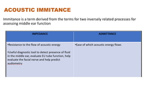
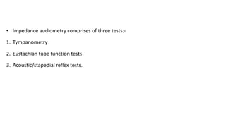
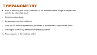
IMPEDANCE
AUDIOMETRY
DR ANKIT (PGT1)
KIMS BHUBNAESWAR
INTRODUCTION
• The first tympanogram was reported by Terkilsen and Thomson in 1959
• It provides wide range of otological and neurological information about
the nature and anatomical site of lesion
USES
a) Objective difference between conductive HL and SNHL
b) Differential diagnosis in cases of conductive deafness
c) Measurement of middle ear pressure and evaluation of eustachian tube function
d) Differential diagnosis of a sensorineural deafness, i.e., whether a lesion is cochlear
or retro-cochlear
e) Identification of site of lesion in facial paralysis and certain brainstem pathologies.
f) To examine the functional or structural integrity of the 7th and 8th cranial nerves,
brainstem lesions and other pathologies related to auditory dysfunction.
ACOUSTIC IMMITANCE
Immitance is a term derived from the terms for two inversely related processes for
assessing middle ear function
• Impedance audiometry comprises of three tests:-
1. Tympanometry
2. Eustachian tube function tests
3. Acoustic/stapedial reflex tests.
TYMPANOMETRY
• A way of measuring how acoustic immittance of the middle ear system changes as air pressure is
varied in the external ear canal.
• Gives information about:-
1. Air pressure status of the middle ear
2. Static acoustic immitance (establishing parameters of stiffness or flaccidity at the ear drum)
3. The integrity and mobility of the eardrum and ossicular chain
4. Resonance point of the middle ear system
TYMPANOMETRY
PRINCIPLE:-
It is defined as measurement of change of impedance of middle ear
at the plane of tympanic membrane as a result of changes in
pressure in external auditory meatus
• Impedance of the medium:- Stiffness, Mass and Friction
• Air has low impedance, but cochlear fluid has high impedance.
• Impedance matching device of auditory system- Middle ear
• ELECTRO-ACOUSTIC BRIDGE:- Efficiency of middle ear to perform impedance
matching.
• Middle ear pathology- Conductive deafness
• Impedance character:- Amount of sound reflected from tympanic membrane depend
upon stiffness (principle of Acoustic Reflex Test)
INSTRUMENTATION
• Major components:-
1. Probe tone oscillator and loudspeaker
2. Monitor microphone
3. Pressure pump and manometer
4. Ipsilateral reflex oscillator and loudspeaker
5. Probe tip
IMPEDANCE AUDIOMETER
• PROBE – 3 aperture
1. Probe tone- 220 or 226 Hz
2. Calculate amount of sound reflecting back
3. Air-pump manometer(300-600mm of water pressure)-
to change stiffness of tympanic membrane
PROCEDURE
• Probe to be applied to the ear.
• Increase the pressure from + 200 mm of H2O to -600 mm of
H2O
• Draw a graph with pressure on the X-axis and compliance
on the Y-axis.
STATIC COMPLIANCE
• Compliance of auditory conductive apparatus as measured
at tympanic membrane.
• In normal middle ear conditions, maximum eardrum
compliance occurs when atmospheric pressure is equal on
both sides of TM (O mm H2O).
PVT(Physical Volume Test):-
• Compliance at +200 mm H2O C1 (TM is completely
stiffened).
• Volume of EAC.
Tympanogram Types:-
• Absolute
• Relative
• Compensated
INTERPRETATION OF TYMPANOGRAM
Measurement of static compliance:-
• Scale of measurement:- 0 to 5.00
• Unit- cc/ml or millimho
• Colour code:- Rd (Rt) Blue (Lt)
• Normal complaince-0.35 to 1.40 ml.
• Normal pressure- +50dkpa to -50dkpa
MEASURMENT OF MIDDLE EAR PRESSURE
• The pressure at which the tympanogram shows highest compliance
(peak of tympanogram).
• Unit-mm of water/ Deca Pascal
• Normal pressure :- +50 to -50 mm of water
• In Paediatric patient:- +25 to -25 mm of water
TYPE AND SHAPE OF TYMPANOGRAM
• TYPE A
• Sharp maximum at 0 mm of water
• Normal ears
2.Type B
• Shows little or no compliance.
• The tympanogram is totally flat.
• Found in:-
• Otitis media with effusion
• Adhesive otitis media
• Perforation of the Tympanic membrane
3. Type C
• Significantly negative middle ear pressure with normal compliance.
• Found in uncomplicated Eustachian tube obstruction.
4. Type As
• Normal middle ear pressure low compliance
tympanogram
• Found in otosclerosis or thickened tympanic
membrane
5. Type Ad
• Normal middle ear pressure with high
compliance.
• Found in ossicular chain discontinuity or
scarring of the tympanic membrane.
GRADIENT OF TYMPANOGRAM
• The half amplitude point on the negative and positive side of tympanogram
peak is determined. This is done by dividing the total amplitude on each side
by 2.
• This midpoint of the amplitude on both sides of the tympanogram peak
marked on the tympanogram and the middle ear pressure corresponding to
that point is noted. The gradient of the tympanogram is the difference
between these two pressure.
EUSTACHIAN TUBE FUNCTION TEST
METHODS:-
• Sonotubometry
• Radiological studies
• Photoelectric method to measure patency
• Assessment of the ventilatory capacity using a sound proof chamber.
• Fluorescien/saccharine to measure mucocillary clearance time
• Impedance audiometers
• The physiological functions of the Eustachian tube
are:-
a) Maintenance of equality of air pressure between
the middle ear and the ambient atmosphere
(ventilatory function).
b) Drainage of the mucus from the ear to the
nasopharynx (mucociliary clearance function).
• The neutralization of the negative middle ear pressure is
brought about by intermittently opening the Eustachian
tube (which normally remains closed) and allowing air to
pass through it to the same level as that of the
atmospheric air pressure.
• The intermittent opening of the eustachian tube is
caused by contraction of the tensor palatine and the
levetor veli palalini muscles which contract during
swallowing.
WILLIAM’S TEST
• Done in intact TM
• Middle ear pressure at 3 conditions:-
• Start of the test (resting pressure)- atmospheric air
pressure (approx. 0 mm of water pressure)
• After the patient swallows (with the nose and mouth
closed), Negative
• After performing valsalva, Positive
Partially impaired tubal function
Grossly impaired tubal function
TOYNBE’S TEST
• Done in perforated TM.
• Done on a pressure difference (i.e., positive pressure
at the tympanic end of the tube and ambient or
atmosphere pressure at the nasopharyngeal end of
the tube).
• The test is carried out for a fixed duration of time,
e.g., 40 seconds (min.) or 160 seconds (max.).
ACOUSTIC/STAPEDIAL REFLEX TEST
• Defined as the lowest intensity required to
elicit a stapedial muscle contraction.
• Objective test
• Non-invasive ,simple
• The time delay of acoustic reflex is thought to
be 10 ms
Acoustic reflex thresholds for tones in patients
with normal hearing are usually 70 to 80 db
above their tone thresholds and about 5db
greater for the contralateral threshold.
• CHARACTERISTICS:-
• Elimination of middle ear pathology
• Differentiation of cochlear and retrocochlear pathology
• Detection of some cases of brain-stem pathologies
• Objective estimation of average hearing threshold level
• Detection of non-organic hearing loss
• Identifying the level of lesion in facial nerve paralysis.
ACOUSTIC/STAPEDIAL REFLEX TEST
• Disorders on the afferent side which abolish reflex are
a) Disease in ipsilateral middle ear causing a moderate to severe degree of
conductive deafness which prevents adequate sound from reaching ipsilateral
cochlea
b) Lesion in ipsilateral cochlea or 8th cranial nerve, i.e., severe sensorineural
deafness which prevents adequate sound from reaching the cochlear nucleus,
c) Lesion in cochlear nucleus or superior olivary complex in the brainstem like
multiple sclerosis, etc.
• Disorders on the efferent side which abolish reflex:-
a. Lesion in the facial nerve nucleus in the brain stem
b. Facial nerve paralysis at a level proximal to the nerve to stapedius like
Ramsay Hunt syndrome
c. Disease of the stapedius muscle like myasthenia gravis
d. Any lesion in the middle ear like otosclerosis, ossicufJr discontinuity or
atelectasis.
INTERPRETATION
IDENTIFICATION OF THE SITE OF LESION IN
FACIAL NERVE PARALYSIS
• Acoustic reflex is present in the ear on the side of facial
paralysis, it indicates the the site of lesion in the facial
nerve is at a point distal to the origin of the nerve to
stapedius in the vertical part of the facial nerve e.g.Bell's
palsy
• Acoustic reflex is absent it indicates the site of lesion is
proximal to the point where the nerve to stapedius
originates e.g. Ramsay Hunt Syndrome.
MALINGERERS
• If acoustic reflexes are present in a so-callad deaf ear
on ipsilateral stimulation or in the other ear on
contralateral stimulation, it indicates that the ear is not
completely deaf and the adequate hearing is present in
that ear.
THANK YOU
Referance:-
Anirban Biswas

IMPEDANCE AUDIOMETRY.pptx

  • 1.
  • 2.
    INTRODUCTION • The firsttympanogram was reported by Terkilsen and Thomson in 1959 • It provides wide range of otological and neurological information about the nature and anatomical site of lesion
  • 3.
    USES a) Objective differencebetween conductive HL and SNHL b) Differential diagnosis in cases of conductive deafness c) Measurement of middle ear pressure and evaluation of eustachian tube function d) Differential diagnosis of a sensorineural deafness, i.e., whether a lesion is cochlear or retro-cochlear e) Identification of site of lesion in facial paralysis and certain brainstem pathologies. f) To examine the functional or structural integrity of the 7th and 8th cranial nerves, brainstem lesions and other pathologies related to auditory dysfunction.
  • 4.
    ACOUSTIC IMMITANCE Immitance isa term derived from the terms for two inversely related processes for assessing middle ear function
  • 6.
    • Impedance audiometrycomprises of three tests:- 1. Tympanometry 2. Eustachian tube function tests 3. Acoustic/stapedial reflex tests.
  • 7.
    TYMPANOMETRY • A wayof measuring how acoustic immittance of the middle ear system changes as air pressure is varied in the external ear canal. • Gives information about:- 1. Air pressure status of the middle ear 2. Static acoustic immitance (establishing parameters of stiffness or flaccidity at the ear drum) 3. The integrity and mobility of the eardrum and ossicular chain 4. Resonance point of the middle ear system
  • 8.
    TYMPANOMETRY PRINCIPLE:- It is definedas measurement of change of impedance of middle ear at the plane of tympanic membrane as a result of changes in pressure in external auditory meatus
  • 9.
    • Impedance ofthe medium:- Stiffness, Mass and Friction • Air has low impedance, but cochlear fluid has high impedance. • Impedance matching device of auditory system- Middle ear • ELECTRO-ACOUSTIC BRIDGE:- Efficiency of middle ear to perform impedance matching. • Middle ear pathology- Conductive deafness • Impedance character:- Amount of sound reflected from tympanic membrane depend upon stiffness (principle of Acoustic Reflex Test)
  • 10.
    INSTRUMENTATION • Major components:- 1.Probe tone oscillator and loudspeaker 2. Monitor microphone 3. Pressure pump and manometer 4. Ipsilateral reflex oscillator and loudspeaker 5. Probe tip
  • 11.
    IMPEDANCE AUDIOMETER • PROBE– 3 aperture 1. Probe tone- 220 or 226 Hz 2. Calculate amount of sound reflecting back 3. Air-pump manometer(300-600mm of water pressure)- to change stiffness of tympanic membrane
  • 12.
    PROCEDURE • Probe tobe applied to the ear. • Increase the pressure from + 200 mm of H2O to -600 mm of H2O • Draw a graph with pressure on the X-axis and compliance on the Y-axis.
  • 13.
    STATIC COMPLIANCE • Complianceof auditory conductive apparatus as measured at tympanic membrane. • In normal middle ear conditions, maximum eardrum compliance occurs when atmospheric pressure is equal on both sides of TM (O mm H2O). PVT(Physical Volume Test):- • Compliance at +200 mm H2O C1 (TM is completely stiffened). • Volume of EAC.
  • 14.
    Tympanogram Types:- • Absolute •Relative • Compensated
  • 16.
  • 17.
    Measurement of staticcompliance:- • Scale of measurement:- 0 to 5.00 • Unit- cc/ml or millimho • Colour code:- Rd (Rt) Blue (Lt) • Normal complaince-0.35 to 1.40 ml. • Normal pressure- +50dkpa to -50dkpa
  • 19.
    MEASURMENT OF MIDDLEEAR PRESSURE • The pressure at which the tympanogram shows highest compliance (peak of tympanogram). • Unit-mm of water/ Deca Pascal • Normal pressure :- +50 to -50 mm of water • In Paediatric patient:- +25 to -25 mm of water
  • 21.
    TYPE AND SHAPEOF TYMPANOGRAM • TYPE A • Sharp maximum at 0 mm of water • Normal ears
  • 22.
    2.Type B • Showslittle or no compliance. • The tympanogram is totally flat. • Found in:- • Otitis media with effusion • Adhesive otitis media • Perforation of the Tympanic membrane
  • 23.
    3. Type C •Significantly negative middle ear pressure with normal compliance. • Found in uncomplicated Eustachian tube obstruction.
  • 24.
    4. Type As •Normal middle ear pressure low compliance tympanogram • Found in otosclerosis or thickened tympanic membrane
  • 25.
    5. Type Ad •Normal middle ear pressure with high compliance. • Found in ossicular chain discontinuity or scarring of the tympanic membrane.
  • 26.
    GRADIENT OF TYMPANOGRAM •The half amplitude point on the negative and positive side of tympanogram peak is determined. This is done by dividing the total amplitude on each side by 2. • This midpoint of the amplitude on both sides of the tympanogram peak marked on the tympanogram and the middle ear pressure corresponding to that point is noted. The gradient of the tympanogram is the difference between these two pressure.
  • 28.
    EUSTACHIAN TUBE FUNCTIONTEST METHODS:- • Sonotubometry • Radiological studies • Photoelectric method to measure patency • Assessment of the ventilatory capacity using a sound proof chamber. • Fluorescien/saccharine to measure mucocillary clearance time • Impedance audiometers
  • 29.
    • The physiologicalfunctions of the Eustachian tube are:- a) Maintenance of equality of air pressure between the middle ear and the ambient atmosphere (ventilatory function). b) Drainage of the mucus from the ear to the nasopharynx (mucociliary clearance function).
  • 30.
    • The neutralizationof the negative middle ear pressure is brought about by intermittently opening the Eustachian tube (which normally remains closed) and allowing air to pass through it to the same level as that of the atmospheric air pressure. • The intermittent opening of the eustachian tube is caused by contraction of the tensor palatine and the levetor veli palalini muscles which contract during swallowing.
  • 31.
    WILLIAM’S TEST • Donein intact TM • Middle ear pressure at 3 conditions:- • Start of the test (resting pressure)- atmospheric air pressure (approx. 0 mm of water pressure) • After the patient swallows (with the nose and mouth closed), Negative • After performing valsalva, Positive
  • 32.
    Partially impaired tubalfunction Grossly impaired tubal function
  • 33.
    TOYNBE’S TEST • Donein perforated TM. • Done on a pressure difference (i.e., positive pressure at the tympanic end of the tube and ambient or atmosphere pressure at the nasopharyngeal end of the tube). • The test is carried out for a fixed duration of time, e.g., 40 seconds (min.) or 160 seconds (max.).
  • 34.
    ACOUSTIC/STAPEDIAL REFLEX TEST •Defined as the lowest intensity required to elicit a stapedial muscle contraction. • Objective test • Non-invasive ,simple • The time delay of acoustic reflex is thought to be 10 ms Acoustic reflex thresholds for tones in patients with normal hearing are usually 70 to 80 db above their tone thresholds and about 5db greater for the contralateral threshold.
  • 35.
    • CHARACTERISTICS:- • Eliminationof middle ear pathology • Differentiation of cochlear and retrocochlear pathology • Detection of some cases of brain-stem pathologies • Objective estimation of average hearing threshold level • Detection of non-organic hearing loss • Identifying the level of lesion in facial nerve paralysis.
  • 36.
  • 37.
    • Disorders onthe afferent side which abolish reflex are a) Disease in ipsilateral middle ear causing a moderate to severe degree of conductive deafness which prevents adequate sound from reaching ipsilateral cochlea b) Lesion in ipsilateral cochlea or 8th cranial nerve, i.e., severe sensorineural deafness which prevents adequate sound from reaching the cochlear nucleus, c) Lesion in cochlear nucleus or superior olivary complex in the brainstem like multiple sclerosis, etc.
  • 38.
    • Disorders onthe efferent side which abolish reflex:- a. Lesion in the facial nerve nucleus in the brain stem b. Facial nerve paralysis at a level proximal to the nerve to stapedius like Ramsay Hunt syndrome c. Disease of the stapedius muscle like myasthenia gravis d. Any lesion in the middle ear like otosclerosis, ossicufJr discontinuity or atelectasis.
  • 39.
  • 40.
    IDENTIFICATION OF THESITE OF LESION IN FACIAL NERVE PARALYSIS • Acoustic reflex is present in the ear on the side of facial paralysis, it indicates the the site of lesion in the facial nerve is at a point distal to the origin of the nerve to stapedius in the vertical part of the facial nerve e.g.Bell's palsy • Acoustic reflex is absent it indicates the site of lesion is proximal to the point where the nerve to stapedius originates e.g. Ramsay Hunt Syndrome.
  • 41.
    MALINGERERS • If acousticreflexes are present in a so-callad deaf ear on ipsilateral stimulation or in the other ear on contralateral stimulation, it indicates that the ear is not completely deaf and the adequate hearing is present in that ear.
  • 42.