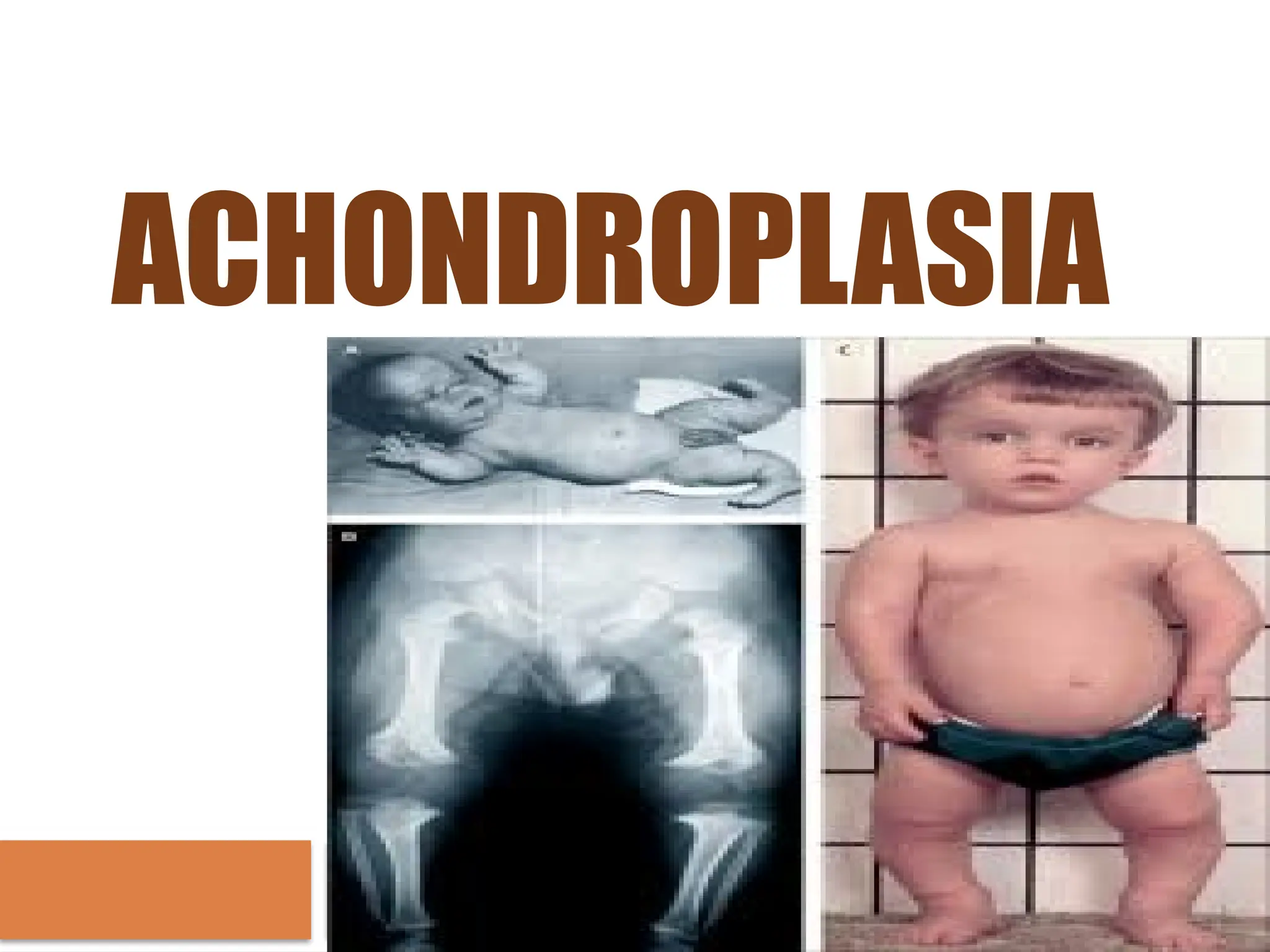
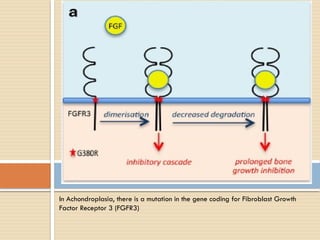
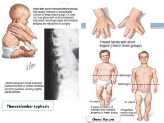
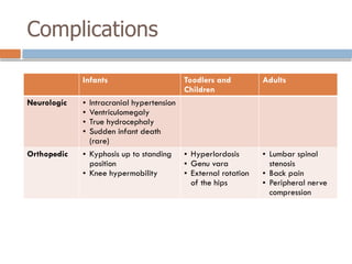
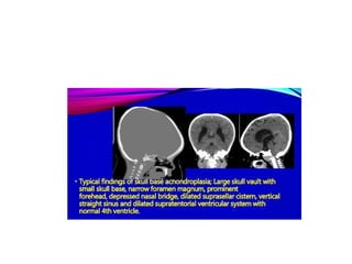
ACHONDROPLASIA
Introduction
 Structural abnormality in the bone leads to
disturbances in growth of the trunk or
extremities.
 More than 300 bone dysplasias have been
described, most of which are extremely rare
 Most of the bone dysplasias result in short
stature
 Some of the dysplasias are genetically
inherited, whereas others are not.
Anatomy of epiphysis
The Stages of Long Bone Development
 During ossification, osteoblasts transform into
osteoid, which progressively calcified, also by
osteoblast action. As the bone grows, the
osteoblasts become trapped in the matrix of
their own making and become osteocytes.
 In mature bone, a fine balance of osteoblast
and osteoclast activity maintains normal bone
structure
Definition
 “Achondroplasia” was first used by Parrot in 1878 to
describe a rhizomelic form of short-limbed dwarfism
associated with enlarged head, depressed nasal bridge,
short stubby trident hands, lordotic lumbar spine,
prominent buttocks, and protuberant abdomen.
 According to Salter Harris, Achondroplasia is the failure of
longitudinal growth in the cartilage of epiphyseal plate.
 Achondroplasia is inherited as an autosomal dominant trait
 Ninety percent of cases are the result of spontaneous
mutation
Epidemiology
 Worldwide, achondroplasia is the most common skeletal
dysplasia and dwarfism-form
 Incidence range between 1.3 per 100.000 live births and
1.5 per 10.000 live births
 Approximately 10,000 individuals are estimated to have
achondroplasia in the United States.
 About 80% of all "little people" have achondroplasia.
 The frequency is equal in males and females and all
races
Ossification
Enchondral Ossification
• Bone replace a cartilage template
• Osteoclast removes the cartilage, and
osteoblasts make the new matrix, which is then
mineralized
• Longitudinal bone growth
Netter’s Concise Orthopaedic Anatomy 2nd
Edition. 2010
Intramembranous Ossification
• Bone develops directly from mesenchymal
cell, then differentiate into osteoblast
• Flat Bone formation : Cranium, Pelvic, Clavicle
Ossification
Netter’s Concise Orthopaedic Anatomy 2nd
Edition. 2010
Ossification
Appositional
• Periosteal –mediated long bone growth (width)
• Osteoblasts make new matrix/bone on top of
existing bone.
Miller’s review of Orthopaedics Seventh Edition. 2016
The physis provides
longitudinal growth in
long bones.
In Proliferative zone,
chondrocytes divide and
stack into columns
In Achondroplasia, there is a mutation in the gene coding for Fibroblast Growth
Factor Receptor 3 (FGFR3)
Diagnosis
 Anamnesis
 Physical examination
 Laboratory examination
 Radiographic findings
Anamnesis
 Previous disease
 Family history
 History of development and growth
 Pregnancy history
 Uterine trauma
 Infection while pregnant
 Teratogenic events
 History during labour
 Nutritional status
 Previous trauma
Physical Examination
Penting untuk menentukan potensi tinggi genetik anak
dan dihitung dengan cara sebagai berikut:
Physical Examination
Craniofacial features
are as follows:
-Large calvarial bones in
contrast to the small
cranial base and facial
bones
-True megalencephaly
(large head) with frontal
bossing
-Midface hypoplasia
Physical Examination
Skeletal features are as follows:
 Disproportionate short stature: Male average adult
height 131 ± 5.6 cm; female average adult height
124 ± 5.9 cm; average adult height for both
approximately 4 feet
Thoracolumbar kyphosis
Genu Varum
Physical Examination
Neurologic
findings are as
follows:
-Hypotonia in
infancy and early
childhood
-Delayed motor
milestones
Laboratory Examination
 Direct DNA analysis of FGFR3 mutations is
used for prenatal screening in families at risk
(ie, parents who are heterozygous for either
the G1138A or the G1138C mutation).
Radiology Examination
 Fetal USG → Early diagnosis, skeletal anomalies
and measure long bones (fetal femoral length)
and evaluate the skull.
Plain X-Ray:
- Os cranium is relatively
enlarged compared to the face
accompanied by frontal
protrusion.
- Foramen magnum shrinks and
irregular funnel-shapped shape.
Plain X-Ray:
- Chest anteroposterior diameter
decreases with anterior rib
shortening.
-Bones of bone from the
hands and feet look short
and wide
-Trident-shaped hands are
often found
lumbar kifosis
- its length
decreases,
especially in the
proximal limb
segment
- The fibula
extends and
bends
Radiology Examination
Head CT Scan → neuroanatomic
abnormalities consistent with arrested
hydrocephalus, enlarged cranial ventricles,
changes in the corpus callosum are seen.
MRI → narrowing of the foramen
magnum and lumbar canal stenosis
MRI Brain
A large cranial vault with relatively short skull base with prominent forehead,
depressed nasal bridge Narrowing of the foramen magnum with cervicomedullary
kinking.
Differential Diagnosis
 Hypochondroplasia (skeletal displasia)
 Thanatophoric dysplasia
 SADDAN Syndrome → Severe Achondroplasia
with Development Delay and Acanthosis
Nigricans(skin pigmentation)
 Pseudoachondroplasia
Treatments and Managements
Based on clinical manifestations
 Short stature → GH therapy, extended limb
lengthening (complications are frequent)
 Kyphosis → bracing support to prevent
persistence of thoracolumbar kyphosis
 Hydrocephalus → know the symptomps of
increased ICP → ventriculoperitoneal shunting
Thoracolumbar Spinal Brace
Complications
Infants Toodlers and
Children
Adults
Neurologic • Intracranial hypertension
• Ventriculomegaly
• True hydrocephaly
• Sudden infant death
(rare)
Orthopedic • Kyphosis up to standing
position
• Knee hypermobility
• Hyperlordosis
• Genu vara
• External rotation
of the hips
• Lumbar spinal
stenosis
• Back pain
• Peripheral nerve
compression
THANK
YOU
Achondroplasia.pptx.......................

Achondroplasia.pptx.......................

  • 1.
  • 2.
    Introduction  Structural abnormalityin the bone leads to disturbances in growth of the trunk or extremities.  More than 300 bone dysplasias have been described, most of which are extremely rare  Most of the bone dysplasias result in short stature  Some of the dysplasias are genetically inherited, whereas others are not.
  • 3.
  • 4.
    The Stages ofLong Bone Development  During ossification, osteoblasts transform into osteoid, which progressively calcified, also by osteoblast action. As the bone grows, the osteoblasts become trapped in the matrix of their own making and become osteocytes.  In mature bone, a fine balance of osteoblast and osteoclast activity maintains normal bone structure
  • 5.
    Definition  “Achondroplasia” wasfirst used by Parrot in 1878 to describe a rhizomelic form of short-limbed dwarfism associated with enlarged head, depressed nasal bridge, short stubby trident hands, lordotic lumbar spine, prominent buttocks, and protuberant abdomen.  According to Salter Harris, Achondroplasia is the failure of longitudinal growth in the cartilage of epiphyseal plate.  Achondroplasia is inherited as an autosomal dominant trait  Ninety percent of cases are the result of spontaneous mutation
  • 6.
    Epidemiology  Worldwide, achondroplasiais the most common skeletal dysplasia and dwarfism-form  Incidence range between 1.3 per 100.000 live births and 1.5 per 10.000 live births  Approximately 10,000 individuals are estimated to have achondroplasia in the United States.  About 80% of all "little people" have achondroplasia.  The frequency is equal in males and females and all races
  • 7.
    Ossification Enchondral Ossification • Bonereplace a cartilage template • Osteoclast removes the cartilage, and osteoblasts make the new matrix, which is then mineralized • Longitudinal bone growth Netter’s Concise Orthopaedic Anatomy 2nd Edition. 2010
  • 8.
    Intramembranous Ossification • Bonedevelops directly from mesenchymal cell, then differentiate into osteoblast • Flat Bone formation : Cranium, Pelvic, Clavicle Ossification Netter’s Concise Orthopaedic Anatomy 2nd Edition. 2010
  • 9.
    Ossification Appositional • Periosteal –mediatedlong bone growth (width) • Osteoblasts make new matrix/bone on top of existing bone. Miller’s review of Orthopaedics Seventh Edition. 2016
  • 10.
    The physis provides longitudinalgrowth in long bones. In Proliferative zone, chondrocytes divide and stack into columns
  • 11.
    In Achondroplasia, thereis a mutation in the gene coding for Fibroblast Growth Factor Receptor 3 (FGFR3)
  • 12.
    Diagnosis  Anamnesis  Physicalexamination  Laboratory examination  Radiographic findings
  • 13.
    Anamnesis  Previous disease Family history  History of development and growth  Pregnancy history  Uterine trauma  Infection while pregnant  Teratogenic events  History during labour  Nutritional status  Previous trauma
  • 14.
    Physical Examination Penting untukmenentukan potensi tinggi genetik anak dan dihitung dengan cara sebagai berikut:
  • 15.
    Physical Examination Craniofacial features areas follows: -Large calvarial bones in contrast to the small cranial base and facial bones -True megalencephaly (large head) with frontal bossing -Midface hypoplasia
  • 16.
    Physical Examination Skeletal featuresare as follows:  Disproportionate short stature: Male average adult height 131 ± 5.6 cm; female average adult height 124 ± 5.9 cm; average adult height for both approximately 4 feet
  • 17.
  • 18.
    Physical Examination Neurologic findings areas follows: -Hypotonia in infancy and early childhood -Delayed motor milestones
  • 19.
    Laboratory Examination  DirectDNA analysis of FGFR3 mutations is used for prenatal screening in families at risk (ie, parents who are heterozygous for either the G1138A or the G1138C mutation).
  • 20.
    Radiology Examination  FetalUSG → Early diagnosis, skeletal anomalies and measure long bones (fetal femoral length) and evaluate the skull.
  • 21.
    Plain X-Ray: - Oscranium is relatively enlarged compared to the face accompanied by frontal protrusion. - Foramen magnum shrinks and irregular funnel-shapped shape. Plain X-Ray: - Chest anteroposterior diameter decreases with anterior rib shortening.
  • 22.
    -Bones of bonefrom the hands and feet look short and wide -Trident-shaped hands are often found lumbar kifosis
  • 23.
    - its length decreases, especiallyin the proximal limb segment - The fibula extends and bends
  • 24.
    Radiology Examination Head CTScan → neuroanatomic abnormalities consistent with arrested hydrocephalus, enlarged cranial ventricles, changes in the corpus callosum are seen. MRI → narrowing of the foramen magnum and lumbar canal stenosis
  • 25.
    MRI Brain A largecranial vault with relatively short skull base with prominent forehead, depressed nasal bridge Narrowing of the foramen magnum with cervicomedullary kinking.
  • 26.
    Differential Diagnosis  Hypochondroplasia(skeletal displasia)  Thanatophoric dysplasia  SADDAN Syndrome → Severe Achondroplasia with Development Delay and Acanthosis Nigricans(skin pigmentation)  Pseudoachondroplasia
  • 27.
    Treatments and Managements Basedon clinical manifestations  Short stature → GH therapy, extended limb lengthening (complications are frequent)  Kyphosis → bracing support to prevent persistence of thoracolumbar kyphosis  Hydrocephalus → know the symptomps of increased ICP → ventriculoperitoneal shunting
  • 30.
  • 31.
    Complications Infants Toodlers and Children Adults Neurologic• Intracranial hypertension • Ventriculomegaly • True hydrocephaly • Sudden infant death (rare) Orthopedic • Kyphosis up to standing position • Knee hypermobility • Hyperlordosis • Genu vara • External rotation of the hips • Lumbar spinal stenosis • Back pain • Peripheral nerve compression
  • 32.