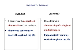
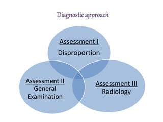
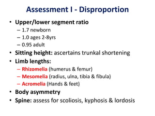
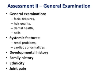
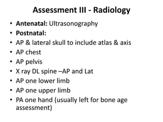
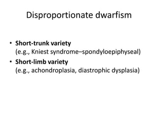
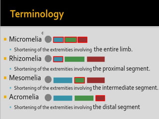
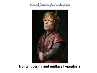
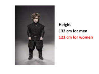
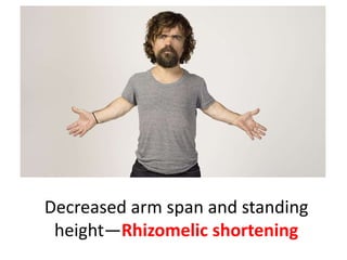
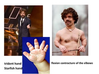
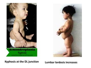
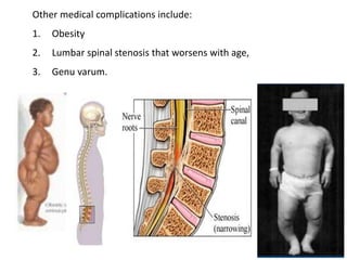
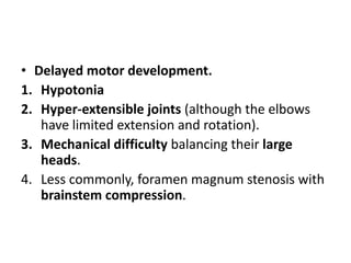
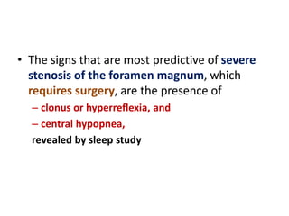
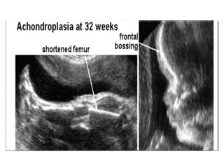
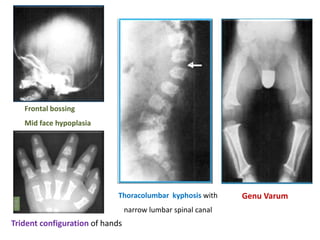
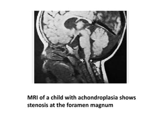
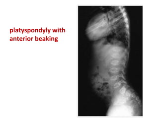
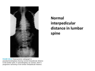
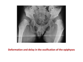
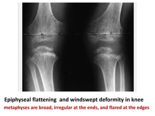
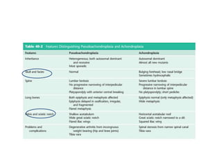
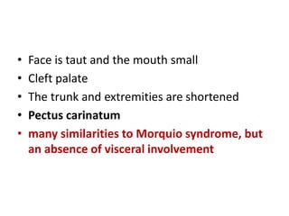
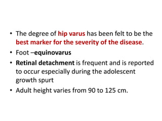
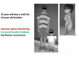
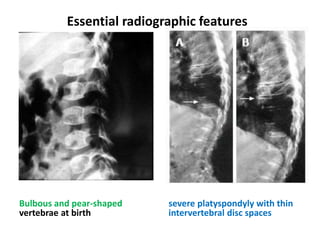
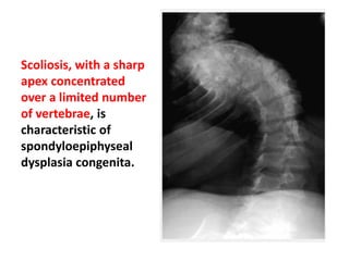
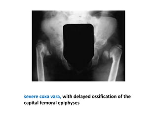
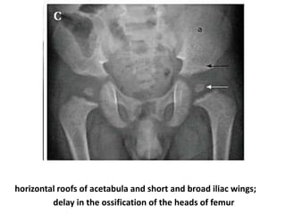
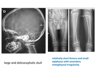
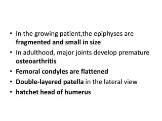
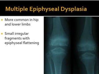
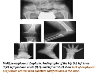
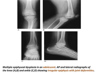
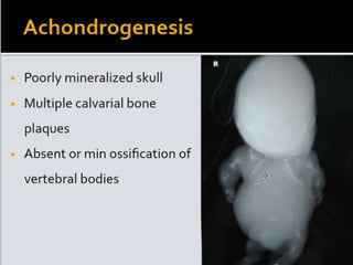
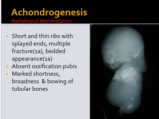
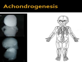
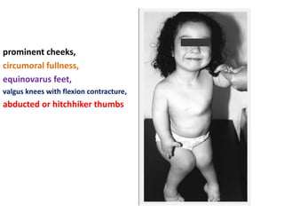
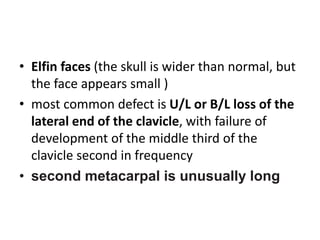
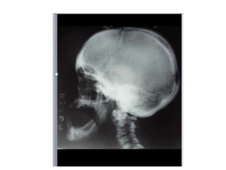
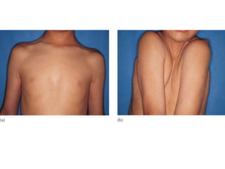
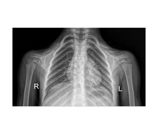
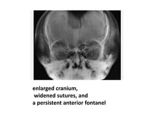
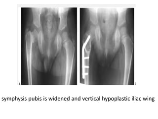
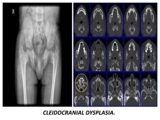
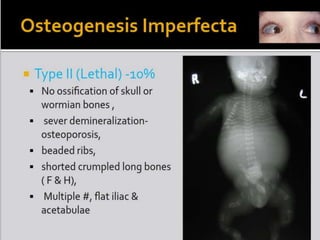
Skeletal dysplasias and dwarfism
Dr G Praveen Kumar Reddy
Introduction
• Bone dysplasias ( Dwarfism)
• Greek word – Dys- disordered
Plasia – development
• Shortening of the growing bones in different
proportions due to multiple gene defects
• >200 disorders, abnormal growth of bones
and cartilage
• Height less than 147cms, <3rd percentile
Dysplasia
• Disorders with generalized
abnormality of the skeleton.
• Phenotype continues to
evolve throughout the life.
Dysostosis
• Disorders with
abnormality of a single or
multiple bones.
• Phenotypically remains
static throughout the life.
Nomenclature and Classification
• Sir Thomas Fairbank was among the first to
try to classify the skeletal dysplasias, in his
Atlas of General Affections of the Skeleton.
• In Dynamic Classification of Bone Dysplasias,
Rubin further refined the classification
schemes, grouping the dysplasias according to
the anatomic distribution of bone changes
Assessment I
Disproportion
Assessment III
Radiology
Assessment II
General
Examination
Assessment I - Disproportion
• Upper/lower segment ratio
– 1.7 newborn
– 1.0 ages 2-8yrs
– 0.95 adult
• Sitting height: ascertains trunkal shortening
• Limb lengths:
– Rhizomelia (humerus & femur)
– Mesomelia (radius, ulna, tibia & fibula)
– Acromelia (Hands & feet)
• Body asymmetry
• Spine: assess for scoliosis, kyphosis & lordosis
Assessment II – General Examination
• General examination:
– facial features,
– hair quality,
– dental health,
– nails
• Systemic features:
– renal problems,
– cardiac abnormalities
• Developmental history
• Family history
• Ethnicity
• Joint pain
Assessment III - Radiology
• Antenatal: Ultrasonography
• Postnatal:
• AP & lateral skull to include atlas & axis
• AP chest
• AP pelvis
• X ray DL spine –AP and Lat
• AP one lower limb
• AP one upper limb
• PA one hand (usually left for bone age
assessment)
Dwarfism
Proportionate Disproportionate
Symmetrical decrease
in both trunk & limb
Short trunk
variety
Short limb
variety
Rhizomelia
(Proximal part)
Mesomelia
(Middle part)
Acromelia
(Distal part)
Micromelia
(entire limb)
• Proportionate-
symmetric decrease in both trunk and limb length.
• Examples
– Constitutional
– Familial
– Endocrinopathies
– mucopolysaccharidoses
Disproportionate dwarfism
• Short-trunk variety
(e.g., Kniest syndrome–spondyloepiphyseal)
• Short-limb variety
(e.g., achondroplasia, diastrophic dysplasia)
Source
• chondro = of cartilage
• osteo = of bone
• spondylo = of the vertebrae
• 1 in 30,000 to 1 in 50,000
• Most common type of dwarfism.
• Autosomal dominant disorder
• 75-80% patients have de novo mutation.
• Gain-of-function mutation in the gene for FGFR3
slows endochondral growth
• paternal age----- chances of achondroplasia
• Limit endochondral bone formation in the
proliferative zone of the physis.
• Intramembranous and periosteal ossification
processes are normal
frontal bossing and midface hypoplasia
Abnormal growth of the skull and facial bones results in:
1. Midface hypoplasia
2. A small cranial base
3. Small cranial foramina.
*The mid face hypoplasia causes :
1. Dental crowding-Maxillary hypoplasia
2. Obstructive apnea-
3. Otitis media-eustachian tubes may not function normally
4. Flat nose at bridge
*Narrowing of the jugular foramina is believed to increase
intracranial venous pressure and thereby to cause hydrocephalus.
Narrowing of the foramen magnum often causes compression of the
brainstem at the craniocervical junction in approximately 10% of
patients and results in an increased frequency of :
1. Hypotonia
2. Quadriparesis
3. Failure to thrive
4. Central apnea
5. Sudden death.
Between 3% and 7% of patients die unexpected during their first year of
life because of brainstem compression (central apnea) or obstructive
apnea.
Height
132 cm for men
122 cm for women
trunk length is within the lower range of normal
Decreased arm span and standing
height—Rhizomelic shortening
fingertips usually reach only to the
tops of the greater trochanters
trident hand
Starfish hand
flexion contracture of the elbows
Kyphosis at the DL junction Lumbar lordosis increases
Other medical complications include:
1. Obesity
2. Lumbar spinal stenosis that worsens with age,
3. Genu varum.
Intelligence is normal
• The overall functional health score is not
dramatically lower.
• Life expectancy is not significantly diminished
• Ligamentous laxity is common at the knees
and ankles
• Delayed motor development.
1. Hypotonia
2. Hyper-extensible joints (although the elbows
have limited extension and rotation).
3. Mechanical difficulty balancing their large
heads.
4. Less commonly, foramen magnum stenosis with
brainstem compression.
• The signs that are most predictive of severe
stenosis of the foramen magnum, which
requires surgery, are the presence of
– clonus or hyperreflexia, and
– central hypopnea,
revealed by sleep study
• The diagnosis of achondroplasia is usually confirmed by
radiographic findings.
• The features of achondroplasia cannot be detected by
prenatal ultrasonography before 24 week’s gestation.
• DNA testing can be performed when both of the
parents are affected and ambiguous cases but is
usually not necessary for the diagnosis to be made.
• double homozygous--stillborn or die
shortly after birth.
• Prenatal diagnosis before 20 weeks of
gestation is available only by molecular
testing of fetal DNA.
• Short tubular long bones, with a relative increase in
bony diameter & density.
• Metaphyses - widened & flared,
• Epiphyses - uninvolved.
• Growth plates - U or V shaped (best seen at distal
femur).
• Metacarpal, metatarsal, & phalangeal bones - short &
thick.
• Widened proximal femoral metaphyses, short femoral
necks - abnormalities in longitudinal growth.
• Ossification of proximal femoral epiphysis is delayed to
more than 1 year of age.
• Greater trochanter is normal in size as formed by
periosteal ossification - decrease in articulotrochanteric
distance.
• True coxa vara is absent, but overgrowth of greater
trochanter - appearance of varus.
• Cervical instability-not usually seen in this
Frontal bossing
Mid face hypoplasia
Thoracolumbar kyphosis with
narrow lumbar spinal canal
Trident configuration of hands
Genu Varum
Antero-posterior radiograph
showing V shaped distal
femoral physis
Radial head dislocation
Lateral radiograph showing
posterior scalloping
of vertebral bodies
& shortened pedicles
short & broad iliac wings,
champagne glass shaped pelvic inlet,
flat acetabula
decreasing interpedicular distance in distal lumbar spine
MRI of a child with achondroplasia shows
stenosis at the foramen magnum
• Treatment of the complications of achondroplasia.
• During infancy and early childhood, patients must be
monitored for :
- Chronic otitis media
- Hydrocephalus
- Brainstem compression
- Obstructive apnea and treated as necessary.
• If stenosis is suspected, sleep studies should
be used for evaluating brain stem functions
• If the diagnosis of foramen magnum stenosis
is made and the clinical picture persists
decompression of the brain stem
• Early hearing screening
• Obstructive sleep apnea –adenotonsillectomy or
more advanced procedures to enlarge the airway.
• Clinical hydrocephalus--ventriculoperitoneal shunt
• During later childhood and through early adulthood, patients
must be monitored for symptomatic :
- Spinal stenosis
- Genu varum
- Obesity.
- Dental complications
- Chronic otitis media and treated as necessary.
• Treatment of the spinal stenosis usually requires surgical
decompression and stabilization of the spine.
• Obesity-more common than in the general
population.
– followed up using
• triceps skinfold thickness
• weight/height squared
• Weight control measures should be instituted
when these values exceed 95% of those for the
general population
• Both growth hormone therapy and surgical
lengthening of the lower legs have been
promoted for treatment of the short stature.
• Both therapies remain controversial.
• Four per million- one of the more common skeletal
dysplasias
• First described in 1959 by Maroteaux and Lamy as a
form of SED.
• Mutation in COMP
• Premature death of physeal chondrocytes results in
decreased linear growth
• Late-onset physical findings and milder spinal
involvement-distinct from SED
• Growth tapers- falls to less than the fifth
percentile by the age of 2 years
• This pattern of progressive involvement is
typical of storage disorders, which is
essentially the nature of this condition.
• Usually, the diagnosis is made by the age of 2
to 4 years.
• skull and facies in pseudoachondroplasia are
normal
• Rhizomelic shortening- both the epiphyses
and metaphyses are involved
• Platyspondyly-spine involved
• Ligamentous laxity
• Knees are most commonly in excessive valgus
or windswept
• waddling gait-hips are dysplastic
• hands are not trident-shaped
• The mean adult height is 119 cm
• normal intelligence, and a normal life
expectancy
• Normal interpedicular distance in LS
head and trunk are
normal
rhizomelic shortening
hands and feet are short
and broad
platyspondyly with
anterior beaking
Normal
interpedicular
distance in lumbar
spine
Deformation and delay in the ossification of the epiphyses
Epiphyseal flattening and windswept deformity in knee
metaphyses are broad, irregular at the ends, and flared at the edges
• Characterized by disproportionate dwarfism with
progressive involvement of the spine and epiphyses
of the long bones.
• There are two major types of SED.
– The congenita type is detectable at birth
– tarda type manifests later in childhood
Spondyloepiphyseal Dysplasia
Congenita
• Obvious short-trunk dwarfism and variable degrees
of coxa vara, accompanied by abnormal epiphyses
and vertebral flattening
• Autosomal dominant
• Chromosome 12
• Defect in type II collagen(mutation in COL2A1 gene)
• Femoral heads are not apparent on radiographs
until the patient is approximately 5 years of age
• Face is taut and the mouth small
• Cleft palate
• The trunk and extremities are shortened
• Pectus carinatum
• many similarities to Morquio syndrome, but
an absence of visceral involvement
• The degree of hip varus has been felt to be the
best marker for the severity of the disease.
• Foot –equinovarus
• Retinal detachment is frequent and is reported
to occur especially during the adolescent
growth spurt
• Adult height varies from 90 to 125 cm.
12-year-old boy is with his
14-year-old brother
extreme spinal shortening,
increased lumbar lordosis,
hip flexion contracture
Bulbous and pear-shaped
vertebrae at birth
severe platyspondyly with thin
intervertebral disc spaces
Essential radiographic features
Scoliosis, with a sharp
apex concentrated
over a limited number
of vertebrae, is
characteristic of
spondyloepiphyseal
dysplasia congenita.
severe coxa vara, with delayed ossification of the
capital femoral epiphyses
horizontal roofs of acetabula and short and broad iliac wings;
delay in the ossification of the heads of femur
large and dolicocephalic skull
relatively short femurs and small
epiphyses with secondary
metaphyseal irregularity
Spondyloepiphyseal Dysplasia Tarda
• Later age at diagnosis and milder features(clinical
attention when the child is approximately 4 years
old, at the earliest)
• Autosomal recessive or X-linked recessive
• The spine and only the larger joints are affected.
• Height is affected only minimally
• Adult height may be 152 cm or more
• Armspan is significantly longer than height
• The condition may be first diagnosed as
bilateral Perthes syndrome
• SED involvement is symmetric, whereas in
bilateral Legg-Calvé-Perthes disease
involvement is discordant, with one hip more
radiographically affected than the other
• Coxa magna
Platyspondyly with
heaped-up or hump-shaped
appearance on the posterior
two-thirds of end-plates
Typical mild
flattening of the
vertebral
bodies, but no
scoliosis.
Bilateral flattened femoral heads, short necks and
premature degenerative changes
• 2 types
– Conradi-Hünermann syndrome- XR-normal life
expectancy(arylsulfatase E (ARSE) gene mutation)
– Autosomal recessive- fatal during the first year of
life, rhizomelic form.
Conradi-Hünermann syndrome
• Short stature,
• dry and scaly skin,
• occasional heart defects, and
• cataracts
• Congenital vertebral anomalies - scoliosis or
kyphosis
• normal life spans
Rhizomelic form(AR)
• In the rhizomelic type, Cataracts tend to be bilateral
and symmetric. In the X-linked dominant type, they are
usually asymmetric and often unilateral
• lack of cataracts is characteristic of the autosomal
dominant form of chondrodysplasia punctata.
• Die in infancy of respiratory complications
• Hallmark of the disease is the presence of multiple punctate
opacities(non specific) in the unossified cartilage at the ends
of the long bones, the tarsals, the pelvis, and the vertebrae.
• Early diagnosis is important because the characteristic
punctate calcifications disappear within the first year of life
• Peculiar appearance of a duplicate calcaneus (also in
thanatophoric dysplasia)
• Defect in COL2A1 gene
• Characterized by
– Dwarfism
– Progressive joint stiffness and contractures
– Retinal detachment
– Cleft palate, midface hypoplasia
– hearing loss
– Kyphoscoliosis is a hallmark of the disease.
• Autosomal dominant: detected at birth
• a disorganized physeal growth plate; soft,
crumbly cartilage with a “Swiss cheese”
appearance
• Rhizomelic involvement
• presence of enlarged cartilaginous epiphyses
and sclerosis within the epiphysis differentiates
Kniest dysplasia from SED congenita.
• Kniest dysplasia resembles Morquio syndrome
radiographically, however, two conditions can be
distinguished by laboratory testing of the urine
• Failure of normal mineralization of the zone of
provisional calcification that leads to widened
physes and enlarged knobby metaphyses
• Epiphyses are spared, and thus arthritis rarely
develops
McKusick-type
• cartilage-hair hypoplasia
• Increased risk of viral infection especially
Varicella zoster
• Hirschsprung disease, intestinal
malabsorption, and megacolon may also
develop
• Increased risk of malignancy, such as
lymphoma, sarcoma, and skin cancer
• Life expectancy is decreased
light, sparse hair,
disproportionate short stature
pectus excavatum
varus deformities of the LL
increased lumbar lordosis
flexion contractures of the
elbows
• more shortening and less
varus of the long bones
than is seen in the Schmid
type
• metaphyseal involvement
is more evenly distributed
• Mild coxa vara,
• bowing of the femurs and
tibias with metaphyseal
expansion, and
• irregular zones of
provisional calcification
Schmid-type
• Autosomal dominant
• Mutations in type X collagen
• D/D - nutritional and vitamin D resistant rickets
normal serum chemistry values.
• Presents with
– leg pains
– varus knees and ankles
– short stature
– waddling gait
Jansen type
• Defect in the receptor for parathyroid
hormone and parathyroid hormone-related
protein(PTRP)
• may have hypercalcemia
• more severe metaphyseal changes
• AD inheritance
• heterogeneous disorder
– Mutations in the gene for COMP on chromosome 19
– COL9A2 mutations
– matrilin-3 mutations
• intracytoplasmic inclusions similar to
pseudoachondroplasia
• produces symptoms mainly in those bones
with significant load bearing
• Historically
– Ribbing dysplasia--mild type
– Fairbank dysplasia--more severe type
• Patients typically present later in childhood
– Joint pain in the lower extremities,
– Decreased range of motion,
– Gait disturbance, or
– Angular deformities of the knees
– Flexion contractures of knees or elbows
– Somewhat short stature, ranging from 145 to 170 cm
• The face and spine are normal.
• There is no visceral involvement.
• may be easily confused with those of bilateral Perthes
disease
• In Perthes disease, usually one hip is involved before
the other, so that each hip is in a different stage of the
disease
Metaphyseal cysts are seen in Perthes disease, but not
in MED
• MED is distinguished from SED by the absence of
severe vertebral changes
• In the growing patient,the epiphyses are
fragmented and small in size
• In adulthood, major joints develop premature
osteoarthritis
• Femoral condyles are flattened
• Double-layered patella in the lateral view
• hatchet head of humerus
Multiple epiphyseal dysplasia. Radiographs of the hip (A), left knee
(B,C), left foot and ankle (D,E), and left wrist (F) show lack of epiphyseal
ossification centers with punctate calcifications in the knee.
Multiple epiphyseal dysplasia in an adolescent. AP and lateral radiographs of
the knee (A,B) and ankle (C,D) showing irregular epiphysis with joint deformities.
Thanatophoric dysplasia.
• most numerous, disparate, and severe skeletal abnormalities
• Diastrophic-distorted
• rhizomelic shortening
• autosomal recessive and is extremely rare
• Lack of sulfation of glycosaminoglycans (GAGs) in articular
cartilage renders the cartilage vulnerable to early degeneration
• Intelligence is normal.
• double-layered manubrium, which is specific to this dysplasia
prominent cheeks,
circumoral fullness,
equinovarus feet,
valgus knees with flexion contracture,
abducted or hitchhiker thumbs
Cervical kyphosis
Cleidocranial dysostosis
• Disorder in which the bones formed by
intramembranous ossification (primarily the
clavicles,cranium, and pelvis) are abnormal
• Autosomal dominant
• Chromosome 6
• Identified within the first 2 years of life
• Elfin faces (the skull is wider than normal, but
the face appears small )
• most common defect is U/L or B/L loss of the
lateral end of the clavicle, with failure of
development of the middle third of the
clavicle second in frequency
• second metacarpal is unusually long
enlarged cranium,
widened sutures, and
a persistent anterior fontanel
symphysis pubis is widened and vertical hypoplastic iliac wing
CLEIDOCRANIAL DYSPLASIA.
short, horizontally running rib with widening of
the costochondral connections.
• osteoclast defect
• Three clinical presentations of osteopetrosis
– infantile or autosomal recessive form
– adult form or autosomal dominant form
( Albers-Schönberg Disease = Marble Bone
Disease(type 2 variety)
– An intermediate clinical type
Autosomal Recessive Osteopetrosis
• Infantile autosomal recessive osteopetrosis is the more severe
form that tends to present earlier. Hence, it is referred to as
"infantile" and "malignant“, compared to the autosomal
dominant osteopetrosis.
• The natural history of the condition means that by age 6, 70% of
the affected will die.
• Most of the remainder have a very poor quality of life with death
resulting by the age of ≈ 10.
Clinical Features
• Those who survive childbirth present with :
• failure to thrive
• cranial nerve entrapment
• optic nerve compression and blindness,
• facial nerve dysfunction, and
• sensorineural deafness
• snuffling (nasal sinus architecture abnormalities)
• hypercalcaemia
• pancytopenia (anemia, leukopenia and thrombocytopenia)
• hepatosplenomegaly (extramedullary haemopoesis)
• infections of the bones or mandibles
• intracerebral hemorrhage (thrombocytopenia)
• Lymphadenopathy
• A prominent forehead and broad upper skull with
hypertelorism
• One of the commonest presentations is ocular disturbance:
failure to establish fixation, nystagmus or strabismus.
(Searching nystagmus )
Lateral radiograph of the skull reveals diffuse thickening of the calvarium, most
significant in the region of the occiput. The partially visualized upper cervical
vertebrae and maxilla are also dense and thickened.
Severe bilateral optic canal narrowing (arrows) in a 4-yr-old patient with
complete loss of vision in the left eye and 20/80 visual acuity in the right eye.
Axial T2-weighted FSE image shows optic canal stenosis and optic nerve atrophy
Autosomal dominant osteopetrosis
• less severe than its autosomal recessive mate.
• "benign" or "adult" since patients survive into
adulthood.
• 50% patients are asymptomatic
• Recurrent fractures
• Mild anemia
• Rarely cranial nerve palsy
.
• X-ray findings
• Diffuse osteosclerosis: Bone-within-bone appearance
• Cortical thickening with medullary encroachment
• Erlenmeyer flask deformity = clublike long bones due to
lack of tubulization + flaring of ends
• "Sandwich" vertebrae = alternating sclerotic + radiolucent
transverse metaphyseal lines (phalanges, iliac bones)
indicate fluctuating course of disease
sandwich vertebrae /
rugger jersey appearance
increased density of osseous structures due to
accumulation of immature bone.
Generalized increased density of bones &
alternating areas of increased & decreased density
in metaphyses (bone-within-bone appearance).
Densely sclerotic bone with Erlenmeyer flask deformity of the femurs with
under-tubulularization (lack of trabeculation) block femoral meatphyses.
• Fractures are treated by conventional methods, but slow to heal
• Intraoperative fractures can occur because the osteopetrotic
bone is difficult to drill or ream
• Coxa vara is the most common orthopaedic deformity-valgus
proximal femoral osteotomy
• Cervical and lumbar spondylolysis are commonly seen
• Dwarfism
• Pectus excavatum
• short fingers from acro-osteolysis
• hypoplasia of the facial bones, causing dysmorphic facial
features.
• Radiographically, there is generalized osteosclerosis similar
to osteopetrosis, but with preservation of the medullary
canal of long bones.
• Dense vertebral bodies with characteristic
sparing of the transverse processes is seen.
• Because the medullary cavities are preserved, patients
with pyknodysostosis (unlike patients with
osteopetrosis) tend not to have concurrent anemia.
• On the other hand, as with osteopetrosis, the bones
are brittle and prone to recurrent fractures.
Left tibia and fibula show
hyperostosis with preservation of
medullary cavity. A subacute
fracture is noted in mid-diaphysis of
tibia.
Overproduction of Bone by Osteoblasts
• Melorheostosis
• Camurati-Engelmann disease
• Sclerosteosis
typically affects one limb or one side
Anteroposterior radiographs
demonstrate the classic dripping
candle wax appearance of cortical
and medullary hyperostosis involving
the lateral aspect of the femur
new periosteal and endosteal bone
• Hyperostosis within the medullary canal and the
diaphyseal region, and also in the skull
• Clinical features
– enlarged head,
– proptosis,
– thin limbs,
– weak proximal muscle sometimes resulting in a
waddling gait, and musculoskeletal pain
• Spontaneous improvement at puberty has been
described
Sclerosteosis
• Characterized by skeletal overgrowth,
particularly of the skull and of the mandible
• Sclerotic long bones and gigantism
• Bony syndactyly is a characteristic clinical
marker
• characterized by striationsn in the metaphyseal
regions of cancellous bone, with sclerosis of the
base and vault of the skull
• symmetric involvement
• striations are radiodense and parallel to the long
axis of the bone
• pathognomonic feature of osteopathia striata is
sclerosis of the skull base
• bone striations do not change with age
• no orthopaedic complications
Chondroectodermal dysplasia
• A/K/A Ellis-van Creveld syndrome
• AR
• Mutation in novel gene, EVC, on Chromososme 4
• Generalized defect of maturation of endochondral
ossification.
• Abnormalities in the
– Mouth
– Teeth(appear early and loose early)
– Limbs
– Heart (ASD/Single atrium)
• Sparse, absent, or fine textured hair
• Heart (ASD/Single atrium)
• Teeth(appear early and loose early)
• Acromesomelic limb shortening
• Nails are hypoplastic
• Hypospadias and epispadias
• normal spine
• chest is narrow
• ligaments are lax
• often significant genu valgum
• external rotation of the femur and internal
rotation of the tibia
• Postaxial polydactyly (quite common in the
hands)
characteristic pronounced
hypoplasia of the lateral
proximal tibial epiphysis
with marked genu valgus.
The carpal bones have delayed maturation but the
maturation of the phalanges is accelerated.
Skeletal dysplasias and dwarfism

Skeletal dysplasias and dwarfism

  • 1.
    Skeletal dysplasias anddwarfism Dr G Praveen Kumar Reddy
  • 2.
    Introduction • Bone dysplasias( Dwarfism) • Greek word – Dys- disordered Plasia – development • Shortening of the growing bones in different proportions due to multiple gene defects • >200 disorders, abnormal growth of bones and cartilage • Height less than 147cms, <3rd percentile
  • 3.
    Dysplasia • Disorders withgeneralized abnormality of the skeleton. • Phenotype continues to evolve throughout the life. Dysostosis • Disorders with abnormality of a single or multiple bones. • Phenotypically remains static throughout the life.
  • 4.
    Nomenclature and Classification •Sir Thomas Fairbank was among the first to try to classify the skeletal dysplasias, in his Atlas of General Affections of the Skeleton. • In Dynamic Classification of Bone Dysplasias, Rubin further refined the classification schemes, grouping the dysplasias according to the anatomic distribution of bone changes
  • 6.
  • 7.
    Assessment I -Disproportion • Upper/lower segment ratio – 1.7 newborn – 1.0 ages 2-8yrs – 0.95 adult • Sitting height: ascertains trunkal shortening • Limb lengths: – Rhizomelia (humerus & femur) – Mesomelia (radius, ulna, tibia & fibula) – Acromelia (Hands & feet) • Body asymmetry • Spine: assess for scoliosis, kyphosis & lordosis
  • 8.
    Assessment II –General Examination • General examination: – facial features, – hair quality, – dental health, – nails • Systemic features: – renal problems, – cardiac abnormalities • Developmental history • Family history • Ethnicity • Joint pain
  • 9.
    Assessment III -Radiology • Antenatal: Ultrasonography • Postnatal: • AP & lateral skull to include atlas & axis • AP chest • AP pelvis • X ray DL spine –AP and Lat • AP one lower limb • AP one upper limb • PA one hand (usually left for bone age assessment)
  • 10.
    Dwarfism Proportionate Disproportionate Symmetrical decrease inboth trunk & limb Short trunk variety Short limb variety Rhizomelia (Proximal part) Mesomelia (Middle part) Acromelia (Distal part) Micromelia (entire limb)
  • 11.
    • Proportionate- symmetric decreasein both trunk and limb length. • Examples – Constitutional – Familial – Endocrinopathies – mucopolysaccharidoses
  • 12.
    Disproportionate dwarfism • Short-trunkvariety (e.g., Kniest syndrome–spondyloepiphyseal) • Short-limb variety (e.g., achondroplasia, diastrophic dysplasia)
  • 14.
    Source • chondro =of cartilage • osteo = of bone • spondylo = of the vertebrae
  • 18.
    • 1 in30,000 to 1 in 50,000 • Most common type of dwarfism. • Autosomal dominant disorder • 75-80% patients have de novo mutation. • Gain-of-function mutation in the gene for FGFR3 slows endochondral growth • paternal age----- chances of achondroplasia
  • 19.
    • Limit endochondralbone formation in the proliferative zone of the physis. • Intramembranous and periosteal ossification processes are normal
  • 20.
    frontal bossing andmidface hypoplasia
  • 21.
    Abnormal growth ofthe skull and facial bones results in: 1. Midface hypoplasia 2. A small cranial base 3. Small cranial foramina. *The mid face hypoplasia causes : 1. Dental crowding-Maxillary hypoplasia 2. Obstructive apnea- 3. Otitis media-eustachian tubes may not function normally 4. Flat nose at bridge *Narrowing of the jugular foramina is believed to increase intracranial venous pressure and thereby to cause hydrocephalus.
  • 22.
    Narrowing of theforamen magnum often causes compression of the brainstem at the craniocervical junction in approximately 10% of patients and results in an increased frequency of : 1. Hypotonia 2. Quadriparesis 3. Failure to thrive 4. Central apnea 5. Sudden death. Between 3% and 7% of patients die unexpected during their first year of life because of brainstem compression (central apnea) or obstructive apnea.
  • 23.
    Height 132 cm formen 122 cm for women
  • 24.
    trunk length iswithin the lower range of normal
  • 25.
    Decreased arm spanand standing height—Rhizomelic shortening
  • 26.
    fingertips usually reachonly to the tops of the greater trochanters
  • 27.
    trident hand Starfish hand flexioncontracture of the elbows
  • 28.
    Kyphosis at theDL junction Lumbar lordosis increases
  • 29.
    Other medical complicationsinclude: 1. Obesity 2. Lumbar spinal stenosis that worsens with age, 3. Genu varum.
  • 30.
  • 31.
    • The overallfunctional health score is not dramatically lower. • Life expectancy is not significantly diminished • Ligamentous laxity is common at the knees and ankles
  • 32.
    • Delayed motordevelopment. 1. Hypotonia 2. Hyper-extensible joints (although the elbows have limited extension and rotation). 3. Mechanical difficulty balancing their large heads. 4. Less commonly, foramen magnum stenosis with brainstem compression.
  • 33.
    • The signsthat are most predictive of severe stenosis of the foramen magnum, which requires surgery, are the presence of – clonus or hyperreflexia, and – central hypopnea, revealed by sleep study
  • 34.
    • The diagnosisof achondroplasia is usually confirmed by radiographic findings. • The features of achondroplasia cannot be detected by prenatal ultrasonography before 24 week’s gestation. • DNA testing can be performed when both of the parents are affected and ambiguous cases but is usually not necessary for the diagnosis to be made.
  • 35.
    • double homozygous--stillbornor die shortly after birth. • Prenatal diagnosis before 20 weeks of gestation is available only by molecular testing of fetal DNA.
  • 36.
    • Short tubularlong bones, with a relative increase in bony diameter & density. • Metaphyses - widened & flared, • Epiphyses - uninvolved. • Growth plates - U or V shaped (best seen at distal femur). • Metacarpal, metatarsal, & phalangeal bones - short & thick. • Widened proximal femoral metaphyses, short femoral necks - abnormalities in longitudinal growth.
  • 37.
    • Ossification ofproximal femoral epiphysis is delayed to more than 1 year of age. • Greater trochanter is normal in size as formed by periosteal ossification - decrease in articulotrochanteric distance. • True coxa vara is absent, but overgrowth of greater trochanter - appearance of varus. • Cervical instability-not usually seen in this
  • 39.
    Frontal bossing Mid facehypoplasia Thoracolumbar kyphosis with narrow lumbar spinal canal Trident configuration of hands Genu Varum
  • 40.
    Antero-posterior radiograph showing Vshaped distal femoral physis Radial head dislocation Lateral radiograph showing posterior scalloping of vertebral bodies & shortened pedicles
  • 41.
    short & broadiliac wings, champagne glass shaped pelvic inlet, flat acetabula
  • 42.
    decreasing interpedicular distancein distal lumbar spine
  • 43.
    MRI of achild with achondroplasia shows stenosis at the foramen magnum
  • 44.
    • Treatment ofthe complications of achondroplasia. • During infancy and early childhood, patients must be monitored for : - Chronic otitis media - Hydrocephalus - Brainstem compression - Obstructive apnea and treated as necessary.
  • 45.
    • If stenosisis suspected, sleep studies should be used for evaluating brain stem functions • If the diagnosis of foramen magnum stenosis is made and the clinical picture persists decompression of the brain stem
  • 46.
    • Early hearingscreening • Obstructive sleep apnea –adenotonsillectomy or more advanced procedures to enlarge the airway. • Clinical hydrocephalus--ventriculoperitoneal shunt
  • 47.
    • During laterchildhood and through early adulthood, patients must be monitored for symptomatic : - Spinal stenosis - Genu varum - Obesity. - Dental complications - Chronic otitis media and treated as necessary. • Treatment of the spinal stenosis usually requires surgical decompression and stabilization of the spine.
  • 48.
    • Obesity-more commonthan in the general population. – followed up using • triceps skinfold thickness • weight/height squared • Weight control measures should be instituted when these values exceed 95% of those for the general population
  • 49.
    • Both growthhormone therapy and surgical lengthening of the lower legs have been promoted for treatment of the short stature. • Both therapies remain controversial.
  • 50.
    • Four permillion- one of the more common skeletal dysplasias • First described in 1959 by Maroteaux and Lamy as a form of SED. • Mutation in COMP • Premature death of physeal chondrocytes results in decreased linear growth • Late-onset physical findings and milder spinal involvement-distinct from SED
  • 51.
    • Growth tapers-falls to less than the fifth percentile by the age of 2 years • This pattern of progressive involvement is typical of storage disorders, which is essentially the nature of this condition. • Usually, the diagnosis is made by the age of 2 to 4 years.
  • 52.
    • skull andfacies in pseudoachondroplasia are normal • Rhizomelic shortening- both the epiphyses and metaphyses are involved • Platyspondyly-spine involved • Ligamentous laxity • Knees are most commonly in excessive valgus or windswept
  • 53.
    • waddling gait-hipsare dysplastic • hands are not trident-shaped • The mean adult height is 119 cm • normal intelligence, and a normal life expectancy • Normal interpedicular distance in LS
  • 54.
    head and trunkare normal rhizomelic shortening hands and feet are short and broad
  • 56.
  • 57.
  • 58.
    Deformation and delayin the ossification of the epiphyses
  • 59.
    Epiphyseal flattening andwindswept deformity in knee metaphyses are broad, irregular at the ends, and flared at the edges
  • 61.
    • Characterized bydisproportionate dwarfism with progressive involvement of the spine and epiphyses of the long bones. • There are two major types of SED. – The congenita type is detectable at birth – tarda type manifests later in childhood
  • 62.
    Spondyloepiphyseal Dysplasia Congenita • Obviousshort-trunk dwarfism and variable degrees of coxa vara, accompanied by abnormal epiphyses and vertebral flattening • Autosomal dominant • Chromosome 12 • Defect in type II collagen(mutation in COL2A1 gene) • Femoral heads are not apparent on radiographs until the patient is approximately 5 years of age
  • 64.
    • Face istaut and the mouth small • Cleft palate • The trunk and extremities are shortened • Pectus carinatum • many similarities to Morquio syndrome, but an absence of visceral involvement
  • 65.
    • The degreeof hip varus has been felt to be the best marker for the severity of the disease. • Foot –equinovarus • Retinal detachment is frequent and is reported to occur especially during the adolescent growth spurt • Adult height varies from 90 to 125 cm.
  • 66.
    12-year-old boy iswith his 14-year-old brother extreme spinal shortening, increased lumbar lordosis, hip flexion contracture
  • 67.
    Bulbous and pear-shaped vertebraeat birth severe platyspondyly with thin intervertebral disc spaces Essential radiographic features
  • 68.
    Scoliosis, with asharp apex concentrated over a limited number of vertebrae, is characteristic of spondyloepiphyseal dysplasia congenita.
  • 69.
    severe coxa vara,with delayed ossification of the capital femoral epiphyses
  • 70.
    horizontal roofs ofacetabula and short and broad iliac wings; delay in the ossification of the heads of femur
  • 71.
    large and dolicocephalicskull relatively short femurs and small epiphyses with secondary metaphyseal irregularity
  • 72.
    Spondyloepiphyseal Dysplasia Tarda •Later age at diagnosis and milder features(clinical attention when the child is approximately 4 years old, at the earliest) • Autosomal recessive or X-linked recessive • The spine and only the larger joints are affected. • Height is affected only minimally • Adult height may be 152 cm or more • Armspan is significantly longer than height
  • 73.
    • The conditionmay be first diagnosed as bilateral Perthes syndrome • SED involvement is symmetric, whereas in bilateral Legg-Calvé-Perthes disease involvement is discordant, with one hip more radiographically affected than the other • Coxa magna
  • 74.
    Platyspondyly with heaped-up orhump-shaped appearance on the posterior two-thirds of end-plates
  • 75.
    Typical mild flattening ofthe vertebral bodies, but no scoliosis.
  • 76.
    Bilateral flattened femoralheads, short necks and premature degenerative changes
  • 77.
    • 2 types –Conradi-Hünermann syndrome- XR-normal life expectancy(arylsulfatase E (ARSE) gene mutation) – Autosomal recessive- fatal during the first year of life, rhizomelic form.
  • 78.
    Conradi-Hünermann syndrome • Shortstature, • dry and scaly skin, • occasional heart defects, and • cataracts • Congenital vertebral anomalies - scoliosis or kyphosis • normal life spans
  • 80.
    Rhizomelic form(AR) • Inthe rhizomelic type, Cataracts tend to be bilateral and symmetric. In the X-linked dominant type, they are usually asymmetric and often unilateral • lack of cataracts is characteristic of the autosomal dominant form of chondrodysplasia punctata. • Die in infancy of respiratory complications
  • 81.
    • Hallmark ofthe disease is the presence of multiple punctate opacities(non specific) in the unossified cartilage at the ends of the long bones, the tarsals, the pelvis, and the vertebrae. • Early diagnosis is important because the characteristic punctate calcifications disappear within the first year of life • Peculiar appearance of a duplicate calcaneus (also in thanatophoric dysplasia)
  • 84.
    • Defect inCOL2A1 gene • Characterized by – Dwarfism – Progressive joint stiffness and contractures – Retinal detachment – Cleft palate, midface hypoplasia – hearing loss – Kyphoscoliosis is a hallmark of the disease.
  • 85.
    • Autosomal dominant:detected at birth • a disorganized physeal growth plate; soft, crumbly cartilage with a “Swiss cheese” appearance • Rhizomelic involvement
  • 86.
    • presence ofenlarged cartilaginous epiphyses and sclerosis within the epiphysis differentiates Kniest dysplasia from SED congenita. • Kniest dysplasia resembles Morquio syndrome radiographically, however, two conditions can be distinguished by laboratory testing of the urine
  • 89.
    • Failure ofnormal mineralization of the zone of provisional calcification that leads to widened physes and enlarged knobby metaphyses • Epiphyses are spared, and thus arthritis rarely develops
  • 91.
    McKusick-type • cartilage-hair hypoplasia •Increased risk of viral infection especially Varicella zoster • Hirschsprung disease, intestinal malabsorption, and megacolon may also develop • Increased risk of malignancy, such as lymphoma, sarcoma, and skin cancer • Life expectancy is decreased
  • 92.
    light, sparse hair, disproportionateshort stature pectus excavatum varus deformities of the LL increased lumbar lordosis flexion contractures of the elbows
  • 93.
    • more shorteningand less varus of the long bones than is seen in the Schmid type • metaphyseal involvement is more evenly distributed • Mild coxa vara, • bowing of the femurs and tibias with metaphyseal expansion, and • irregular zones of provisional calcification
  • 94.
    Schmid-type • Autosomal dominant •Mutations in type X collagen • D/D - nutritional and vitamin D resistant rickets normal serum chemistry values. • Presents with – leg pains – varus knees and ankles – short stature – waddling gait
  • 96.
    Jansen type • Defectin the receptor for parathyroid hormone and parathyroid hormone-related protein(PTRP) • may have hypercalcemia • more severe metaphyseal changes
  • 97.
    • AD inheritance •heterogeneous disorder – Mutations in the gene for COMP on chromosome 19 – COL9A2 mutations – matrilin-3 mutations • intracytoplasmic inclusions similar to pseudoachondroplasia
  • 98.
    • produces symptomsmainly in those bones with significant load bearing • Historically – Ribbing dysplasia--mild type – Fairbank dysplasia--more severe type
  • 99.
    • Patients typicallypresent later in childhood – Joint pain in the lower extremities, – Decreased range of motion, – Gait disturbance, or – Angular deformities of the knees – Flexion contractures of knees or elbows – Somewhat short stature, ranging from 145 to 170 cm • The face and spine are normal. • There is no visceral involvement.
  • 100.
    • may beeasily confused with those of bilateral Perthes disease • In Perthes disease, usually one hip is involved before the other, so that each hip is in a different stage of the disease Metaphyseal cysts are seen in Perthes disease, but not in MED • MED is distinguished from SED by the absence of severe vertebral changes
  • 101.
    • In thegrowing patient,the epiphyses are fragmented and small in size • In adulthood, major joints develop premature osteoarthritis • Femoral condyles are flattened • Double-layered patella in the lateral view • hatchet head of humerus
  • 103.
    Multiple epiphyseal dysplasia.Radiographs of the hip (A), left knee (B,C), left foot and ankle (D,E), and left wrist (F) show lack of epiphyseal ossification centers with punctate calcifications in the knee.
  • 104.
    Multiple epiphyseal dysplasiain an adolescent. AP and lateral radiographs of the knee (A,B) and ankle (C,D) showing irregular epiphysis with joint deformities.
  • 115.
  • 116.
    • most numerous,disparate, and severe skeletal abnormalities • Diastrophic-distorted • rhizomelic shortening • autosomal recessive and is extremely rare • Lack of sulfation of glycosaminoglycans (GAGs) in articular cartilage renders the cartilage vulnerable to early degeneration • Intelligence is normal. • double-layered manubrium, which is specific to this dysplasia
  • 117.
    prominent cheeks, circumoral fullness, equinovarusfeet, valgus knees with flexion contracture, abducted or hitchhiker thumbs
  • 118.
  • 121.
    Cleidocranial dysostosis • Disorderin which the bones formed by intramembranous ossification (primarily the clavicles,cranium, and pelvis) are abnormal • Autosomal dominant • Chromosome 6 • Identified within the first 2 years of life
  • 122.
    • Elfin faces(the skull is wider than normal, but the face appears small ) • most common defect is U/L or B/L loss of the lateral end of the clavicle, with failure of development of the middle third of the clavicle second in frequency • second metacarpal is unusually long
  • 127.
    enlarged cranium, widened sutures,and a persistent anterior fontanel
  • 128.
    symphysis pubis iswidened and vertical hypoplastic iliac wing
  • 130.
  • 132.
    short, horizontally runningrib with widening of the costochondral connections.
  • 138.
    • osteoclast defect •Three clinical presentations of osteopetrosis – infantile or autosomal recessive form – adult form or autosomal dominant form ( Albers-Schönberg Disease = Marble Bone Disease(type 2 variety) – An intermediate clinical type
  • 139.
    Autosomal Recessive Osteopetrosis •Infantile autosomal recessive osteopetrosis is the more severe form that tends to present earlier. Hence, it is referred to as "infantile" and "malignant“, compared to the autosomal dominant osteopetrosis. • The natural history of the condition means that by age 6, 70% of the affected will die. • Most of the remainder have a very poor quality of life with death resulting by the age of ≈ 10.
  • 140.
    Clinical Features • Thosewho survive childbirth present with : • failure to thrive • cranial nerve entrapment • optic nerve compression and blindness, • facial nerve dysfunction, and • sensorineural deafness • snuffling (nasal sinus architecture abnormalities) • hypercalcaemia • pancytopenia (anemia, leukopenia and thrombocytopenia) • hepatosplenomegaly (extramedullary haemopoesis)
  • 141.
    • infections ofthe bones or mandibles • intracerebral hemorrhage (thrombocytopenia) • Lymphadenopathy • A prominent forehead and broad upper skull with hypertelorism • One of the commonest presentations is ocular disturbance: failure to establish fixation, nystagmus or strabismus. (Searching nystagmus )
  • 143.
    Lateral radiograph ofthe skull reveals diffuse thickening of the calvarium, most significant in the region of the occiput. The partially visualized upper cervical vertebrae and maxilla are also dense and thickened.
  • 144.
    Severe bilateral opticcanal narrowing (arrows) in a 4-yr-old patient with complete loss of vision in the left eye and 20/80 visual acuity in the right eye.
  • 145.
    Axial T2-weighted FSEimage shows optic canal stenosis and optic nerve atrophy
  • 146.
    Autosomal dominant osteopetrosis •less severe than its autosomal recessive mate. • "benign" or "adult" since patients survive into adulthood. • 50% patients are asymptomatic • Recurrent fractures • Mild anemia • Rarely cranial nerve palsy .
  • 147.
    • X-ray findings •Diffuse osteosclerosis: Bone-within-bone appearance • Cortical thickening with medullary encroachment • Erlenmeyer flask deformity = clublike long bones due to lack of tubulization + flaring of ends • "Sandwich" vertebrae = alternating sclerotic + radiolucent transverse metaphyseal lines (phalanges, iliac bones) indicate fluctuating course of disease
  • 148.
    sandwich vertebrae / ruggerjersey appearance
  • 149.
    increased density ofosseous structures due to accumulation of immature bone. Generalized increased density of bones & alternating areas of increased & decreased density in metaphyses (bone-within-bone appearance).
  • 150.
    Densely sclerotic bonewith Erlenmeyer flask deformity of the femurs with under-tubulularization (lack of trabeculation) block femoral meatphyses.
  • 152.
    • Fractures aretreated by conventional methods, but slow to heal • Intraoperative fractures can occur because the osteopetrotic bone is difficult to drill or ream • Coxa vara is the most common orthopaedic deformity-valgus proximal femoral osteotomy • Cervical and lumbar spondylolysis are commonly seen
  • 153.
    • Dwarfism • Pectusexcavatum • short fingers from acro-osteolysis • hypoplasia of the facial bones, causing dysmorphic facial features. • Radiographically, there is generalized osteosclerosis similar to osteopetrosis, but with preservation of the medullary canal of long bones.
  • 154.
    • Dense vertebralbodies with characteristic sparing of the transverse processes is seen. • Because the medullary cavities are preserved, patients with pyknodysostosis (unlike patients with osteopetrosis) tend not to have concurrent anemia. • On the other hand, as with osteopetrosis, the bones are brittle and prone to recurrent fractures.
  • 155.
    Left tibia andfibula show hyperostosis with preservation of medullary cavity. A subacute fracture is noted in mid-diaphysis of tibia.
  • 158.
    Overproduction of Boneby Osteoblasts • Melorheostosis • Camurati-Engelmann disease • Sclerosteosis
  • 161.
    typically affects onelimb or one side
  • 163.
    Anteroposterior radiographs demonstrate theclassic dripping candle wax appearance of cortical and medullary hyperostosis involving the lateral aspect of the femur
  • 164.
    new periosteal andendosteal bone
  • 167.
    • Hyperostosis withinthe medullary canal and the diaphyseal region, and also in the skull • Clinical features – enlarged head, – proptosis, – thin limbs, – weak proximal muscle sometimes resulting in a waddling gait, and musculoskeletal pain • Spontaneous improvement at puberty has been described
  • 169.
    Sclerosteosis • Characterized byskeletal overgrowth, particularly of the skull and of the mandible • Sclerotic long bones and gigantism • Bony syndactyly is a characteristic clinical marker
  • 170.
    • characterized bystriationsn in the metaphyseal regions of cancellous bone, with sclerosis of the base and vault of the skull • symmetric involvement • striations are radiodense and parallel to the long axis of the bone • pathognomonic feature of osteopathia striata is sclerosis of the skull base • bone striations do not change with age • no orthopaedic complications
  • 174.
    Chondroectodermal dysplasia • A/K/AEllis-van Creveld syndrome • AR • Mutation in novel gene, EVC, on Chromososme 4 • Generalized defect of maturation of endochondral ossification. • Abnormalities in the – Mouth – Teeth(appear early and loose early) – Limbs – Heart (ASD/Single atrium)
  • 175.
    • Sparse, absent,or fine textured hair • Heart (ASD/Single atrium) • Teeth(appear early and loose early) • Acromesomelic limb shortening • Nails are hypoplastic • Hypospadias and epispadias • normal spine
  • 176.
    • chest isnarrow • ligaments are lax • often significant genu valgum • external rotation of the femur and internal rotation of the tibia • Postaxial polydactyly (quite common in the hands)
  • 177.
    characteristic pronounced hypoplasia ofthe lateral proximal tibial epiphysis with marked genu valgus.
  • 180.
    The carpal boneshave delayed maturation but the maturation of the phalanges is accelerated.