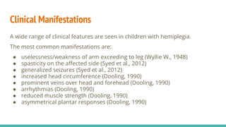
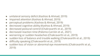
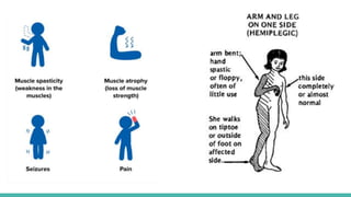
INFANTILE HEMIPLEGIA
APOORVA BALODHI
MPT NEUROLOGY (1ST YEAR)
2021-517-004
Definition
Infantile hemiplegia, commonly called acute infantile hemiplegia, defines
cases of hemiplegia of sudden onset in children, of a few weeks to 6 or more
years of age.
The hemiplegia may be quickly or gradually lethal, permanent with mental
impairment, slow and partial recovery, or rapid or complete restoration to
normal health.
Extensive literature was published on infantile hemiplegia between 1840 and
the 1920s to collect cases theorize on their causation.
It is most commonly seen during the first three years of infant life. (Wyllie W.,
1948)
Etiology
(Dooling, 1990)
Pathophysiology
The most common cause for
infantile hemiplegia is occlusion
due to a thrombus or embolus.
Unlike adult hemiplegia,
occlusion takes place in the
venous territory rather than the
arterial typically of the Great
vein of Galen.
In others destruction of grey
matter is seen. (Wyllie W., 1948)
Clinical Manifestations
A wide range of clinical features are seen in children with hemiplegia.
The most common manifestations are:
● uselessness/weakness of arm exceeding to leg (Wyllie W., 1948)
● spasticity on the affected side (Syed et al., 2012)
● generalized seizures (Syed et al., 2012)
● increased head circumference (Dooling, 1990)
● prominent veins over head and forehead (Dooling, 1990)
● arrhythmias (Dooling, 1990)
● reduced muscle strength (Dooling, 1990)
● asymmetrical plantar responses (Dooling, 1990)
● unilateral sensory deficit (Kashoo & Ahmad, 2019)
● impaired attention (Kashoo & Ahmad, 2019)
● perceptual problems (Kashoo & Ahmad, 2019)
● decreased cognitive ability (Kashoo & Ahmad, 2019)
● decreased postural control (Chakravarthi et al., 2019)
● decreased reaction time (Palomo-Carrión et al., 2021)
● worsening or sudden headaches (Chakravarthi et al., 2019)
● sudden loss of balance and trouble walking (Chakravarthi et al., 2019)
● difficulty speaking (Chakravarthi et al., 2019)
● sudden loss of vision or abnormal eye movements (Chakravarthi et al.,
2019)
Assessment
● History
The patient presents with a history of recurrent seizures, set in by a mild and
innocuous febrile or afebrile illness leading to persistent weakness or
paralysis. (Dooling, 1990)
A history of abnormal labor (Syed et al., 2012; Wyllie W.,1948), delayed cry
(Goyal et al., 2022), or preterm birth (Kashoo & Ahmad, 2006) may also be
present.
● Clinical Examination
● General Examination
The skin should be examined carefully. Any discrepancy in the head
circumference should be looked for. So should be suppleness of neck or
signs of meningeal irritation.
Prominence and tortuous patterns of veins should be looked for on the
head and forehead.
Heart should be auscultated for murmurs, clicks, and arrhythmias. Radial
and femoral pulses should be compared.
Nail beds should be checked for cyanosis. (Dooling, 1990)
Poor hygiene may be noted.
Visual field neglect and slight
flattening of the nasolabial fold
may be present.
(Syed et al., 2012)
● Mental Status Screening
IQ may be lower than normal with reduced verbal ability and dysphasia.
Outbursts of severe temper tantrums may be present.
(Syed et al., 2012)
● Motor Examination
Upper Extremity
Attention is paid to the asymmetry of posture, tone, muscle power, and
movements. Hand grip is tested for grasp. (Mercuri E. et al, 1999)
There may also be impaired voluntary movements. (Syed et al., 2012)
Lower Extremity
Tone of the lower limbs is checked. Gait pattern is assessed. There
maybe a difference in the limb length.
(Mercuri E. et al, 1999)
● Sensory Examination
Both upper and lower extremities are compared for tests of
pinprick, temperature, vibration, and proprioception.
(Mercuri E. et al, 1999)
● Reflexes
Reflexes may be exaggerated with
positive Babinski’s sign. Deep tendon
reflexes and pain sensation may or may
not be affected.
(Syed et al., 2012)
Investigations
● Cranial ultrasonography (to exclude hematoma)
● CT/MRI scan
● Lumbar puncture
● EEG
● EKG and Echocardiogram
● CBC
● Blood culture
● Lipid profile
● Antinuclear antibody
(Dooling, 1990)
Management
● Medical Management
● Mannitol (to reduce oedema)
● Antibiotics (if required)
● Anti-convulsants (phenobarbital, phenytoin)
● Fluid restriction
(Dooling, 1990)
● Surgical Management
● Embolization
● Hemispherectomy
● Radiation (photon or proton beam)
● Intracranial-extracranial arterial by-passes
● Dental procedures
(Dooling, 1990)
● Physiotherapy (PT) Management
The main goal of physiotherapeutic treatment is to restore muscle power so
as to improve the activities of daily living. This could be done by various
means. The ways to do so cited in literature are:
● Automatic or confused motion theory by Phelps
This method is based on the principle of ‘pathological overflow.’ It says
that the muscular contraction of a weak muscle can be elicited by
resisting the contraction of another muscle or muscle group which then
passes on the impetus to the weak muscle.
The resistance is reduced gradually until the mere thought of resistances
suffices to produce a contraction of the weak muscle.
● Kabat and Knott approach
They recommend heavy resistance to the contraction of the weak muscle
itself so as to help fire more motor units.
● Method of Fay
The method is based on the knowledge of postural reflexes, defence
reactions, and pattern movements. Fay also remarks about the possibility
of initiating a crossed pattern movement by training the coordinated
existing responses.
● Faradism
It aims at eliciting a volitional contraction of a weak muscle using faradic
current.
● Synkinetic method
This method appoints an unintentional contralateral and mirror-like
symmetrical movement elicited by a volitional movement from the
unaffected side to cause a movement in the affected limb.
(Obholzer, 1951)
Recent advances in PT Management
References
Chakravarthi, V., Meera, R., Arunachalam, R., Sujatha, B., & Abraham, M. M. (2019). Effectiveness of modified trunk dissociation retrainer in
improving trunk control in subjects with infantile hemiplegia. Research Journal of Pharmacy and Technology, 12(3), 1141–1144.
https://doi.org/10.5958/0974-360X.2019.00188.4
Dooling, E. C. (1990). Acute hemiplegia in infancy. The Indian Journal of Pediatrics, 57(3), 325–335. https://doi.org/10.1007/BF02727908
Goyal, C., Vardhan, V., & Naqvi, W. M. (2022). Haptic Feedback-Based Virtual Reality Intervention for a Child With Infantile Hemiplegia: A Case
Report. Cureus, 14(3), 3–7. https://doi.org/10.7759/cureus.23489
Kashoo, F. Z., & Ahmad, M. (2019). Effect of sensory integration on attention span among children with infantile hemiplegia. International Journal of
Health Sciences, 13(3), 29–33.
http://www.ncbi.nlm.nih.gov/pubmed/31123437%0Ahttp://www.pubmedcentral.nih.gov/articlerender.fcgi?artid=PMC6512152
Mercuri E. et al. (1999). Congenital Hemiplegia in Children at School Age: Assessment of Hand Function in the Non-Hemiplegic Hand and
Correlation with MRI. Neuropediatrics. https://www.thieme-connect.com/products/ejournals/abstract/10.1055/s-2007-973449
Obholzer, A. (1951). Synkineses in neuromuscular re-education. 741–744.
Palomo-Carrión, R., Bravo-Esteban, E., Ando-La Fuente, S., López-Muñoz, P., Martínez-Galán, I., & Romay-Barrero, H. (2021). Efficacy of the use of
unaffected hand containment in unimanual intensive therapy to increase visuomotor coordination in children with hemiplegia: a randomized
controlled pilot study. Therapeutic Advances in Chronic Disease, 12, 1–12. https://doi.org/10.1177/20406223211001280
Syed, G., Benni, D., Naik, S., & Surendra, P. (2012). Infantile hemiplegia in pediatric dental set-up. Dental Research Journal, 9(5), 651.
https://doi.org/10.4103/1735-3327.104890
Wyllie W. (1948). Section of Paediatrics Acute Infantile Hemiplegia. 459–466.

Infantile Hemiplegia.pptx

  • 1.
    INFANTILE HEMIPLEGIA APOORVA BALODHI MPTNEUROLOGY (1ST YEAR) 2021-517-004
  • 2.
    Definition Infantile hemiplegia, commonlycalled acute infantile hemiplegia, defines cases of hemiplegia of sudden onset in children, of a few weeks to 6 or more years of age. The hemiplegia may be quickly or gradually lethal, permanent with mental impairment, slow and partial recovery, or rapid or complete restoration to normal health. Extensive literature was published on infantile hemiplegia between 1840 and the 1920s to collect cases theorize on their causation. It is most commonly seen during the first three years of infant life. (Wyllie W., 1948)
  • 3.
  • 4.
    Pathophysiology The most commoncause for infantile hemiplegia is occlusion due to a thrombus or embolus. Unlike adult hemiplegia, occlusion takes place in the venous territory rather than the arterial typically of the Great vein of Galen. In others destruction of grey matter is seen. (Wyllie W., 1948)
  • 5.
    Clinical Manifestations A widerange of clinical features are seen in children with hemiplegia. The most common manifestations are: ● uselessness/weakness of arm exceeding to leg (Wyllie W., 1948) ● spasticity on the affected side (Syed et al., 2012) ● generalized seizures (Syed et al., 2012) ● increased head circumference (Dooling, 1990) ● prominent veins over head and forehead (Dooling, 1990) ● arrhythmias (Dooling, 1990) ● reduced muscle strength (Dooling, 1990) ● asymmetrical plantar responses (Dooling, 1990)
  • 6.
    ● unilateral sensorydeficit (Kashoo & Ahmad, 2019) ● impaired attention (Kashoo & Ahmad, 2019) ● perceptual problems (Kashoo & Ahmad, 2019) ● decreased cognitive ability (Kashoo & Ahmad, 2019) ● decreased postural control (Chakravarthi et al., 2019) ● decreased reaction time (Palomo-Carrión et al., 2021) ● worsening or sudden headaches (Chakravarthi et al., 2019) ● sudden loss of balance and trouble walking (Chakravarthi et al., 2019) ● difficulty speaking (Chakravarthi et al., 2019) ● sudden loss of vision or abnormal eye movements (Chakravarthi et al., 2019)
  • 8.
    Assessment ● History The patientpresents with a history of recurrent seizures, set in by a mild and innocuous febrile or afebrile illness leading to persistent weakness or paralysis. (Dooling, 1990) A history of abnormal labor (Syed et al., 2012; Wyllie W.,1948), delayed cry (Goyal et al., 2022), or preterm birth (Kashoo & Ahmad, 2006) may also be present.
  • 9.
    ● Clinical Examination ●General Examination The skin should be examined carefully. Any discrepancy in the head circumference should be looked for. So should be suppleness of neck or signs of meningeal irritation. Prominence and tortuous patterns of veins should be looked for on the head and forehead. Heart should be auscultated for murmurs, clicks, and arrhythmias. Radial and femoral pulses should be compared. Nail beds should be checked for cyanosis. (Dooling, 1990)
  • 10.
    Poor hygiene maybe noted. Visual field neglect and slight flattening of the nasolabial fold may be present. (Syed et al., 2012)
  • 11.
    ● Mental StatusScreening IQ may be lower than normal with reduced verbal ability and dysphasia. Outbursts of severe temper tantrums may be present. (Syed et al., 2012) ● Motor Examination Upper Extremity Attention is paid to the asymmetry of posture, tone, muscle power, and movements. Hand grip is tested for grasp. (Mercuri E. et al, 1999) There may also be impaired voluntary movements. (Syed et al., 2012)
  • 12.
    Lower Extremity Tone ofthe lower limbs is checked. Gait pattern is assessed. There maybe a difference in the limb length. (Mercuri E. et al, 1999) ● Sensory Examination Both upper and lower extremities are compared for tests of pinprick, temperature, vibration, and proprioception. (Mercuri E. et al, 1999)
  • 13.
    ● Reflexes Reflexes maybe exaggerated with positive Babinski’s sign. Deep tendon reflexes and pain sensation may or may not be affected. (Syed et al., 2012)
  • 14.
    Investigations ● Cranial ultrasonography(to exclude hematoma) ● CT/MRI scan ● Lumbar puncture ● EEG ● EKG and Echocardiogram ● CBC ● Blood culture ● Lipid profile ● Antinuclear antibody (Dooling, 1990)
  • 15.
    Management ● Medical Management ●Mannitol (to reduce oedema) ● Antibiotics (if required) ● Anti-convulsants (phenobarbital, phenytoin) ● Fluid restriction (Dooling, 1990)
  • 16.
    ● Surgical Management ●Embolization ● Hemispherectomy ● Radiation (photon or proton beam) ● Intracranial-extracranial arterial by-passes ● Dental procedures (Dooling, 1990)
  • 17.
    ● Physiotherapy (PT)Management The main goal of physiotherapeutic treatment is to restore muscle power so as to improve the activities of daily living. This could be done by various means. The ways to do so cited in literature are: ● Automatic or confused motion theory by Phelps This method is based on the principle of ‘pathological overflow.’ It says that the muscular contraction of a weak muscle can be elicited by resisting the contraction of another muscle or muscle group which then passes on the impetus to the weak muscle. The resistance is reduced gradually until the mere thought of resistances suffices to produce a contraction of the weak muscle.
  • 18.
    ● Kabat andKnott approach They recommend heavy resistance to the contraction of the weak muscle itself so as to help fire more motor units. ● Method of Fay The method is based on the knowledge of postural reflexes, defence reactions, and pattern movements. Fay also remarks about the possibility of initiating a crossed pattern movement by training the coordinated existing responses.
  • 19.
    ● Faradism It aimsat eliciting a volitional contraction of a weak muscle using faradic current. ● Synkinetic method This method appoints an unintentional contralateral and mirror-like symmetrical movement elicited by a volitional movement from the unaffected side to cause a movement in the affected limb. (Obholzer, 1951)
  • 20.
    Recent advances inPT Management
  • 21.
    References Chakravarthi, V., Meera,R., Arunachalam, R., Sujatha, B., & Abraham, M. M. (2019). Effectiveness of modified trunk dissociation retrainer in improving trunk control in subjects with infantile hemiplegia. Research Journal of Pharmacy and Technology, 12(3), 1141–1144. https://doi.org/10.5958/0974-360X.2019.00188.4 Dooling, E. C. (1990). Acute hemiplegia in infancy. The Indian Journal of Pediatrics, 57(3), 325–335. https://doi.org/10.1007/BF02727908 Goyal, C., Vardhan, V., & Naqvi, W. M. (2022). Haptic Feedback-Based Virtual Reality Intervention for a Child With Infantile Hemiplegia: A Case Report. Cureus, 14(3), 3–7. https://doi.org/10.7759/cureus.23489 Kashoo, F. Z., & Ahmad, M. (2019). Effect of sensory integration on attention span among children with infantile hemiplegia. International Journal of Health Sciences, 13(3), 29–33. http://www.ncbi.nlm.nih.gov/pubmed/31123437%0Ahttp://www.pubmedcentral.nih.gov/articlerender.fcgi?artid=PMC6512152 Mercuri E. et al. (1999). Congenital Hemiplegia in Children at School Age: Assessment of Hand Function in the Non-Hemiplegic Hand and Correlation with MRI. Neuropediatrics. https://www.thieme-connect.com/products/ejournals/abstract/10.1055/s-2007-973449 Obholzer, A. (1951). Synkineses in neuromuscular re-education. 741–744. Palomo-Carrión, R., Bravo-Esteban, E., Ando-La Fuente, S., López-Muñoz, P., Martínez-Galán, I., & Romay-Barrero, H. (2021). Efficacy of the use of unaffected hand containment in unimanual intensive therapy to increase visuomotor coordination in children with hemiplegia: a randomized controlled pilot study. Therapeutic Advances in Chronic Disease, 12, 1–12. https://doi.org/10.1177/20406223211001280 Syed, G., Benni, D., Naik, S., & Surendra, P. (2012). Infantile hemiplegia in pediatric dental set-up. Dental Research Journal, 9(5), 651. https://doi.org/10.4103/1735-3327.104890 Wyllie W. (1948). Section of Paediatrics Acute Infantile Hemiplegia. 459–466.