
Download to read offline
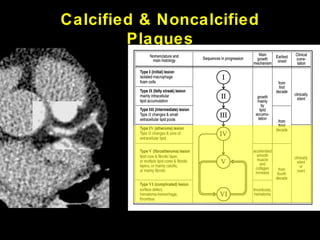
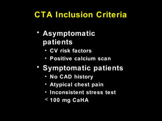
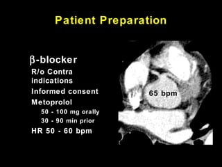
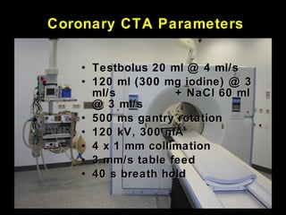
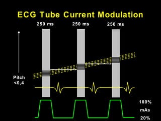
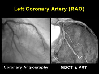
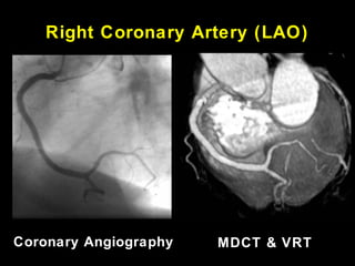
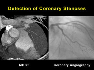
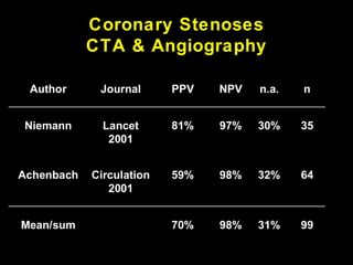
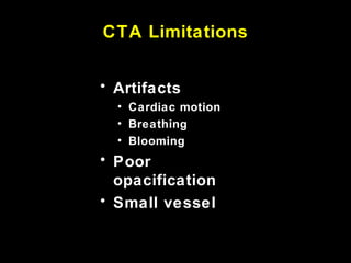
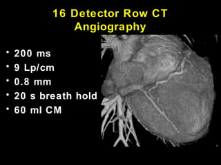
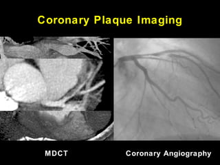
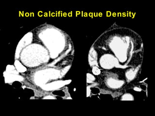
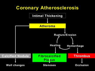
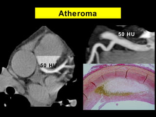
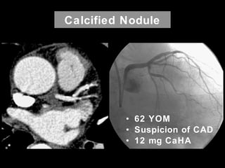
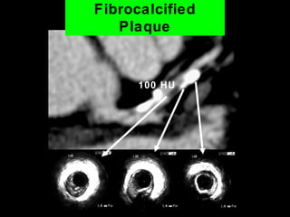
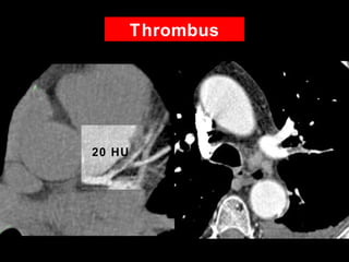
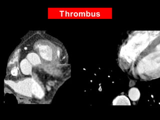
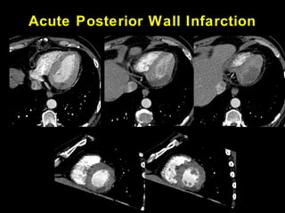
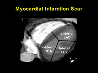
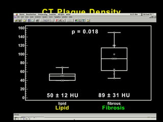
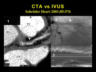
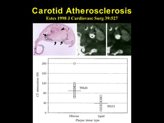
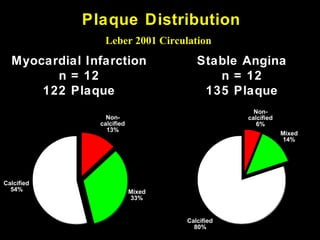
This document discusses the use of coronary CT angiography (CTA) to detect and characterize coronary atherosclerosis beyond just detecting coronary stenoses. CTA can identify calcified plaques, non-calcified plaques, and mixed plaques. It can detect atheromas and characterize plaque density. CTA can also identify intracoronary thrombi and myocardial infarction scars. The document outlines the CTA scanning parameters and techniques used to minimize motion artifacts and optimize image quality for plaque detection and characterization.


