Downloaded 453 times
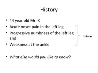


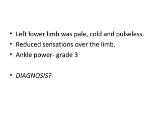


A 44-year-old man presented with acute onset left leg pain, numbness, and weakness. On examination, his left lower limb was pale, cold, pulseless with reduced sensation. Imaging showed a thrombus in his aorta and left iliac and femoral arteries. He was diagnosed with acute limb ischemia and underwent aortic endarterectomy, femoral embolectomy and patch angioplasty, and fasciotomy to restore blood flow and prevent compartment syndrome. Post-operatively, he required monitoring for reperfusion injuries and long-term anticoagulation to prevent recurrence.


