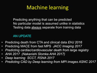
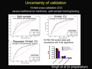
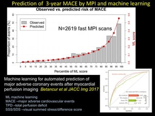
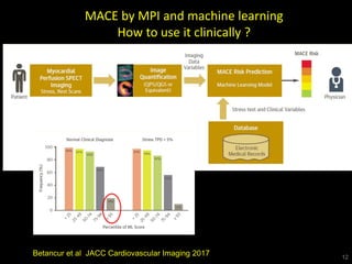
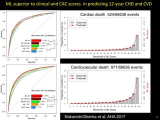
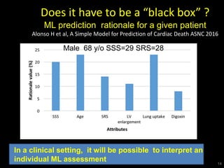
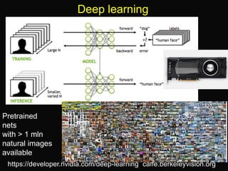
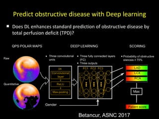
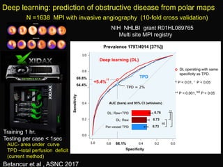
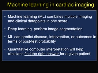
Piotr J. Slomka, PhD, FACC
Scientist , Artificial Intelligence in Medicine
Cedars Sinai Medical Center
Professor of Medicine, UCLA school of Medicine
Cedars-Sinai Medical Center
Machine learning improves image analysis, diagnosis and
prognostic risk assessment in cardiac imaging: an update”
Piotr.Slomka@cshs.org
Some software described in this presentation is
owned by Cedars-Sinai Medical Center, which
receives or may receive royalties from its licensing.
A minority portion of those royalties is shared by the
authors
Machine learning
Predicitng anything that can be predicted
No particular model is assumed unlike in statistics
Testing data always separate from training data
• Predicting death from CTA and clinical data EHJ 2016
• Predicting MACE from fast MPS JACC Imaging 2017
• Predicting cardiac/cardiovascular death from large registry
• AHA 2017 (Nakanishi Slomka AHA 2017)
• Deep learning SCCT, RSNA 2017
• Predicting CAD by Deep learning from MPI images ASNC 2017
AN UPDATE
Simple machine learning
The k-nearest neighbor algorithm assigns outcome based
on the most similar training examples
MI -myocardial infarction
Deo RC Circulation 2015
Ensemble (boosted) learning
http://magizbox.com
Similar to
consensus
human
reading
Motwani M, EHJ 2016
Feature selection and learning
(ensemble
Motwani M et al EHJ 2016
Death prediction by ensemble machine
learning over standard risk scores
ML-B -boosted machine learning
FRS –Framingham risk score
SSS –segmental stenosis score
SIS –segmental involvement score
DI -Duke index
AUC –area under curve
N=10,030
Motwani M et al, EHJ 2016
Uncertainty of validation
10-fold cross-validation (CV)
versus traditional (in medicine) split sample training/testing
Singh et al (in preparation)
Split sample
N=700 100 random data split
simulations with 4 ML algorithms
10-fold CV
Repeated 10-fold CV
min/max min/max
min/max
Average of 100 split sample divisions
Average of 10 split sample divisionsOne arbitrary split sample division
Prediction of 3-year MACE by MPI and machine learning
Stress TPD< 4 %
Stress TPD≥ 4 %
ML score ≥0.15
N= 1965
SSS ≥1
SSS= 0
N= 653
N=1993
N= 625
N= 1956
Machine learning for automated prediction of
major adverse coronary events after myocardial
perfusion imaging Betancur et al JACC Img 2017
N=2619 fast MPI scans
ML machine learning
MACE –major adverse cardiovascular events
TPD –total perfusion deficit
SSS/SDS –visual summed stress/difference score
12
MACE by MPI and machine learning
How to use it clinically ?
Betancur et al JACC Cardiovascular Imaging 2017
ML superior to clinical and CAC scores in predicting 12-year CHD and CVD
13
Cardiovascular death 971/66636 events
Cardiac death 524/66636 events
Nakanishi/Slomka et al. AHA 2017
Does it have to be a “black box” ?
ML prediction rationale for a given patient
Alonso H et al, A Simple Model for Prediction of Cardiac Death ASNC 2016
14
In a clinical setting, it will be possible to interpret an
individual ML assessment
Male 68 y/o SSS=29 SRS=28
Deep learning
https://developer.nvidia.com/deep-learning caffe.berkeleyvision.org
Pretrained
nets
with > 1 mln
natural images
available
Deep learning for adipose tissue segmentation
from non-contrast CT
Dey D R01HL133616
>7000 patients
Deep learning for image analysis
non-contrast CT pericardial fat segmentation
Ground truth by expertDeep learning
Dey D NIH RO1:1R01HL133616
Caffe:4 layers convolution/activation output per-pixel probability
Training 1100 slices: few hours on 2x Nvidia GTX1080. Testing < 5 sec per patient
Commandeur et al SCCT, RSNA 2017
R=0.93
R=0.95
Predict obstructive disease with Deep learning
 Does DL enhances standard prediction of obstructive disease by
total perfusion deficit (TPD)?
Betancur, ASNC 2017
LAD
LCX
RCA
Patient score
Deep learning: prediction of obstructive disease from polar maps
AUC- area under curve
TPD –total perfusion deficit
(current method)
N =1638 MPI with invasive angiography (10-fold cross validation)
Betancur et al , ASNC 2017
NIH NHLBI grant R01HL089765
Multi site MPI registry
Training 1 hr.
Testing per case < 1sec
Collected to date > 2,000 cases with cath, 18,500 with MACE follow up
Site System Protocols
Cedars-Sinai , Los Angeles
Slomka, Berman
D-SPECT Sestamibi rest/stress
supine/upright
Aspire, Kansas City
Bateman
DSPECT
Siemens Symbia SPECT/CT
Low-dose sestamibi stress or stress/rest supine/upright
~50% stress-only AC for Symbia
Columbia, New York
Einstein
GE 530c Sestamibi prone/supine
Assuta, Israel
Sharir
GE 530c Low dose sestamibi
supine/prone
Oregon Heart/Vascular Institute
Fish
D-SPECT Sestamibi rest supine
stress supine/upright
Zurich University Hospital
Kaufmann
GE 570c SPECT/CT
GE Ventri/VCT SPECT/CT
Low-dose tetrofosmin stress/rest
AC for all
Ottawa Heart Institute Canada
Ruddy
GE 530c
GE Infinia SPECT/CT
Low-dose tetrofosmin
AC for GE Infinia
Yale University, New Haven,
Miller, Sinusas
GE570c SPECT/CT
GE Infinia SPECT/CT
Stress/rest sestamibi protocols.
AC for all
Brigham and Women’s Boston
Dorbala, DiCarli
D-SPECT
Siemens Symbia SPECT/CT
(IQ∙SPECT)
Same-day, dose-dependent rest/stress sestamibi.
Supine/upright for D-SPECT
AC for Symbia
NIH NHLBI grant R01HL089765
REgistry of Fast Myocardial Perfusion Imaging
with NExt generation SPECT: ReFINe SPECT
NIH sponsored diagnostic/prognostic image registry, 9 national/international sites
All images automatically processed!
Multimodality machine learning
CTA + coronary PET for prognosis of heart attacks in high risk patients ?
Coronary 18F-NaF PET +CTA
Coronary PET quantification
Prediction of Recurrent Events With
18F-Fluoride (PREFFIR)
ClinicalTrials.gov NCT02278211
Joshi et al Lancet 2014
Rubeaux et al JNM 2016
R01 HL135557
Software vs physician?
“Patients whose lives and medical
histories shape the algorithms,
will emerge the biggest winners
as machine learning transforms
clinical medicine”
Obermayer and Emanuel New England Journal of Medicine 2016
Machine learning in cardiac imaging
• Machine learning (ML) combines multiple imaging
and clinical datapoints in one score.
• Deep learning perform image segmentation
• ML can predict disease, intervention, or outcomes in
terms of post-test probability
• Quantitative computer interpretation will help
clinicians find the right answer for a given patient

AHA SHAPE Symposium 2017 Dr. Slomka Presentation

  • 1.
    Piotr J. Slomka,PhD, FACC Scientist , Artificial Intelligence in Medicine Cedars Sinai Medical Center Professor of Medicine, UCLA school of Medicine Cedars-Sinai Medical Center Machine learning improves image analysis, diagnosis and prognostic risk assessment in cardiac imaging: an update” Piotr.Slomka@cshs.org Some software described in this presentation is owned by Cedars-Sinai Medical Center, which receives or may receive royalties from its licensing. A minority portion of those royalties is shared by the authors
  • 2.
    Machine learning Predicitng anythingthat can be predicted No particular model is assumed unlike in statistics Testing data always separate from training data • Predicting death from CTA and clinical data EHJ 2016 • Predicting MACE from fast MPS JACC Imaging 2017 • Predicting cardiac/cardiovascular death from large registry • AHA 2017 (Nakanishi Slomka AHA 2017) • Deep learning SCCT, RSNA 2017 • Predicting CAD by Deep learning from MPI images ASNC 2017 AN UPDATE
  • 3.
    Simple machine learning Thek-nearest neighbor algorithm assigns outcome based on the most similar training examples MI -myocardial infarction Deo RC Circulation 2015
  • 4.
  • 5.
  • 6.
    Feature selection andlearning (ensemble Motwani M et al EHJ 2016
  • 7.
    Death prediction byensemble machine learning over standard risk scores ML-B -boosted machine learning FRS –Framingham risk score SSS –segmental stenosis score SIS –segmental involvement score DI -Duke index AUC –area under curve N=10,030 Motwani M et al, EHJ 2016
  • 8.
    Uncertainty of validation 10-foldcross-validation (CV) versus traditional (in medicine) split sample training/testing Singh et al (in preparation) Split sample N=700 100 random data split simulations with 4 ML algorithms 10-fold CV Repeated 10-fold CV min/max min/max min/max Average of 100 split sample divisions Average of 10 split sample divisionsOne arbitrary split sample division
  • 9.
    Prediction of 3-yearMACE by MPI and machine learning Stress TPD< 4 % Stress TPD≥ 4 % ML score ≥0.15 N= 1965 SSS ≥1 SSS= 0 N= 653 N=1993 N= 625 N= 1956 Machine learning for automated prediction of major adverse coronary events after myocardial perfusion imaging Betancur et al JACC Img 2017 N=2619 fast MPI scans ML machine learning MACE –major adverse cardiovascular events TPD –total perfusion deficit SSS/SDS –visual summed stress/difference score
  • 10.
    12 MACE by MPIand machine learning How to use it clinically ? Betancur et al JACC Cardiovascular Imaging 2017
  • 11.
    ML superior toclinical and CAC scores in predicting 12-year CHD and CVD 13 Cardiovascular death 971/66636 events Cardiac death 524/66636 events Nakanishi/Slomka et al. AHA 2017
  • 12.
    Does it haveto be a “black box” ? ML prediction rationale for a given patient Alonso H et al, A Simple Model for Prediction of Cardiac Death ASNC 2016 14 In a clinical setting, it will be possible to interpret an individual ML assessment Male 68 y/o SSS=29 SRS=28
  • 13.
  • 14.
    Deep learning foradipose tissue segmentation from non-contrast CT Dey D R01HL133616 >7000 patients
  • 15.
    Deep learning forimage analysis non-contrast CT pericardial fat segmentation Ground truth by expertDeep learning Dey D NIH RO1:1R01HL133616 Caffe:4 layers convolution/activation output per-pixel probability Training 1100 slices: few hours on 2x Nvidia GTX1080. Testing < 5 sec per patient Commandeur et al SCCT, RSNA 2017 R=0.93 R=0.95
  • 16.
    Predict obstructive diseasewith Deep learning  Does DL enhances standard prediction of obstructive disease by total perfusion deficit (TPD)? Betancur, ASNC 2017 LAD LCX RCA Patient score
  • 17.
    Deep learning: predictionof obstructive disease from polar maps AUC- area under curve TPD –total perfusion deficit (current method) N =1638 MPI with invasive angiography (10-fold cross validation) Betancur et al , ASNC 2017 NIH NHLBI grant R01HL089765 Multi site MPI registry Training 1 hr. Testing per case < 1sec
  • 18.
    Collected to date> 2,000 cases with cath, 18,500 with MACE follow up Site System Protocols Cedars-Sinai , Los Angeles Slomka, Berman D-SPECT Sestamibi rest/stress supine/upright Aspire, Kansas City Bateman DSPECT Siemens Symbia SPECT/CT Low-dose sestamibi stress or stress/rest supine/upright ~50% stress-only AC for Symbia Columbia, New York Einstein GE 530c Sestamibi prone/supine Assuta, Israel Sharir GE 530c Low dose sestamibi supine/prone Oregon Heart/Vascular Institute Fish D-SPECT Sestamibi rest supine stress supine/upright Zurich University Hospital Kaufmann GE 570c SPECT/CT GE Ventri/VCT SPECT/CT Low-dose tetrofosmin stress/rest AC for all Ottawa Heart Institute Canada Ruddy GE 530c GE Infinia SPECT/CT Low-dose tetrofosmin AC for GE Infinia Yale University, New Haven, Miller, Sinusas GE570c SPECT/CT GE Infinia SPECT/CT Stress/rest sestamibi protocols. AC for all Brigham and Women’s Boston Dorbala, DiCarli D-SPECT Siemens Symbia SPECT/CT (IQ∙SPECT) Same-day, dose-dependent rest/stress sestamibi. Supine/upright for D-SPECT AC for Symbia NIH NHLBI grant R01HL089765 REgistry of Fast Myocardial Perfusion Imaging with NExt generation SPECT: ReFINe SPECT NIH sponsored diagnostic/prognostic image registry, 9 national/international sites All images automatically processed!
  • 19.
    Multimodality machine learning CTA+ coronary PET for prognosis of heart attacks in high risk patients ? Coronary 18F-NaF PET +CTA Coronary PET quantification Prediction of Recurrent Events With 18F-Fluoride (PREFFIR) ClinicalTrials.gov NCT02278211 Joshi et al Lancet 2014 Rubeaux et al JNM 2016 R01 HL135557
  • 20.
    Software vs physician? “Patientswhose lives and medical histories shape the algorithms, will emerge the biggest winners as machine learning transforms clinical medicine” Obermayer and Emanuel New England Journal of Medicine 2016
  • 21.
    Machine learning incardiac imaging • Machine learning (ML) combines multiple imaging and clinical datapoints in one score. • Deep learning perform image segmentation • ML can predict disease, intervention, or outcomes in terms of post-test probability • Quantitative computer interpretation will help clinicians find the right answer for a given patient