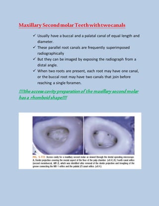
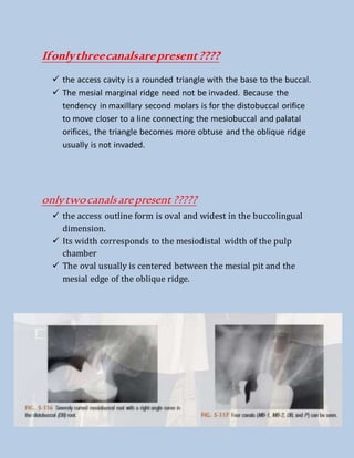
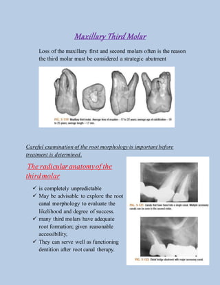
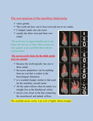
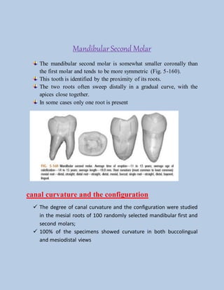
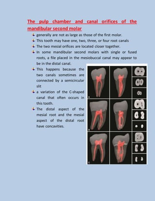
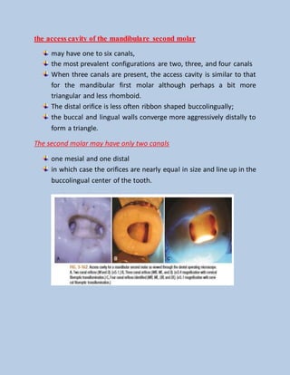
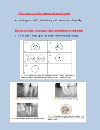
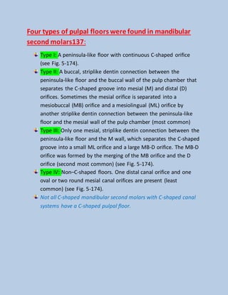
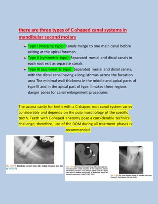
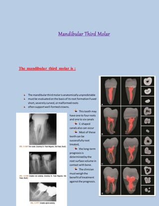
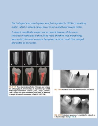
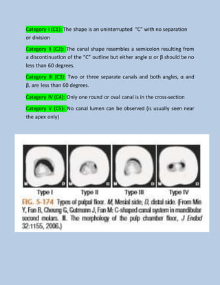
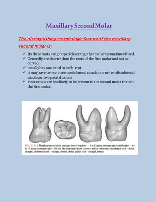
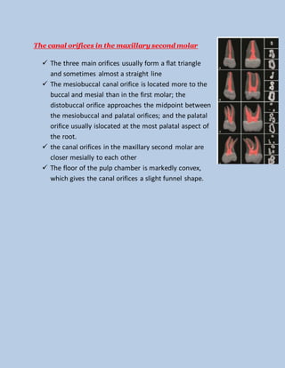
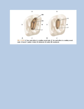
Endodontics
Accessopeningofsecondandthirdmolars
Presentedby: StephanieChahrouk
MaxillarySecondMolar
The distinguishing morphologic feature of the maxillary
second molar is:
 Its three roots aregrouped closer together and aresometimesfused.
 Generally are shorter thanthe roots of thefirst molar and not as
curved.
 usually has one canalin each root
 it may have two or three mesiobuccalcanals, one or two distobuccal
canals, or twopalatalcanals
 Four canalsare less likely to be present in the second molar thanin
the first molar.
The canal orifices in the maxillary second molar
 The three main orifices usually form a flat triangle
and sometimes almost a straight line
 The mesiobuccal canal orifice is located more to the
buccal and mesial than in the first molar; the
distobuccal orifice approaches the midpoint between
the mesiobuccal and palatal orifices; and the palatal
orifice usually islocated at the most palatal aspect of
the root.
 the canal orifices in the maxillary second molar are
closer mesially to each other
 The floor of the pulp chamber is markedly convex,
which gives the canal orifices a slight funnel shape.
Maxillary SecondmolarTeethwithtwocanals
 Usually have a buccal and a palatal canal of equal length and
diameter.
 These parallel root canals are frequently superimposed
radiographically
 But they can be imaged by exposing the radiograph from a
distal angle.
 When two roots are present, each root may have one canal,
or the buccal root may have two canals that join before
reaching a single foramen.
!!!!the access cavitypreparationof the maxillary second molar
has a rhomboid shape!!!!
Ifonlythreecanalsarepresent????
 the access cavity is a rounded triangle with the base to the buccal.
 The mesial marginal ridge need not be invaded. Because the
tendency in maxillary second molars is for the distobuccal orifice
to move closer to a line connecting the mesiobuccal and palatal
orifices, the triangle becomes more obtuse and the oblique ridge
usually is not invaded.
onlytwocanalsarepresent?????
 the access outline form is oval and widest in the buccolingual
dimension.
 Its width corresponds to the mesiodistal width of the pulp
chamber
 The oval usually is centered between the mesial pit and the
mesial edge of the oblique ridge.
Maxillary Third Molar
Loss of the maxillary first and second molars often is the reason
the third molar must be considered a strategic abutment
Careful examination of the root morphology is important before
treatment is determined.
The radicular anatomyof the
thirdmolar
 is completely unpredictable
 May be advisable to explore the root
canal morphology to evaluate the
likelihood and degree of success.
 many third molars have adequate
root formation; given reasonable
accessibility,
 They can serve well as functioning
dentition after root canal therapy.
The root anatomy of the maxillary third molar
 varies greatly.
 This tooth can have one to four roots and one to six canals,
 C-shaped canals also can occur.
 usually has three roots and three root
canals
The tooth may be tipped significantly to the
distal, the buccal, or both, which creates an
even greater access problem than with the
second molar.
The access cavity form for the third molar
can vary greatly
 Because the tooth typically has one to
three canals,
 the access preparation can be anything
from an oval that is widest in the
buccolingual dimension
 to a rounded triangle similar to that used
for the maxillary second molar.
 All the canal orifices often lie nearly in a
straight line as the distobuccal orifice
 moves even closer to the line connecting
the mesiobuccal and palatal orifices.
The resultant access cavity is an oval or highly obtuse triangle
Mandibular Second Molar
The mandibular second molar is somewhat smaller coronally than
the first molar and tends to be more symmetric (Fig. 5-160).
This tooth is identified by the proximity of its roots.
The two roots often sweep distally in a gradual curve, with the
apices close together.
In some cases only one root is present
canal curvature and the configuration
 The degree of canal curvature and the configuration were studied
in the mesial roots of 100 randomly selected mandibular first and
second molars;
 100% of the specimens showed curvature in both buccolingual
and mesiodistal views
The pulp chamber and canal orifices of the
mandibular second molar
generally are not as large as those of the first molar.
This tooth may have one, two, three, or four root canals
The two mesial orifices are located closer together.
In some mandibular second molars with single or fused
roots, a file placed in the mesiobuccal canal may appear to
be in the distal canal.
This happens because the
two canals sometimes are
connected by a semicircular
slit
a variation of the C-shaped
canal that often occurs in
this tooth.
The distal aspect of the
mesial root and the mesial
aspect of the distal root
have concavities.
the access cavity of the mandibulare second molar
may have one to six canals,
the most prevalent configurations are two, three, and four canals
When three canals are present, the access cavity is similar to that
for the mandibular first molar although perhaps a bit more
triangular and less rhomboid.
The distal orifice is less often ribbon shaped buccolingually;
the buccal and lingual walls converge more aggressively distally to
form a triangle.
The second molar may have only two canals
one mesial and one distal
in which case the orifices are nearly equal in size and line up in the
buccolingual center of the tooth.
The access cavity for a two-canal second molar
is rectangular, wide mesiodistally and narrow buccolingually
The access cavity for a single-canal mandibular second molar
is oval and is lined up in the center of the occlusal surface.
Four types of pulpal floors were found in mandibular
second molars137:
Type I: A peninsula-like floor with continuous C-shaped orifice
(see Fig. 5-174).
Type II: A buccal, striplike dentin connection between the
peninsula-like floor and the buccal wall of the pulp chamber that
separates the C-shaped groove into mesial (M) and distal (D)
orifices. Sometimes the mesial orifice is separated into a
mesiobuccal (MB) orifice and a mesiolingual (ML) orifice by
another striplike dentin connection between the peninsula-like
floor and the mesial wall of the pulp chamber (most common)
Type III: Only one mesial, striplike dentin connection between the
peninsula-like floor and the M wall, which separates the C-shaped
groove into a small ML orifice and a large MB-D orifice. The MB-D
orifice was formed by the merging of the MB orifice and the D
orifice (second most common) (see Fig. 5-174).
Type IV: Non–C-shaped floors. One distal canal orifice and one
oval or two round mesial canal orifices are present (least
common) (see Fig. 5-174).
Not all C-shaped mandibular second molars with C-shaped canal
systems have a C-shaped pulpal floor.
there are three types of C-shaped canal systems in
mandibular second molars
Type I (merging type): Canals merge to one main canal before
exiting at the apical foramen
Type II (symmetric type): Separated mesial and distal canals in
each root exit as separate canals
Type III (asymmetric type): Separated mesial and distal canals,
with the distal canal having a long isthmus across the furcation
area The minimal wall thickness in the middle and apical parts of
type III and in the apical part of type II makes these regions
danger zones for canal enlargement procedures
The access cavity for teeth with a C-shaped root canal system varies
considerably and depends on the pulp morphology of the specific
tooth. Teeth with C-shaped anatomy pose a considerable technical
challenge; therefore, use of the DOM during all treatment phases is
recommended.
Mandibular Third Molar
The mandibular third molar is :
The mandibularthirdmolarisanatomicallyunpredictable
mustbe evaluatedonthe basisof itsroot formationFused
short,severelycurved,ormalformedroots
often supportwell-formedcrowns.
Thistooth may
have one to fourroots
and one to six canals
C-shaped
canalsalso can occur
Most of these
teethcan be
successfullyroot
treated,
the long-term
prognosisis
determinedbythe
root surface volume in
contact with bone.
The clinician
mustweighthe
benefitof treatment
againstthe prognosis.
The anatomy of the mandibular third molar
is unpredictable
the access cavity can take any of several
shapes.
When three or more canals are present, a
traditional rounded triangle or rhomboid
shape is typical.
When two canals are present, a rectangular
shape is used.
For single-canal molars, an oval shape is
customary.
Teeth with C-Shaped Root Canal Systems
The main cause for C-shaped roots and canals is the failure of
Hertwig’s epithelial root sheath to fuse on either the buccal or
lingual root surface
The C-shaped canal system can assume many variations in its
morphology
Further cross section of types I, II, and III
The C-shaped canal configuration can vary along the root depth so
that the appearance of the orifices may not be good predictors of
the actual canal anatomy.
The C-shaped root canal system was first reported in 1979 in a maxillary
molar. Most C-shaped canals occur in the mandibular second molar.
C-shaped mandibular molars are so named because of the cross-
sectional morphology of their fused roots and their root morphology
were noted, the most common being two or three canals that merged
and exited as one canal.
Category I (C1): The shape is an uninterrupted “C” with no separation
or division
Category II (C2): The canal shape resembles a semicolon resulting from
a discontinuation of the “C” outline but either angle α or β should be no
less than 60 degrees.
Category III (C3): Two or three separate canals and both angles, α and
β, are less than 60 degrees.
Category IV (C4): Only one round or oval canal is in the cross-section
Category V (C5): No canal lumen can be observed (is usually seen near
the apex only)
Access opening of second and third molars

Access opening of second and third molars

  • 1.
  • 2.
    MaxillarySecondMolar The distinguishing morphologicfeature of the maxillary second molar is:  Its three roots aregrouped closer together and aresometimesfused.  Generally are shorter thanthe roots of thefirst molar and not as curved.  usually has one canalin each root  it may have two or three mesiobuccalcanals, one or two distobuccal canals, or twopalatalcanals  Four canalsare less likely to be present in the second molar thanin the first molar.
  • 3.
    The canal orificesin the maxillary second molar  The three main orifices usually form a flat triangle and sometimes almost a straight line  The mesiobuccal canal orifice is located more to the buccal and mesial than in the first molar; the distobuccal orifice approaches the midpoint between the mesiobuccal and palatal orifices; and the palatal orifice usually islocated at the most palatal aspect of the root.  the canal orifices in the maxillary second molar are closer mesially to each other  The floor of the pulp chamber is markedly convex, which gives the canal orifices a slight funnel shape.
  • 5.
    Maxillary SecondmolarTeethwithtwocanals  Usuallyhave a buccal and a palatal canal of equal length and diameter.  These parallel root canals are frequently superimposed radiographically  But they can be imaged by exposing the radiograph from a distal angle.  When two roots are present, each root may have one canal, or the buccal root may have two canals that join before reaching a single foramen. !!!!the access cavitypreparationof the maxillary second molar has a rhomboid shape!!!!
  • 6.
    Ifonlythreecanalsarepresent????  the accesscavity is a rounded triangle with the base to the buccal.  The mesial marginal ridge need not be invaded. Because the tendency in maxillary second molars is for the distobuccal orifice to move closer to a line connecting the mesiobuccal and palatal orifices, the triangle becomes more obtuse and the oblique ridge usually is not invaded. onlytwocanalsarepresent?????  the access outline form is oval and widest in the buccolingual dimension.  Its width corresponds to the mesiodistal width of the pulp chamber  The oval usually is centered between the mesial pit and the mesial edge of the oblique ridge.
  • 7.
    Maxillary Third Molar Lossof the maxillary first and second molars often is the reason the third molar must be considered a strategic abutment Careful examination of the root morphology is important before treatment is determined. The radicular anatomyof the thirdmolar  is completely unpredictable  May be advisable to explore the root canal morphology to evaluate the likelihood and degree of success.  many third molars have adequate root formation; given reasonable accessibility,  They can serve well as functioning dentition after root canal therapy.
  • 8.
    The root anatomyof the maxillary third molar  varies greatly.  This tooth can have one to four roots and one to six canals,  C-shaped canals also can occur.  usually has three roots and three root canals The tooth may be tipped significantly to the distal, the buccal, or both, which creates an even greater access problem than with the second molar. The access cavity form for the third molar can vary greatly  Because the tooth typically has one to three canals,  the access preparation can be anything from an oval that is widest in the buccolingual dimension  to a rounded triangle similar to that used for the maxillary second molar.  All the canal orifices often lie nearly in a straight line as the distobuccal orifice  moves even closer to the line connecting the mesiobuccal and palatal orifices. The resultant access cavity is an oval or highly obtuse triangle
  • 9.
    Mandibular Second Molar Themandibular second molar is somewhat smaller coronally than the first molar and tends to be more symmetric (Fig. 5-160). This tooth is identified by the proximity of its roots. The two roots often sweep distally in a gradual curve, with the apices close together. In some cases only one root is present canal curvature and the configuration  The degree of canal curvature and the configuration were studied in the mesial roots of 100 randomly selected mandibular first and second molars;  100% of the specimens showed curvature in both buccolingual and mesiodistal views
  • 10.
    The pulp chamberand canal orifices of the mandibular second molar generally are not as large as those of the first molar. This tooth may have one, two, three, or four root canals The two mesial orifices are located closer together. In some mandibular second molars with single or fused roots, a file placed in the mesiobuccal canal may appear to be in the distal canal. This happens because the two canals sometimes are connected by a semicircular slit a variation of the C-shaped canal that often occurs in this tooth. The distal aspect of the mesial root and the mesial aspect of the distal root have concavities.
  • 11.
    the access cavityof the mandibulare second molar may have one to six canals, the most prevalent configurations are two, three, and four canals When three canals are present, the access cavity is similar to that for the mandibular first molar although perhaps a bit more triangular and less rhomboid. The distal orifice is less often ribbon shaped buccolingually; the buccal and lingual walls converge more aggressively distally to form a triangle. The second molar may have only two canals one mesial and one distal in which case the orifices are nearly equal in size and line up in the buccolingual center of the tooth.
  • 12.
    The access cavityfor a two-canal second molar is rectangular, wide mesiodistally and narrow buccolingually The access cavity for a single-canal mandibular second molar is oval and is lined up in the center of the occlusal surface.
  • 13.
    Four types ofpulpal floors were found in mandibular second molars137: Type I: A peninsula-like floor with continuous C-shaped orifice (see Fig. 5-174). Type II: A buccal, striplike dentin connection between the peninsula-like floor and the buccal wall of the pulp chamber that separates the C-shaped groove into mesial (M) and distal (D) orifices. Sometimes the mesial orifice is separated into a mesiobuccal (MB) orifice and a mesiolingual (ML) orifice by another striplike dentin connection between the peninsula-like floor and the mesial wall of the pulp chamber (most common) Type III: Only one mesial, striplike dentin connection between the peninsula-like floor and the M wall, which separates the C-shaped groove into a small ML orifice and a large MB-D orifice. The MB-D orifice was formed by the merging of the MB orifice and the D orifice (second most common) (see Fig. 5-174). Type IV: Non–C-shaped floors. One distal canal orifice and one oval or two round mesial canal orifices are present (least common) (see Fig. 5-174). Not all C-shaped mandibular second molars with C-shaped canal systems have a C-shaped pulpal floor.
  • 14.
    there are threetypes of C-shaped canal systems in mandibular second molars Type I (merging type): Canals merge to one main canal before exiting at the apical foramen Type II (symmetric type): Separated mesial and distal canals in each root exit as separate canals Type III (asymmetric type): Separated mesial and distal canals, with the distal canal having a long isthmus across the furcation area The minimal wall thickness in the middle and apical parts of type III and in the apical part of type II makes these regions danger zones for canal enlargement procedures The access cavity for teeth with a C-shaped root canal system varies considerably and depends on the pulp morphology of the specific tooth. Teeth with C-shaped anatomy pose a considerable technical challenge; therefore, use of the DOM during all treatment phases is recommended.
  • 15.
    Mandibular Third Molar Themandibular third molar is : The mandibularthirdmolarisanatomicallyunpredictable mustbe evaluatedonthe basisof itsroot formationFused short,severelycurved,ormalformedroots often supportwell-formedcrowns. Thistooth may have one to fourroots and one to six canals C-shaped canalsalso can occur Most of these teethcan be successfullyroot treated, the long-term prognosisis determinedbythe root surface volume in contact with bone. The clinician mustweighthe benefitof treatment againstthe prognosis.
  • 16.
    The anatomy ofthe mandibular third molar is unpredictable the access cavity can take any of several shapes. When three or more canals are present, a traditional rounded triangle or rhomboid shape is typical. When two canals are present, a rectangular shape is used. For single-canal molars, an oval shape is customary. Teeth with C-Shaped Root Canal Systems The main cause for C-shaped roots and canals is the failure of Hertwig’s epithelial root sheath to fuse on either the buccal or lingual root surface The C-shaped canal system can assume many variations in its morphology Further cross section of types I, II, and III The C-shaped canal configuration can vary along the root depth so that the appearance of the orifices may not be good predictors of the actual canal anatomy.
  • 17.
    The C-shaped rootcanal system was first reported in 1979 in a maxillary molar. Most C-shaped canals occur in the mandibular second molar. C-shaped mandibular molars are so named because of the cross- sectional morphology of their fused roots and their root morphology were noted, the most common being two or three canals that merged and exited as one canal.
  • 18.
    Category I (C1):The shape is an uninterrupted “C” with no separation or division Category II (C2): The canal shape resembles a semicolon resulting from a discontinuation of the “C” outline but either angle α or β should be no less than 60 degrees. Category III (C3): Two or three separate canals and both angles, α and β, are less than 60 degrees. Category IV (C4): Only one round or oval canal is in the cross-section Category V (C5): No canal lumen can be observed (is usually seen near the apex only)